The mid-term effects on quality of life and foot
functions following pilon fracture
Yüksel Uğur Yaradılmış, M.D.,1 Mustafa Caner Okkaoğlu, M.D.,1 Alparslan Kılıç, M.D.,1 Bahtiyar Haberal, M.D.,2 İsmail Demirkale, M.D.,1 Murat Altay, M.D.1
1Department of Orthopaedics and Traumatology, University of Health Sciences Keçiören Training and Research Center, Ankara-Turkey 2Department of Orthopaedics and Traumatology, Başkent University Faculty of Medicine, Ankara-Turkey
ABSTRACT
BACKGROUND: Although pilon fractures are uncommon, they are of importance to orthopaedic surgeons because of the difficulty of treatment. Poor outcomes and high complication rates are seen despite various surgical methods. This study aims to examine the changes affecting the quality of life and foot functions in patients applied with open reduction and internal fixation (ORIF) for a pilon fracture.
METHODS: In this study, a total of 45 patients treated with ORIF for a pilon fracture in our clinic between January 2010 and De-cember 2016 were evaluated with AOFAS and SF-12 in a total of 10 categories according to demographic data, fracture classification and surgical technique. In addition to functional values, patient records were examined regarding complications, including infection, soft-tissue defect, malalignment, non-union, arthrosis and Sudeck atrophy. In patients with AOFAS <85 and low SF-12 scores, variables were examined and the relationship with complications was evaluated.
RESULTS: The mean follow-up period was 3.7 years (range 2 to 7). The AOFAS value was determined to fall to <85 when the Ruedi Allgower classification increased (p=0.010), when AO classification increased (p=0.020), when there was a concomitant lateral malleolar fracture (p=0.028), and when the status was non-anatomic according to the Ovadia Bell criteria (p=0.031). The SF-12 PCS value was observed to decrease when the Ruedi Allgower classification increased (p=0.018) and when the status was non-anatomic ac-cording to the Ovadia Bell criteria (p=0.012). A correlation was determined between the SF-12 PCS and the AOFAS values (p=0.000).
CONCLUSION: The reasons for the failure of ORIF in tibia pilon fractures were found to be Ruedi 3 classification, concomitant lateral malleolar fracture, and non-anatomic surgical reduction. Failure in foot functions has a direct effect on quality of life in both the short and mid term.
Keywords: Arthrosis; infection; open reduction; pilon fracture; quality of life.
tures as they are seen as multi-fragmented articular fractures in a weight-bearing joint.[3,4]
The aim of treatment is to obtain anatomic restoration of the joint, appropriate alignment, stable internal fixation and early mobilization.[5,6] Therefore, conservative treatment methods are not currently recommended for these frac-tures. Open reduction and internal fixation (ORIF), two-stage surgery and external fixation methods are used in surgical treatment. Although surgery is the gold standard
INTRODUCTION
Pilon fractures are fractures of the distal tibia joint surface involving the metaphysis. Some authors have described them as including a 5 cm section of the distal tibia.[1] Pilon fractures constitute approximately 1% of all lower extremity fractures and 5%–10% of all tibia fractures.[2] Most pilon fractures oc-cur as a result of a turning or twisting mechanism at varying degrees following axial loading. Although these fractures are seen infrequently, they are labelled as difficult-to-treat
frac-Cite this article as: Yaradılmış YU, Okkaoğlu MC, Kılıç A, Haberal B, Demirkale İ, Altay M. The mid-term effects on quality of life and foot functions following pilon fracture. Ulus Travma Acil Cerrahi Derg 2020;26:798-804.
Address for correspondence: Yüksel Uğur Yaradılmış, M.D.
Sağlık Bilimleri Üniversitesi, Keçiören Eğitim ve Araştırma Hastanesi Ortopedi ve Travmatoloji Anabilim Dalı, Ankara, Turkey Tel: +90 312 - 356 90 00 E-mail: [email protected]
Ulus Travma Acil Cerrahi Derg 2020;26(5):798-804 DOI: 10.14744/tjtes.2020.85601 Submitted: 26.11.2019 Accepted: 13.05.2020 Online: 09.09.2020 Copyright 2020 Turkish Association of Trauma and Emergency Surgery
treatment, low foot function scores and high complication rates may be seen.[7]
Another problem encountered in these fractures is low quality of life due to inability to undertake daily tasks and a late return to work.[8,9] Although this study is short-term and presents different surgical techniques, it may be able to provide some ideas for these deficiencies. It is necessary to know the complications of pilon fractures affecting long-term foot functions and quality of life and the effects of these to be able to predict them and take precautions. In this study, an examination was carried out of mid-term foot functions, quality of life and complications occurring in patients applied with ORIF for pilon fractures.
This study aims to investigate the variables affecting foot functions and quality of life in patients applied with ORIF for pilon fracture and to investigate the relationships with com-plications.
MATERIALS AND METHODS
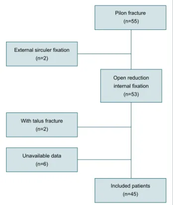
A retrospective cohort study examination was carried out with 55 patients who were operated on because of pilon frac-tures in our clinic between January 2010 and December 2016 (Fig. 1). Patients treated with ORIF and followed up for at least two years were included in this study. A total of 10 (18.2%) patients were excluded from this study: two (3.6%) patients because external circular fixation was used, two (3.6%) pa-tients because adjunct foot fractures were treated, and six (10.9%) patients because of incomplete data. The patients
were grouped according to age, gender, osteoporosis, pre-operative waiting time, postpre-operative follow-up time, Ruedi Allgower classification, AO classification, concomitant later-al mlater-alleolar fracture, Ovadia Bell criteria,[10] plate placement evaluation and statistical distribution. The 45 (81.8%) patients treated with ORIF were evaluated with AOFAS and SF-12 ac-cording to a total of 10 categories acac-cording to demographic data, fracture classification and surgical technique.[11–14] Foot scoring of the patient was made with AOFAS and quality of life was evaluated with the SF-12. The AOFAS values were grouped in four groups as >85, 85-70, 70-50 and <50. Apart from the functional values, a record was made for each pa-tient of complications, including infection, soft-tissue defects, malalignment, non-union, arthrosis and Sudeck atrophy.
Preoperative Evaluation
In the Emergency Department, a splint was applied to the patients, correcting malalignment and providing joint compat-ibility. Preoperatively, a cold compress, non-steroid anti-in-flammatory drugs (NSAID) and antibiotic treatment were applied. Soft tissue was carefully evaluated preoperatively with checks made within the splint three times a day. In cases with a positive “wrinkle test” and the healing of hemorrhagic bullae, surgery was planned with three surgeons’ approval. In two of the current series of patients, skeletal traction was applied from the calcaneus as stability was not obtained with the splint (Fig. 2).
Surgical Technique
Each patient was positioned supine on a radiolucent oper-ating table. A medial or lateral incision was planned accord-ing to the fracture wound. In cases with a lateral malleolar fracture, lateral malleolar reduction and fixation were applied first. The joint surface restoration was performed first, then metaphyseal diaphyseal bone reduction. Full joint reduction and <5˚ coronal sagittal, alignment were accepted. When ap-plying medial or lateral plating, an additional fragment-specific screw was applied when necessary (Fig. 3). Soft tissue layers were sutured appropriately.
External sirculer fixation (n=2)
With talus fracture (n=2) Unavailable data (n=6) Pilon fracture (n=55) Included patients (n=45) Open reduction internal fixation (n=53)
Follow-up
Soft tissue follow-up was made daily postoperatively, and when it was assured, the patient was discharged. A splint was applied for two weeks for soft tissue healing. At the end of the second week, after removal of the splint, joint range of movement exercises were started together with Vitamin C therapy. Partial weight-bearing was permitted at six weeks postoperatively and full weight-bearing at eight weeks.
Ethical Approval
Our study was ethically approved by the Ethical Committee of our hospital and also the University of Health Sciences Keçiören Health Practice and Research Center.
Statistical Analysis
Data obtained in the study were analyzed statistically with SPSS vn 22 software and examined in a 95% confidence in-terval. Qualitative data were stated as frequency distribution and quantitative data as mean, minimum and maximum val-ues. The relationships between AOFAS and variables were analyzed using the Chi-square test. To determine differences in SFC-12 PCS (physical) variables, the analysis was applied using ANOVA tests as normal distribution was shown. As the SF-12 MCS (mental) points did not show normal distri-bution, analyses were applied with the Mann-Whitney and Kruskal Wallis tests. Statistical significance was defined at the 5% (p≤0.05) level.
RESULTS
The cohort consisted of 29 (64.4%) males and 16 (35.6%) female with a mean age of 44.7 years (range 18–77 years). The mean follow-up period was 3.7 years (range 2 to 7). The time from trauma to surgery was mean 4.5 days (range 1 to 16). A preoperative wait of < 7 days was determined in 70.5% of cases and ≥7 days in 29.5%. Patient data are shown with frequencies and percentages in Table 1.
When patients were grouped acording to AOFAS, 40% of patients were evaluated as excellent, 37.7% as good, 17.7% as fair and 4.4% as poor. The SF-12 PCS (physical score) was
Table 1. Demografic data of the patients
Age , n (%) 44.7 (18–77) <50 28 (62.2) >50 17 (37.8) Gender, n (%) Male 29 (64.4%) Female 16 (35.6%) 3.7 (1–7) Follow up (year), n (%) <4 year 26 (57.7) >4 year 19 (42.3) 4.5 (1–16) Preoperatice day, n (%) <7 day 31 (68.8) >7 day 14 (31.2) Ruedi allgower, n (%) Ruedi1 15 (33.3) Ruedi2 18 (40) Ruedi3 12 (26.7) B1 5 (11.1) B2 11 (24.4) B3 6 (13.3) C1 5 (11.1) C2 7 (15.6) C3 7 (15.6)
Ovadia Bells Criteria, n (%)
Anatomic 29 (64.4)
Good 8 (17.7)
Moderate 7 (15.5)
Poor 1 (2.2)
Lateral malleol fracture, n (%)
Yes 25 (55.5) No 20 (44.5) Plate fixation, n (%) Medial 35 (77.7) Lateral 10 (33.3) AOFAS*, n (%) >85 18 (40) 70–85 17 (37.7) 70–50 8 (17.7) 50–30 2 (4.4) SF12-PCS (Physical)* 47.6 (24.2–56.5) SF12-MCS (Mental)* 59.4 (39.9–65.0)
Figure 3. Case example of medial plate fixation (a) and lateral plate fixation (b).
(a)
mean 47.6 (range, 24.2–56.5) and the SF-12 MCS (mental) was mean 59.4 (range, 39.9–65.0).
The AOFAS value was determined to fall to <85 when the Ruedi Allgower classification increased (p=0.010), when AO classification increased (BC1, BC2, BC3) (p=0.020), when there was a concomitant lateral malleolar fracture (p=0.028), and when the status was non-anatomic according to the Ova-dia Bell criteria (p=0.031) (Table 2).
Arthrosis was observed in 11 patients (24%) and in these 11, a significant difference was observed from other patients in
re-spect of SF-12 values (p=0.041). Arthodesis was not request-ed as treatment in any of the patients with arthrosis (Table 3).
Table 2. Analysis of the relationship between AOFAS and variables AOFAS p >85 <85 Age, n (%) <50 10 (37) 17 (63) 0.784 >50 7 (41.2) 10 (58.8) Gender, n (%) Male 8 (28.6) 20 (71.4) 0.068 Female 9 (56.3) 7 (43.8) Follow up (year), n (%) <4 year 12 (46.2) 14 (53.8) 0.436 >4 year 5 (29.4) 12 (70.6) Preoperative day, n (%) <7 day 13 (41.9) 18 (58.1) 0.437 >7 day 4 (33.3) 8 (66.7) Ruedi allgower, n (%) Ruedi1 10 (66.7) 5 (33.3) 0.010* Ruedi2 6 (33.3) 12 (66.7) Ruedi3 1 (9.1) 10 (90.9) AO clasification, n (%) B1-C1 7 (70) 3 (30) 0.020* B2-C2 6 (33.3) 12 (66.7) B3-C3 1 (8.3) 11 (91.7) Intact fibula, n (%) No 11 (57.9) 8 (42.1) 0.028* Yes 6 (25) 18 (75)
Ovadia Bells Criteria, n (%)
Anatomic 15 (51.7) 14 (48.3) 0.031* Other 2 (13.3) 13 (86.7)
Plate fixation, n (%)
Medial 15 (42.9) 20 (57.1) 0.304 Lateral 2 (25) 6 (75)
*p<0.05. AOFS: American Orthopedic Foot and Ankle Society.
Table 3. Analyse of the relationship between SF12-PCS and variables SF12-PCS (Physical) p Age <50 48.08±6.59 0.593 >50 46.82±9.04 Gender Male 47.15±6.22 0.640 Female 48.42±9.65 Follow up (year) <4 year 47.5±8.51 0.907 >4 year 47.78±6.35 Preoperaative time (day)
<7 day 48.52±6.71 0.229 >7 day 45.46±9.41 Ruedi Allgower Ruedi1 51.59±5.16 0.018* Ruedi2 46.89±7.01 Ruedi3 43.68±8.82 Intact fibula Yes 49.63±6.01 0.119 No 46±8.43
Ovadia Bells Criteria
Anatomic 49.81±6.45 0.012* Good 41.43±9.75 Moderate/poor 45.78±5.3 Plate fixatio Medial 47.95±6.94 0.765 Lateral 47.58±6.78 B1-C1 50.84±5.45 AO clasification B2-C2 47.22±6.86 0.059 B3-C3 43.47±8.47 AOFAS >85 53.94±3.83 0.000* 70-85 46.82±4.42 <70 38.75±6.96 Infection Superfisial/deep 40.20±4.29 No 49.26±7.23 0.002* Artrosis Yes 43.92±6.56 No 48.79±7.53 0.041*
Infection was observed in eight patients (17%), all of whom sustained the fracture in high-energy trauma (Ruedi 2: n=2, Ruedi 3: n=6). Patients with superficial infection were treated with oral antibiotics and for those with deep infection, the implant was removed and debridement was treated. Delayed union was observed in one of the patients with deep infec-tion. Arthrosis developed in three patients with superficial infection and in one with deep infection. A significant differ-ence was determined between the patients with infection and the other patients regarding the long-term scores (p=0.002). Sudeck’s atrophy was observed in four patients (9%), of whom three patients had C3 fractures and one patient had a C2 fracture. Treatment was applied with a physical therapy program.
Malalignment was observed in five patients (11%), all of whom had a concomitant lateral malleolar fracture.
Full union of the fractures was obtained at a high rate (95.5%). In two patients with delayed union, one patient had a B2 ture with deep infection and the other patient had a C1 frac-ture with superficial infection.
DISCUSSION
Pilon fractures are always accepted as problematic fractures by orthopaedic surgeons because they are injuries of large di-mensions with damage to the cartilage joint surface and soft tissue oedema.[15] High complication rates are seen, which may include skin necrosis and infection in the early period, and non-union and arthrosis in the long-term.[16–19] Howev-er, they are infrequently seen fractures constituting approxi-mately 1% of all lower extremity fracture.[2]
In the current study, the severity of the injury was deter-mined as Ruedi 1 in 33.3%, Ruedi 2 in 40.0% and Ruedi 3 in 26.7%. There were seen to be similar rates of low-energy and high-energy traumas.
The time from trauma to surgery was mean 4.5 days (range, 1–16 days) and this period was seen to be prolonged in cases evaluated as Ruedi 3. The approach of early surgery before soft tissue swelling has formed has been abandoned as some studies have reported worse results from surgery performed in the first five days.[20] This period varies according to the severity of the injury. When the soft tissue status has been ensured, surgery should be applied.
Successful operations with ORIF came to prominence with a study by Rüedi and Allgöwer[21] in 1969, which reported 74% success in 84 patients treated with ORIF. Anatomic reduction according to AO rules and early mobilization were applied. The weakest point of that study was that the majority of pa-tients had low-energy skiing injuries. In a second series by the same team, the majority of cases arose from by high-energy
injuries (traffic accidents). The same level of success could not be achieved with a good result obtained in 69%. Other studies conducted according to the mechanism of injury have reported that the success rate has fallen to 50%.[22]
When the patients in the current study were grouped acord-ing to AOFAS, 40% of the patients were evaluated as excellent, 37.7% as good, 17.7% as fair and 4.4% as poor. Rubio-Suarez et al.[19] treated 90 patients with ORIF and reported 30.5% as excellent results (>85 AOFAS), 46.7% as good, 13.1% as fair and 9.7% as poor. In that study, Rubio-Suarez et al.[19] exam-ined complications and emphasized the risk of medial plating. When the patients with AOFAS <85 (60%) were examined in the current study, they were seen to be those with Ruedi type 3 (p=0.010) and AO type C injuries (p=0.020), those with lateral malleolar fractures (p=0.028) and those who were non-anatomic according to Ovadia Bell criteria (p=0.031). No difference was seen regarding age, gender, preoperative waiting time, follow-up period, and plate placement. Infection and arthrosis were determined to decrease AOFAS values. Ilizarov fixation was applied to two patients because of both metaphyseal and tibial defect, and these cases were excluded from this study. Single-stage ORIF was applied to the pilon fractures externally. Rather than two-stage surgery the cases were transferred to the ward with a splint. In two patients (4.4%) where stability could not be achieved with a splint, skeletal traction was applied from the calcaneus and they were followed up in the ward.
When the single-stage ORIF of the current study was com-pared with literature, the results of single-stage were found to be the same as those for two-stage.[23] In a similar study that compared three methods, low AOFAS scores were ob-tained with external fixators.[24] However, the use of exter-nal fixators in more severe injuries could explain the worse results of external fixators in these studies. In a comparison of limited ORIF and external fixation with ORIF, no shortfall was found in the limited approach.[25] In Ruedi Allgower type 1 and type 2 injuries, results have been shown to be better irrespective of the treatment, and treatment problems have not yet been resolved in Ruedi type 3 injuries.[26] In the light of these data, the effects of classic ORIF treatment were ex-amined in the current study rather than the surgical methods. The SF-12 PCS value in the current study was found to be mean 47.6 (range, 24.2–56.5), which was lower than the SF-12 value of the Turkish population in general. However, the SF-12 MCS value of mean 59.4 (range, 39.9–65.0) was observed to be similar to that of the general population. When the SF-12 PCS was examined, the most significant relationship was seen to be in parallel with the AOFAS values (p=0.000). Cutil-las-Ybarra et al.[9] examined short-term quality of life with the SF36 and reported that patients were affected both physically and psychosocially compared to the general population. In
the current study, foot function in the long-term was found to have no mental effect (SF-12 MCS evaluation). The SF-12 PCS was seen to be most affected by foot functions. In a similar study in Australia of a 12-month examination of pilon fractures, lower SF-12 PCS values were obtained.[8]
A concomitant lateral malleolar fracture was observed to re-duce the functional results in the current study (p=0.028). In a study by Liangjun et al.,[27] higher functional results were obtained in pilon fractures with an intact lateral malleolus. Medial plate placement has been said to be associated with skin necrosis, more severe arthrosis and non-union.7 In the current study, flap was applied to one patient with medial plate placement. In addition, of the eight patients who developed infection, there was observed to be medial plate osteosynthe-sis in seven patients. However, no difference was determined regarding arthrosis and non-union. Although there was no sta-tistically significant difference, there was observed to be more arthrosis in cases with lateral plate placement (lateral plate osteosynthesis 44.4%, medial plate osteosynthesis 20%). In a study by Rubio-Suarez et al.,[19] ORIF complications were examined in a series of 137 patients. Skin necrosis was deter-mined in 15%, non-union in 16%, arthrosis in 13% and infec-tion in 8%.
In the current study, infection was observed at the rate of 17.7%. In two (4%) patients with deep infection, the implant was removed and debridement was performed, and the only risk factor was determined to be Ruedi classification (p=0.044). As the severity of the wound increases, care must be taken in respect of infection. Delayed union was seen in two (4.4%) patients. The two patients with non-union were also determined to have infection (1 superficial, 1 deep). Arthrosis was observed in 11 (24.4%) patients of the current study. In Ruedi type 3, AO type C injuries, patients with con-comitant lateral malleolar fracture (p=0.028) and those with non-anatomic fixation according to Ovadia Bell (p=0.031), ar-throsis was observed at a statistically signifcantly higher rate. Patients with arthrosis were seen to have lower AOFAS and SF-12 scores. Arthrodesis was not requested for any of these patients. Similarly, Bonar et al.[28] reported that arthodesis was not required in patients with reduction and malalignment.
Conclusion
The results of this study demonstrated that the reasons for ORIF failure in tibia pilon fractures were Ruedi 3, lateral mal-leolar fracture, and non-anatomic reduction. Failure in foot functions has a direct effect on the quality of life in both the short and mid-term.
Ethics Committee Approval: Approved by the local eth-ics committee.
Peer-review: Internally peer-reviewed.
Authorship Contributions: Concept: Y.U.Y; Design: Y.U.Y., B.H.; Supervision: Y.U.Y, B.H.; Fundings: Y.U.Y., A.K.; Materials: Y.U.Y., A.K.; Data: Y.U.Y., A.K.; Analysis: Y.U.Y., İ.D; Literature search: Y.U.Y., M.C.O; Writing: Y.U.Y., M.C.O.; Critical revision: Y.U.Y., M.A.
Conflict of Interest: None declared.
Financial Disclosure: The authors declared that this study has received no financial support.
REFERENCES
1. Tornetta P 3rd, Weiner L, Bergman M, Watnik N, Steuer J, Kelley M, et al. Pilon fractures: treatment with combined internal and external fixa-tion. J Orthop Trauma 1993;7:489−96. [CrossRef ]
2. Bourne RB, Rorabeck CH, Macnab J. Intra-articular fractures of the dis-tal tibia: the pilon fracture. J Trauma 1983;23:591−6. [CrossRef ]
3. Chowdhry M, Porter K. The pilon fracture. Trauma 2010;12:89−103. 4. Calhoun JH, Li F, Ledbetter BR, Viegas SF. A comprehensive study of
pressure distribution in the ankle joint with inversion and eversion. Foot Ankle Int 1994;15:125−33. [CrossRef ]
5. Borrelli J Jr, Ellis E. Pilon fractures: assessment and treatment. Orthop Clin North Am 2002;33:231−x. [CrossRef ]
6. Rüedi T. Fractures of the lower end of the tibia into the ankle joint: results 9 years after open reduction and internal fixation. Injury 1973;5:130−4. 7. Carbonell-Escobar R, Rubio-Suarez JC, Ibarzabal-Gil A,
Rodriguez-Merchan EC. Analysis of the variables affecting outcome in fractures of the tibial pilon treated by open reduction and internal fixation. J Clin Orthop Trauma 2017;8:332−8. [CrossRef ]
8. Bonato LJ, Edwards ER, Gosling CM, Hau R, Hofstee DJ, Shuen A, et al. Patient reported health related quality of life early outcomes at 12 months after surgically managed tibial plafond fracture. Injury 2017;48:946−53. 9. Cutillas-Ybarra MB, Lizaur-Utrilla A, Lopez-Prats FA. Prognostic fac-tors of health-related quality of life in patients after tibial plafond frac-ture. A pilot study. Injury 2015;46:2253−7. [CrossRef ]
10. Ovadia DN, Beals RK. Fractures of the tibial plafond. J Bone Joint Surg Am 1986;68:543−51. [CrossRef ]
11. Kitaoka HB, Alexander IJ, Adelaar RS, Nunley JA, Myerson MS, San-ders M. Clinical rating systems for the ankle-hindfoot, midfoot, hallux, and lesser toes. Foot Ankle Int 1994;15:349−53. [CrossRef ]
12. Ibrahim T, Beiri A, Azzabi M, Best AJ, Taylor GJ, Menon DK. Reliability and validity of the subjective component of the American Orthopaedic Foot and Ankle Society clinical rating scales. J Foot Ankle Surg 2007;46:65−74. 13. Ware J Jr, Kosinski M, Keller SD. A 12-Item Short-Form Health Survey: construction of scales and preliminary tests of reliability and validity. Med Care 1996;34:220−33.
14. Orthotolkit. SF-12 – OrthoToolKit. Available at: http://orthotoolkit. com/sf-12.
15. Mast JW, Spiegel PG, Pappas JN. Fractures of the tibial pilon. Clin Orthop Relat Res 1988;230:68−82. [CrossRef ]
16. McFerran MA, Smith SW, Boulas HJ, Schwartz HS. Complications encountered in the treatment of pilon fractures. J Orthop Trauma 1992;6:195−200. [CrossRef ]
17. D’Alleyrand JC, Manson TT, Dancy L, Castillo RC, Bertumen JB, Meskey T, et al. Is time to flap coverage of open tibial fractures an in-dependent predictor of flap-related complications?. J Orthop Trauma 2014;28:288−93. [CrossRef ]
18. Lomax A, Singh A, N Jane M, C Senthil K. Complications and early re-sults after operative fixation of 68 pilon fractures of the distal tibia. Scott Med J 2015;60:79−84. [CrossRef ]
19. Rubio-Suarez JC, Carbonell-Escobar R, Rodriguez-Merchan EC, Ibarzabal-Gil A, Gil-Garay E. Fractures of the tibial pilon treated by open reduction and internal fixation (locking compression plate-less invasive stabilising system): Complications and sequelae. Injury 2018;49:S60−4. [CrossRef ]
20. Trumble TE, Benirschke SK, Vedder NB. Use of radial forearm flaps to treat complications of closed pilon fractures. J Orthop Trauma 1992;6:358−65. [CrossRef ]
21. Rüedi TP, Allgöwer M. The operative treatment of intra-articular fractures of the lower end of the tibia. Clin Orthop Relat Res 1979;(138):105−10. 22. Kellam JF, Waddell JP. Fractures of the distal tibial metaphysis with
intra-articular extension--the distal tibial explosion fracture. J Trauma 1979;19:593−601. [CrossRef ]
23. Minator Sajjadi M, Ebrahimpour A, Okhovatpour MA, Karimi A, Zandi R, Sharifzadeh A. The Outcomes of Pilon Fracture Treatment:
Prima-ry Open Reduction and Internal Fixation Versus Two-stage Approach. Arch Bone Jt Surg 2018;6:412−9.
24. Biz C, Angelini A, Zamperetti M, Marzotto F, Sperotto SP, Carniel D, et al. Medium-Long-Term Radiographic and Clinical Outcomes after Surgical Treatment of Intra-Articular Tibial Pilon Fractures by Three Different Techniques. Biomed Res Int 2018;2018:6054021. [CrossRef ]
25. Zhang SB, Zhang YB, Wang SH, Zhang H, Liu P, Zhang W, et al. Clin-ical efficacy and safety of limited internal fixation combined with external fixation for Pilon fracture: A systematic review and meta-analysis. Chin J Traumatol 2017;20:94−8. [CrossRef ]
26. Kemal Aktuğlu, Nadir Özkayın. Surgical modalities in tibial pilon frac-tures TOTBİD Derg 2013;12:142−52. [CrossRef ]
27. Liangjun J, Qiang Z, Hang L, Zhijun P. Injury mechanism, fracture characteristics and clinical treatment of pilon fracture with intact fibu-la-A retrospective study of 23 pilon fractures. J Clin Orthop Trauma 2017;8:S9−15. [CrossRef ]
28. Bonar SK, Marsh JL. Tibial Plafond Fractures: Changing Principles of Treatment. J Am Acad Orthop Surg 1994;2:297−305. [CrossRef ]
OLGU SUNUMU
Pilon kırıkları sonrası ayak fonksiyonları ve hayat kalitesinde orta dönem etkenler
Dr. Yüksel Uğur Yaradılmış,1 Dr. Mustafa Caner Okkaoğlu,1 Dr. Alparslan Kılıç,1
Dr. Bahtiyar Haberal,2 Dr. İsmail Demirkale,1 Dr. Murat Altay1
1Sağlık Bilimleri Üniversitesi, Keçiören Eğitim ve Araştırma Hastanesi, Ortopedi ve Travmatoloji Anabilim Dalı, Ankara 2Başkent Üniversitesi Tıp Fakültesi, Ortopedi ve Travmatoloji Anabilim Dalı, Ankara
AMAÇ: Pilon kırıkları az görülmesine rağmen tedavisi zor olması nedeniyle ortopedik cerrahlar tarafından önemsenmiştir. Değişen cerrahi yöntem-lere rağmen kötü sonuç ve yüksek komplikasyonlar görülmektedir. Bu çalışmanın amacı, pilon kırığı nedeniyle açık redüksiyon ve iç fiksasyon (ORIF) yaptığımız hastalarda ayak fonksiyonları ve hayat kalitesini etkileyen değişkenleri incelemek ve komplikasyonlarla ilişkilendirmek.
GEREÇ VE YÖNTEM: Kliniğimizde Ocak 2010–Aralık 2016 tarihleri arasında, pilon kırığı nedeniyle ORIF uygulanan 45 hasta demografik verileri, kırık sınıflaması ve cerrahi tekniğe göre toplam 10 kategoriye göre AOFAS ve SF-12 ile değerlendirildi. Hastalar fonksiyonel değerler haricinde komplikasyonlar olarak enfeksiyon, yumuşak doku defekti, dizilim bozukluğu, kaynamama, artroz ve Sudeck atrofisi açısından da hastaların kayıtları incelendi. AOFAS 85 altı olan ve düşük SF12 olan hastaların değişkenleri incelendi ve komplikasyonlarla ilişkisi değerlendirildi.
BULGULAR: Ortalama takip süresi 3.7 yıl olup (2–7 yıl). AOFAS değerlerine bakıldığında, Ruedi-Allgower sınıflaması arttığında (p=0.010), AO sınıflaması arttığında (p=0.020), eşlik eden lateral malleol kırığında (p=0.028) ve Ovadia-Bell kriterleri’ne göre non-anatomik olması durumunda (p=0.031) AOFAS değerleri 85’in altına düşmektedir. SF-12 PCS değerlerine bakıldığında, Ruedi-Allgower sınıflaması arttığında (p=0.018), Ovadia-Bell kriterleri’ne göre non-anatomik olması durumunda (p=0.012) SF-12 PCS değerlerinde düşme gözlendi. SF-12 PCS ile AOFAS değerlerinde korelasyon gözlendi (p=0.000).
TARTIŞMA: Tibia pilon kırıklarında ORIF ile başarısızlık nedeni Ruedi 3, lateral malleol kırığı, cerrahi anatomik olmayan redüksiyon olarak bulundu. Ayak fonksiyonlarındaki başarısızlık hem erken dönem hem de orta dönemde hayat kalitesini doğrudan etkilemektedir.
Anahtar sözcükler: Açık cerrahi; artroz, enfeksiyon; hayat kalitesi; pilon kırıkları.
Ulus Travma Acil Cerrahi Derg 2020;26(5):798-804 doi: 10.14744/tjtes.2020.85601