791
Araştırma/Research
Analysis of surgical treatment and outcomes of complicated intraabdominal infections
Sabri Özdaş1
, Safa Önel2, Mehmet Şirik3,
1 Adıyaman Üniversitesi Eğitim ve Araştırma Hastanesi Genel Cerrahi Kliniği 2
Adana Şehir hastanesi Genel Cerrahi Kliniği
3Adıyaman Üniversitesi Eğitim ve Araştırma Hastanesi Radyoloji Bölümü ABSTRACT
Aim: Despite recent advances in treatment, the mortality rate due to complicated intra-abdominal infection (cIAI) still remains high. We aimed to present the clinical characteristics and outcome of patients with cIAI who were treated surgically.
Methods: Seventy-two patients (28 female, 44 male; mean age 44.2 years) who were diagnosed with cIAI and underwent surgical treatment between May 2012 and May 2013 included in this study. The clinical characteristics of patients, origin of infection, microbiology of peritoneal fluid, and outcome of surgery were also recorded.
Results: Sixty-one patients (84.7%) acquired cIAI from community, whereas 11 patients (15.3%) from nosocomial sources. Twenty-four patients (33.3%) had disseminated peritonitis, and the remaining 48 patients (66.6%) had local peritoneal infection or abscesses. Infected fluid from peritoneal cavity was positive for E. coli in 27 patients (38%), and for B. fragilis in 17 patients (24%). Fourteen patients (19.4%) were followed up in the intensive care unit. Overall postoperative mortality rate was 8.3% (6 patients).
Conclusion: The cIAI is an emergency medical condition with high mortality rate. In addition to early diagnosis and preventive measures, advanced surgical techniques and postoperative care are critical to obtain better outcomes.
Keywords: Complicated intra-abdominal infection, Peritonitis, Antibiotic therapy
DOI: 10.30569/adiyamansaglik. 415553
Yazışmadan Sorumlu Yazar Mehmet ŞİRİK
Adıyaman Üniversi Tıp Fakültesi Radyoloji Anabilim Dalı.
Tel : +90 05058303526
Email: [email protected]
Geliş Tarihi: 16.04.2018
792
Komplike İntraabdominal Enfeksiyonların Cerrahi Tedavi ve Sonuçlarının Analizi
ÖZET
Amaç: Bu çalışmamızda kliniğimize akut karın bulguları ile gelen ve opere edilen komplike
karın içi enfeksiyonlu hastaların pre-postop takip ve tedavilerinin mortalite ve morbiditelerini izlemsel olarak ortaya koyulmasını amaçladık.
Yöntemler: Çalışmamız kliniğimizde Mayıs 2012 ile Mayıs 2013 tarihleri arasında opere komplike karın içi enfeksiyonlu hastaların verilerini içermektedir. Bu veriler şunlardır: (i) hasta ve hastalık özellikleri, yani, demografik veriler, enfeksiyon tipi, şiddet kriteri, ameliyat öncesinde 7 gün uygulanan küratif antibiyotik tedavisi; (ii) enfeksiyon kökeni ve uygulanan cerrahi işlemler (iii) mikrobiyolojik veriler ve bakteriyel izolatların antibiyotik duyarlılıklarıdır. Hastaların hastanede toplam kalış süreleri, post-op yoğun bakımda takip edilen hasta sayısı dikkate ve bu verilerin istatiksel analizleri anlatılmıştır.
Bulgular: Çalışmaya 28( % 38,8)’i kadın, 44( % 61,2 )’ü erkek olmak üzere 72 hasta dahil edildi. Hastalar radyolojik olarak ayakta direk batın grafisi(ADBG), Ultrasonografi(USG) ve karın Bilgisayarlı tomografi(BT) ile teyit edilmiş, klinik tam parametreleri ortaya konulmuş ve gelen hastaların % 100’de Karın ağrısı ve karında hassasiyet ön planda olmuştur. Aynı zamanda risk faktörleri skalası ortaya konulmuştur. 24(% 33,3) hastada yaygın periton enfeksiyonu gözlemlenirken 48(% 66,6) hastada lokal periton enfeksiyonu yada apseler bildirilmiştir. 72(% 100) hastadan kültür amaçlı batın içi enfekte sıvı alındı. 14 hasta (% 19,4) durumu kritik olarak kabul edildi ve yoğun bakım ünitesinde takip edildi. Hastaların 66(% 91,6) ilk geliş beyaz küreleri yüksekti, 4(% 5,5) hastada aldıkları kemoterapi nedeniyle beyaz küre’leri normalin altındaydı.
Sonuç: Komplike İntraabdominal Enfeksiyonlar mortalite oranının yüksek olduğu acil bir tıbbi durumdur. Erken tanı ve önleyici tedbirlerin yanı sıra, ileri cerrahi teknikler ve postoperatif bakım daha iyi sonuçların elde edilmesi için kritik öneme sahiptir.
793
Introduction
The complicated intra-abdominal infection (cIAI) is defined as the bacterial invasion of otherwise sterile peritoneal environment through perforation of hollow viscus, such a appendicitis or colonic diverticulitis, or by other irritants, such as bile from a perforated gall bladder or lacerated liver, gastric acid from a perforated ulcer, or an infected fallopian tube or ruptured ovarian cyst in women (1, 2). The cIAI can be localized or extend to other organs causing diffuse peritonitis. In some case it can even lead to sepsis, acute respiratory distress syndrome, or multiple organ failure. Despite the progress in diagnostic methods and surgical and antimicrobial treatments, cIAI is still associated with high mortality rates ranging from 10% to 50% depending on its severity, extent, and presence of concomitant conditions (3-8).
Patients with cIAI present with various clinical characteristics and severity of disease, which makes the diagnosis and treatment difficult. The common clinical symptoms of cIAI are abdominal pain and tenderness, nausea, diminished intestine sounds, high fever, hypotension and tachycardia. However, diagnosis is based on the radiological imaging studies including abdominal and chest radiography, ultrasonography, and computed tomography (1,2,5,7,9). The white blood cell count less than 4000/mm3 or more than 25 000/mm3 is usually associated with high mortality in cIAI (10). The management of cIAI involves parenteral antimicrobial therapy targeted to pathogens and surgery, which often requires sustained hospital stay with high healthcare cost (4,7-9,11). The surgical treatment either can be a single operation to prevent or treat the source of infection, or aggressive surgical techniques to decompress intra-abdominal pressure, especially in case of severe forms of peritonitis (8).
Although, cIAI is the most important intra-abdominal condition requiring emergency surgery worldwide, its clinics and management show geographical differences depending on population-specific conditions and medical facilities (6). Therefore, in order to define health
794
policies against cIAI at national level, the clinical, microbiological, and surgical treatment profiles of cIAIs should be described at local level.
In this study, we aimed to present the clinical characteristics, microbiology and postoperative outcome of patients with cIAI who were diagnosed and surgically treated in our clinic.
MATERIALS AND METHODS
In this descriptive case-series, we studied patients admitted to general surgery clinics of our hospital for acute abdominal pain and tenderness between May 2012 and May 2013. Seventy-two patients (28 female, 44 male; mean age 44.2 years; age range 15-87 years) with post-operative or post-traumatic community-acquired or nosocomial cIAI due to appendicitis, gastroduodenal perforation, small intestinal perforation, colon perforation (iatrogenic, diverticulum, or foreign body), gall bladder perforation, anastomotic leakage or other reasons were included in the study. Exclusion criteria were pancreatitis or primary peritonitis.
The study protocol was approved by the Institutional Ethics Committee and conducted according to the Declaration of Helsinki. All patients signed the informed consent form before participation in the study.
Study procedures
Data on demographics, admission symptoms, surgery indications, operation history, origin of infection, and prior use of antibiotics and other drugs within last 7 days were recorded. Preoperative total blood count, routine biochemical tests, chest radiography and electrocardiography were used to identify comorbidities of the patients. Clinical response was evaluated as the response to clinical management, postoperative need for intensive care, and death attributable to cIAIs.
795
Surgical procedure and pathological diagnosis
Based on medical examination, and laboratory and radiological results at the admission, surgical technique—laparotomy or open surgery—was decided. For patients with appendicitis, appendectomy was performed by the right paramedian or lower abdominal incision using McBurney's technique. All the other patients were operated by upper, lower or midline incisions. The peptic ulcer and small intestinal perforations were primarily sutured. Anastomotic stoma was performed in patients with colon cancer perforation. A drain was placed for elderly patients, those with dirty abdominal fluid or high risk of intra-abdominal abscess.
Intra-abdominal fluid sample was collected during the surgery for microbiological analysis. Routine culture tests were performed for the identification of pathogens, and antibiotic therapy was started accordingly.
Statistical analysis
Statistical analysis was performed by the SPSS software package for Windows (Statistical Package for Social Sciences, version 12.0, SPSS Inc., Chicago, Illinois, USA). Categorical variables were summarized as numbers and percentages and quantitative variables as mean±standard deviation (SD).
RESULTS
Clinical characteristics of the study population
A majority of patients (n=61, 84.7%) acquired infection from community, whereas 11 patients (15.3%) from nosocomial sources. The most common cause of cIAIs was appendicitis (n=27, 37.5%), followed by gastroduodenal perforation (n=9, 12.4%) and cholecystitis (n=9, 12.4%)
796
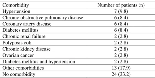
(Table 1). Disseminated peritonitis was observed in 24 patients (33.3%), while local peritoneal infection or abscesses was noted in 48 patients (66.6%). The patients with appendicitis were younger than other diagnoses of cIAIs. The most common comorbidity was hypertension observed in 7 patients (9.8%) followed by with chronic obstructive pulmonary disease, coronary artery disease, and diabetes mellitus (n=6, 8.4% for each) (Table 2). Twenty-four patients (33.2%) had no comorbidities.
Preoperative white blood cell count was above 12 000/mm3 in 66 patients (91.6%) and below 4 000/mm3 in 4 patients (5.5%). High preoperative white blood cell counts returned to normal in 42 patients (58.3%) after surgery.
Table 1. Diagnoses of cIAI with respect to underlying cause.
Cause of cIAI Number of patients (%)
Appendicitis 27 (37.5) Gastroduodenal perforation 9 (12.4) Cholecystitis 9 (12.4) Iatrogenic perforation 7 (9.8) Post-operative perforation 5 (6.9) Post-traumatic perforation 4 (5.5) Other reasons 4 (5.5) Diverticulum perforation 3 (4.3)
Small intestinal perforation 2 (2.8) Post-colonoscopy perforation 2 (2.8)
Total 72 (100)
Table 2. Distribution of comorbidities among the study patients.
Comorbidity Number of patients (n)
Hypertension 7 (9.8)
Chronic obstructive pulmonary disease 6 (8.4)
Coronary artery disease 6 (8.4)
Diabetes mellitus 6 (8.4)
Chronic renal failure 2 (2.8)
Polyposis coli 2 (2.8)
Chronic kidney disease 2 (2.8)
Ovarian cancer 2 (2.8)
Diabetes mellitus and hypertension 2 (2.8)
Other comorbidities 13 (17.9)
797
Surgical treatment
All patients were treated with surgery. The most common surgical procedure for appendicitis was appendectomy by McBurney's technique (24/27, 88.9%), lower abdominal incision (2/27, 7.4%), and right paramedian incision (1/27, 3.7%). Seventeen patients (62.9%) were required appendectomy due to localized infection and abscesses, 8 patients (29.6%) due to generalized peritonitis, and 2 patients (7.4%) due to complicated appendicitis. Primary suturing was the main surgical technique used for the treatment of patients with gastroduodenal perforation (7/9, 77.7%). Eight patients with acute cholecystitis was operated by open cholecystectomy (8/9, 88.8%), the other 1 patient was operated by laparoscopic cholecystectomy (12.2%). All perforated colon carcinoma patients were treated with open laparotomy. The left colon perforation was treated with open Hartmann’s resection (5/7, 71.4%), and right colon
perforation with enlarged right hemicolectomy and protective ileostomy (2/7, 27.6%). Hartmann operation was the surgical procedure used for the treatment of cIAI patients due to diverticulitis (n=3, 100%). Four patients with post-traumatic perforation were operated by open abdominal surgery for the treatment of blunt abdominal trauma and acute abdomen. There were only 2 patients surgically treated for small intestinal perforation; 1 patient operated by small intestine segmental resection and terminal ileostomy due to diffused peritonitis with necrotic microperforations, and the other patient by resection end-to-end jejuno-ileal anastomosis due to tumor perforation in 70 cm distal of the ligament of Treitz. Five patients who had cIAI due to postoperative perforation were treated with percutaneous drainage. One of two post-colonoscopy cIAI cases was surgically treated by Hartmann’s procedure (1/2, 50%), and the other one by sigmoid loop colostomy (1/2, 50%). Four patients who developed cIAIs due to gunshot injuries were treated as war injuries.
798
Microbiological results
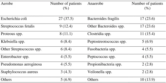
Microbiological analysis of peritoneal fluid identified several aerobic and anaerobic bacteria as pathogens of cIAIs (Table 3). Escherichia coli was the most common bacterial cause of cIAI among aerobic microorganisms as detected in 27 patients (37.5%), followed by Streptococcus fetalis in 9 patients (12.4%). Proteous, Klebsiella, Streptococcus species, and Enterobacter species, Pseudomonas aeruginosa, and Staphylococcus aureus were among the aerobic bacteria identified in infected intra-abdominal fluid. The most common anaerobic microorganisms were Bacteriodes fragilis and other Bacteroides species (n=17, 23.6% for both). The most common microorganisms detected in patients with appendicitis were E. coli and B. fragilis. Similarly, E. coli was the most common cause of intra-abdominal infection in patients with gastroduodenal perforation and acute cholecystitis.
Table 3. Microbiological analysis of intra-abdominal fluid samples of patients.
Aerobe Number of patients
(%)
Anaerobe Number of patients (%)
Escherichia coli 27 (37.5) Bacteroides fragilis 17 (23.6) Streptococcus fetalis 9 (12.4) Other Bacteroides spp. 17 (23.6)
Proteous spp. 8 (11.1) Clostridia spp. 11 (15.4)
Klebsiella spp. 6 (8.4) Peptostreotococcus spp. 5 (6.9) Other Streptococcus spp. 6 (8.4) Fusobacteria spp. 4 (5.5)
Enterobacter spp. 4 (5.5) Peptococcus spp. 4 (5.5)
Pseudomonas aeroginosa 4 (5.5) Propionibacteria spp. 2 (2.8)
Staphylococcus aureus 3 (4.3) Veilonella spp. 2 (2.8)
799
Antibiotic therapy
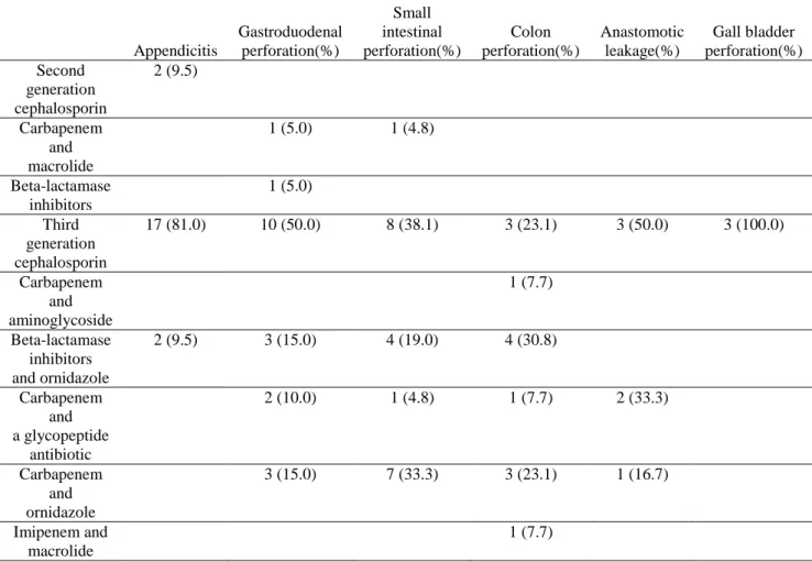
Based on the results of microbiological analyses, antibiotic therapy was given to all patients for a mean of 6.5 days (Table 4). Third generation cephalosporin was the most common antibiotic administered in all diagnostic groups. Beta-lactamase inhibitors with or without ornidazole, and carbapenem with several concomitant antibiotics (macrolide, aminoglycoside, glycopeptides and ornidazole) were used in the treatment of gastroduodenal perforation, small intestinal perforation and colon perforation.
Table 4. Antibiotic therapy given to study patients.
Appendicitis Gastroduodenal perforation(%) Small intestinal perforation(%) Colon perforation(%) Anastomotic leakage(%) Gall bladder perforation(%) Second generation cephalosporin 2 (9.5) Carbapenem and macrolide 1 (5.0) 1 (4.8) Beta-lactamase inhibitors 1 (5.0) Third generation cephalosporin 17 (81.0) 10 (50.0) 8 (38.1) 3 (23.1) 3 (50.0) 3 (100.0) Carbapenem and aminoglycoside 1 (7.7) Beta-lactamase inhibitors and ornidazole 2 (9.5) 3 (15.0) 4 (19.0) 4 (30.8) Carbapenem and a glycopeptide antibiotic 2 (10.0) 1 (4.8) 1 (7.7) 2 (33.3) Carbapenem and ornidazole 3 (15.0) 7 (33.3) 3 (23.1) 1 (16.7) Imipenem and macrolide 1 (7.7)
800
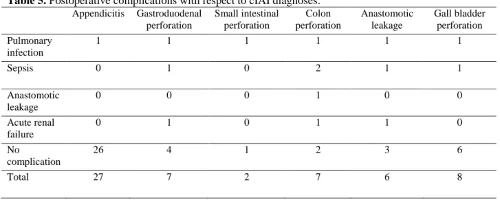
Clinical outcome
Majority of patients did not develop postoperative complications. However, some patients had pulmonary infection, sepsis, anastomotic leakage and acute renal failure postoperatively (Table 5).
Table 5. Postoperative complications with respect to cIAI diagnoses. Appendicitis Gastroduodenal perforation Small intestinal perforation Colon perforation Anastomotic leakage Gall bladder perforation Pulmonary infection 1 1 1 1 1 1 Sepsis 0 1 0 2 1 1 Anastomotic leakage 0 0 0 1 0 0 Acute renal failure 0 1 0 1 1 0 No complication 26 4 1 2 3 6 Total 27 7 2 7 6 8
Fourteen patients (19.4%) with severe sepsis or septic shock were followed up in the intensive care unit. Most of the patients were discharged with recovery (n=66, 91.7%). The overall mortality rate was 8.3% (6 patients)—2 patients with colon perforation, 2 patients with anastomotic leakage, 1 patient with gastroduodenal perforation, and 1 patient with gall bladder perforation.
DISCUSSION
Local epidemiological studies are important to define the causes, treatment approaches and outcomes for efficient diagnosis and management to decrease morbidity and mortality due to cIAIs. Therefore, in this study we aimed to present a series of cIAI patients in Adana province of Turkey with respect to demographics, origin of infection, clinical features, microbiological
801
results, administered antibiotics for the treatment, surgical procedures performed, and clinical outcomes.
The cIAI involves many pathological cases changing from appendicitis to fecal peritonitis. Acute appendicitis was previously defined as the most common cause of cIAIs (1,12). Later, the CIAO Study (“Complicated Intra-Abdominal Infection Observational” Study)
summarized the results of a multicenter investigation performed in 68 medical institutions throughout Europe over the course of a 6-month period (13), and similar to the results of CIAO Study, acute appendicitis was the main intra-abdominal condition leading to cIAI in Adana province of Turkey.
Due to the complex causes of cIAIs, treatment and outcome depends on many factors including the overall health situation of the patient, virulence index of the source pathogen, development and character of the infection, and medical facilities in the hospital (8,11,12,14-17). Antibiotic therapy may be the main treatment option for patients with simple cIAIs; however, acute cases with localized or diffused peritonitis require both surgical intervention and antibiotic therapy for a full recovery (12,16,18). The surgical treatment of cIAIs in this study was mainly performed by the commonly used and usually well-accepted techniques (1,13). The standard approach for the treatment of acute appendicitis is still appendectomy, although several recent studies discuss the requirement of surgery, and advices the treatment with antibiotics (19,22). In the present study, we applied appendectomy in all patients with cIAIs due to acute appendicitis, as it is the routine approach for the surgical treatment of patients with appendicitis in our clinic. The most successful treatment of gastroduodenal, especially peptic ulcer perforation, is accepted to be the urgent surgical intervention (23), and we used primary suturing for the treatment of patients with gastroduodenal perforation. Although the study patients with acute cholecystitis were mainly treated with open
802
cholecystectomy (13), the recommended surgical approach is laparoscopic cholecystectomy when risk factors are accurately evaluated (24,25). We used laparoscopic approach only in 1 patient out of 9 patients. Perforated colon carcinoma patients are among the high-risk group in terms of morbidity and mortality (26). Although no optimal treatment has yet been defined (27), Hartmann’s resection, as performed in the present study, is known to be an effective method for the treatment of perforated colon carcinoma (1,13,28). Antibiotic therapy is the recommended treatment for simple acute diverticulitis (29). However, in this study, we preferred open surgical intervention in the treatment of 3 cIAI patients with acute diverticulitis followed by antibiotic therapy based on the pathogen profile. Similarly, the CIAO Study indicated Hartmann resection as the most frequently performed procedure for the treatment of complicated diverticulitis in Europe (13). The cIAIs developed post-operatively are very hard to treat, and require control of the source of infection by surgery and/or drainage and excessive antibiotic therapy (30). In this study, we treated 5 patients (6.9%) with post-operative cIAIs with different approaches depending on the origin and source of infection, but mainly using the percutaneous drainage as recommended in the literature (31,32).
The causative pathogen of cIAIs depends on the contamination source of sterile peritoneal environment and on the level of disruption of the gastrointestinal tract (1,33). For example, infections derived from stomach, duodenum, and proximal small bowel contain less microorganisms, and mainly enriched in aerobic and facultative bacteria (1,33). The lower gastrointestinal tract, however, contain more bacteria, and therefore, infections derived from distal ileum perforations are enriched in a multiple microorganisms (1,33). Most importantly, aerobic bacteria are more common than anaerobes, with the exception of Bacteroides species(1,33). In this study, microbiological profiles of all patients were analyzed from peritoneal fluid samples taken during surgery. E. coli is accepted as the main causative
803
aerobic microorganism in patients hospitalized for cIAIs (7,17,34), and it was found to be the most common aerobe in the peritoneal fluid samples analyzed in this study (37.5%, Table 3). The CIAO Study reported the common pathogens involved in community-acquired cIAIs as Enterobacteriaceae, Streptococcus species, and certain anaerobes, especially B. fragilis, which are in close agreement with the microbiological profiles obtained from Adana province of Turkey (Table 3).
In the present study, antibiotic therapy of all the patients were initiated after microbiological identification of the pathogens as suggested (35,36), and most patients received single or multiple antibiotic regimens for the treatment of cIAIs. Third-generation cephalosporin was the most commonly used antibiotic in the treatment of cIAI patients in this study, as recently recommended by several studies (3,34,37) for the treatment of community-acquired Enterobacteriaceae in diffuse peritonitis in the presence of other risk factors.
Despite all progress in surgical and antibiotic treatments, the morbidity rate due to cIAIs is still high (4,7,8). The overall mortality rate reported by the CIAO study involving 68 medical institutions in Europe was 7.5% (163/2152 patients) (1). Later, Jean et al. (17) prospectively analyzed cIAI patients in a multicenter study with five countries (Columbia, The Philippines, Portugal, Taiwan and Thaliand) and reported the mortality rate as 11.4% (12/105). The same study indicated the case fatality rate to be significantly lower than previous reports (17). The overall mortality rate in a very recent study performed in southern China with 3233 cIAI patients retrospectively evaluated from 2008-2013 was 1.4%, whereas only 14.3% of the deaths were attributable to cIAI (7). The overall mortality rate in our study was 8.3% (6/72), which is in the range reported by the previous studies (Table 6).
804
Table 6. Clinical outcome and death due to cIAI with respect to cIAI diagnoses.
Discharged (%) Exitus (%) Total
Appendicitis 27 (100.0) 0 27 Gastroduodenal perforation 7 (87.5) 1 (12.5) 8 Small intestinal perforation 2 (100.0) 0 2 Colon perforation 5 (71.4) 2 (28.6) 7 Anastomotic leakage 4 (66.7) 2 (33.3) 6 Gall bladder perforation 7 (87.5) 1 (12.5) 8 Other reasons 14 (100.0) 0 14 Total 66 (91.7) 6 (8.3) 72
The main limitation of the study is the low number of patients included in the study given the fact that it was a single-center study for a year period. In addition, we did not investigate risk factors and the clinical severity of the patients’ upon admission, which are associated with the
overall mortality.
To our best of knowledge, this is the first study describing the epidemiological, clinical, microbiological, and surgical treatment profiles of community-acquired and nosocomial cIAIs in Turkey. We presented our hospital’s surgical approaches for the treatment of cIAIs developed due to different reasons, and microbiological profiles of cIAIs for the benefit of local physicians responsible for cIAI management.
Disclosure: We have no conflict of interests, and the work was not supported or funded by any drug company.
805 REFERENCES
1. Sartelli M. A focus on intra-abdominal infections. World J Emerg Surg 2010; 5: 9.
2. Friedrich AK, Cahan M. Intraabdominal infections in the intensive care unit. J Intensive Care Med 2014; 29: 247-254.
3. Herzog T, Chromik AM, Uhl W. Treatment of complicated intra-abdominal infections in the era of multi-drug resistant bacteria. Eur J Med Res 2010; 15: 525-532.
4. Sartelli M, Viale P, Koike K, Pea F, Tumietto F, van Goor H, Guercioni G, Nespoli A, Tranà C, Catena F, et al. WSES consensus conference: Guidelines for first-line management of intra-abdominal infections. World J Emerg Surg 2011; 6: 2.
5. Skrupky LP, Tellor BR, Mazuski JE. Current strategies for the treatment of complicated intraabdominal infections. Expert Opin Pharmacother 2013; 14: 1933-1947.
6. Sartelli M, Catena F, Ansaloni L, Coccolini F, Corbella D, Moore EE, Malangoni M, Velmahos G, Coimbra R, Koike K, et al. Complicated intra-abdominal infections worldwide: the definitive data of the CIAOW Study. World Journal of Emergency Surgery : WJES 2014; 9: 37.
7. Ouyang W, Xue H, Chen Y, Gao W, Li X, Wei J, Wen Z. Clinical characteristics and antimicrobial patterns in complicated intra-abdominal infections: a 6-year epidemiological study in southern China. Int J Antimicrob Agents 2016; 47: 210-216.
8. van Ruler O, Boermeester MA. Surgical treatment of secondary peritonitis: A continuing problem. Chirurg 2016; 87: 13-19.
9. Esteban A, Frutos-Vivar F, Ferguson ND, Peñuelas O, Lorente JA, Gordo F, Honrubia T, Algora A, Bustos A, García G, Diaz-Regañón IR, de Luna RR. Sepsis incidence and outcome: contrasting the intensive care unit with the hospital ward. Crit Care Med 2007; 35: 1284-1289. 10. Rivers E, Nguyen B, Havstad S, Ressler J, Muzzin A, Knoblich B, Peterson E, Tomlanovich M. Early goal-directed therapy collaborative group: Early goal-directed therapy in the treatment of severe sepsis and septic shock. N Eng J Med 2001; 345: 1368-1377.
11. Solomkin JS, Mazuski JE, Bradley JS, Rodvold KA, Goldstein EJ, Baron EJ, O'Neill PJ, Chow AW, Dellinger EP, Eachempati SR, et al. Diagnosis and management of complicated intra-abdominal infection in adults and children: guidelines by the Surgical Infection Society and the Infectious Diseases Society of America. Surg Infect (Larchmt) 2010; 11: 79-109. 12. Spirt MJ. Complicated intra-abdominal infections: a focus on appendicitis and diverticulitis.
Postgrad Med 2010; 122: 39-51.
13. Sartelli M, Catena F, Ansaloni L, Leppaniemi A, Taviloglu K, van Goor H, Viale P, Lazzareschi DV, Coccolini F, Corbella D, et al. Complicated intra-abdominal infections in Europe: a comprehensive review of the CIAO study. World J Emerg Surg 2012; 7: 36.
14. Brozek JL, Akl EA, Jaeschke R, Lang DM, Bossuyt P, Glasziou P, Helfand M, Ueffing E, Alonso-Coello P, Meerpohl J, et al. Grading quality of evidence and strength of recommendations in clinical practice guidelines: part 2 of 3. The GRADE approach to grading quality of evidence about diagnostic tests and strategies. Allergy 2009; 64: 1109-1116.
15. Inui T, Haridas M, Claridge JA, Malangoni MA. Mortality for intra-abdominal infection is associated with intrinsic risk factors rather than the source of infection. Surgery 2009; 146: 654-661.
806
16. Menichetti F, Sganga G. Definition and classification of intra-abdominal infections. J Chemother 2009; 21: 3-4.
17. Jean SS, Ko WC, Xie Y, Pawar V, Zhang D, Prajapati G, Mendoza M, Kiratisin P, Ramalheira E, Castro AP, et al. Clinical characteristics of patients with community-acquired complicated intra-abdominal infections: a prospective, multicentre, observational study. Int J Antimicrob Agents 2014; 44: 222-228.
18. Pieracci FM, Barie PS. Management of severe sepsis of abdominal origin. Scand J Surg 2007; 96: 184-196.
19. Simillis C, Symeonides P, Shorthouse AJ, Tekkis PP. A meta-analysis comparing conservative treatment versus acute appendectomy for complicated appendicitis (abscess or phlegmon). Surgery 2010; 147: 818-829.
20. Liu K, Fogg L. Use of antibiotics alone for treatment of uncomplicated acute appendicitis: a systematic review and meta-analysis. Surgery 2011; 150: 673-683.
21. Mason RJ, Moazzez A, Sohn H, Katkhouda N. Meta-analysis of randomized trials comparing antibiotic therapy with appendectomy for acute uncomplicated (no abscess or phlegmon) appendicitis. Surg Infect (Larchmt) 2012; 13: 74-84.
22. Varadhan KK, Neal KR, Lobo DN. Safety and efficacy of antibiotics compared with appendicectomy for treatment of uncomplicated acute appendicitis: meta-analysis of randomised controlled trials. BMJ 2012, 344: e2156.
23. Millat B, Fingerhut A, Borie F. Surgical treatment of complicated duodenal ulcers: controlled trials. World J Surg 2000; 24: 299-306.
24. Lujan JA, Parrilla P, Robles R, Marin P, Torralba JA, Garcia-Ayllon J. Laparoscopic cholecystectomy vs open cholecystectomy in the treatment of acute cholecystitis: a prospective study. Arch Surg 1998; 133: 173-175.
25. Hadad SM, Vaidya JS, Baker L, Koh HC, Heron TP, Hussain K, Thompson AM. Delay from symptom onset increases the conversion rate in laparoscopic cholecystectomy for acute cholecystitis. World J Surg 2007; 31: 1298-1301.
26. Kriwanek S, Armbruster C, Dittrich K, Beckerhinn P. Perforated colorectal cancer. Dis Colon Rectum 1996; 39: 1409-1414.
27. Won DY, Lee IK, Lee YS, Cheung DY, Choi SB, Jung H, Oh ST. The indications for nonsurgical management in patients with colorectal perforation after colonoscopy. Am Surg 2012; 78: 550-554.
28. Meyer F, Marusch F, Koch A, Meyer L, Führer S, Köckerling F, Lippert H, Gastinger I. German study group "colorectal carcinoma (primary tumor)". Emergency operation in carcinomas of the left colon: value of Hartmann's procedure. Tech Coloproctol 2004; 8: 226-229.
29. Chabok A, Pahlman L, Hjern F, Haapaniemi S, Smedh K, AVOD Study Group. Randomized clinical trial of antibiotics in acute uncomplicated diverticulitis. Br J Surg 2012; 99: 532-539. 30. Augustin P, Kermarrec N, Muller-Serieys C, Lasocki S, Chosidow D, Marmuse JP, Valin N,
Desmonts JM, Montravers P. Risk factors for multidrug resistant bacteria and optimization of empirical antibiotic therapy in postoperative peritonitis. Crit Care 2010; 14: R20.
807
31. Benoist S, Panis Y, Pannegeon V, Soyer P, Watrin T, Boudiaf M, Valleur P. Can failure of percutaneous drainage of postoperative abdominal abscesses be predicted? Am J Surg 2002; 184: 148-153.
32. Theisen J, Bartels H, Weiss W, Berger H, Stein HJ, Siewert JR. Current concepts of percutaneous abscess drainage in postoperative retention. J Gastrointest Surg 2005; 9: 280-283.
33. Hasper D, Schefold JC, Baumgart DC. Management of severe abdominal infections. Recent Pat Antiinfect Drug Discov 2009; 4: 57-65.
34. Kurup A, Liau KH, Ren J, Lu MC, Navarro NS, Farooka MW, Usman N, Destura RV, Sirichindakul B, Tantawichien T, et al. Antibiotic management of complicated intra-abdominal infections in adults: The Asian perspective. Ann Med Surg (Lond) 2014; 3: 85-91. 35. Solomkin JS, Mazuski JE, Baron EJ, Sawyer RG, Nathens AB, DiPiro JT, Buchman T,
Dellinger EP, Jernigan J, Gorbach S, et al. Infectious Diseases Society of America. Guidelines for the selection of anti-infective agents for complicated intra-abdominal infections. Clin Infect Dis 2003; 37: 997-1005.
36. Blot S, De Waele JJ, Vogelaers D. Essentials for selecting antimicrobial therapy for intra-abdominal infections. Drugs 2012; 72: e17-32.
37. Eckmann C, Dryden M, Montravers P, Kozlov R, Sganga G. Antimicrobial treatment of "complicated" intra-abdominal infections and the new IDSA guidelines? A commentary and an alternative European approach according to clinical definitions. Eur J Med Res 2011; 16: 115-126.