Analysis of Monomer Elution from Bulk-fill and
Nanocomposites Cured with Different Light Curing
Units Using High Performance Liquid
Chromatography
Feridun Hürmüzlü1 and Vahti Kılıç2*
1 Department of Restorative Dentistry, Faculty of Dentistry, Lokman Hekim University,
Çankaya, Ankara 06510, Turkey
2 Department of Restorative Dentistry, Faculty of Dentistry, Frat University, Merkez,
Elazğ 23200, Turkey *[email protected]
The aim of this study was to investigate the effect of different light curing units and light modes on the amount of residual monomers eluted from different resin-based composites. Initially, a total of 96 composite samples (N=24/group) were prepared from 3 bulk-fill composites with different characteristics and a nanohybrid composite using a mold (diameter: 5 mm, height: 4 mm). Then, each group was divided into 4 subgroups (n=6). Polymerization of the resin composites was performed using a halogen light source (Hilux 250), a 2nd generation LED device (Elipar DeepCure –S) and a 3rd generation LED device (Valo, standard and ultra modes). Samples were stored in 75% ethanol solution and residual monomers eluted in the solution were analyzed with HPLC after 1 day and 1 month. Monomer concentrations corresponding to the peak areas in chromatograms were calculated in ppm to obtain data for statistical analysis. The study data were analyzed using One Way ANOVA (p=0.05) and post-hoc Tukey tests. The type of the light-curing unit significantly affected the amount of residual monomer released in all composite groups (p<0.05). Except the Fill-Up composite groups, the least monomer elution was detected in the groups cured with Elipar DeepCure-S. Residual monomer amounts detected after 30 days were significantly increased in comparison to those eluted after 1 day in all groups. In light of these findings, it was concluded that the light curing units might have an impact on the monomer elution from different composites.
Keywords: Bulk-fill composite, Light curing units, Photopolymerization, HPLC, residual monomer
1. Introduction
The use of composite resins for clinical applications has gained popularity in current dental practice. Composite resins consist of a resin matrix, inorganic fillers and coupling agents. The monomers most commonly found in a resin matrix include bisphenol A-glycidyl methacrylate (Bis-GMA), triethylene glycol dimethacrylate (TEGDMA), urethane dimethacrylate (UDMA) and ethoxylated bisphenol A dimethacrylate (Bis-EMA) [1]. Complete polymerization requires all carbon double bonds of these monomers react with and incorporated into the growing polymer chain.
However, in the clinical setting, not all of these monomers are converted into a polymer and a significant amount of residual monomers remain unbound in the polymerized composite resin, which may be released from the composite material over time [2,3].
As monomers make up a large portion of the resin matrix, they pose a great risk for the mechanical properties and biocompatibility of the composite resin when they are degraded [4]. Release of residual monomers not only affects mechanical properties adversely resulting in reduced wear resistance and hardness and increased
January 16, 2020
March 29, 2020
Received Accepted
tendency for discoloration but also causes local reactions including pulpal irritation by leaking into the oral cavity or diffusing into the pulp through dentin tubules [5,6]. Moreover, residual monomers show allergenic, cytotoxic and genotoxic effects on the tissues [5-7]. Previous studies demonstrated that residual monomers HEMA, TEGDMA and Bis-GMA are converted by metabolism in the hepatic microsomes into epoxy compounds which are associated with mutagenesis and cariogenesis [8]. The degree of monomer conversion depends on internal factors including chemical structure and size of the monomers inside the composite, filler ratio, filler particle size and photoinitiator concentration and external factors including conditions and mode of polymerization and application thickness [9-12]. The amount of conversion of carbon double bonds is affected by the types and rates of light-sensitive initiators (photoinitiators) [9]. Using a more sensitive photoinitiator system, the tendency to initiate the polymerization reaction can be increased with more material thickness and less light energy. The light intensity and polymerization effectiveness of the light device used have a substantial effect on the degree of monomer conversion [13]. Another method used to decrease the amount of residual monomers and clinical loss is the layering technique for which a maximum thickness of 2 mm is recommended for sufficient photopolymerization and light penetration [14,15]. Nevertheless, the layering technique has many setbacks including contamination during application, poor adhesion and air bubbles between layers, difficulty of application in small cavities and its time-consuming nature [16].
In recent years, a new generation composite resins known as bulk-fill resin-based composites have been introduced to the market. These composites have been designed to overcome aforementioned limitations of the layering technique, which allows for placement of the resin as a 4 mm monolayer versus 2 mm-increments used in conventional composites [17,18]. Improved polymerization characteristics of these materials including higher translucency have been achieved by adding stress-relief monomers, more reactive photoinitiators, photosensitive materials to their formulation and incorporation of different types of fillers such as prepolymer particles and fiberglass rod segments [19]. While inorganic filler ratio of bulk-fill composites is generally lower in comparison to conventional resins, the size of the
fillers is greater. This is considered as a factor associated with increased polymerization depth [18]. Currently, dual cure bulk-fill composites and giomer-based bulk-fill composites are used to obtain better polymerization and release fluoride in clinical applications.
In parallel with the advances in the composite resins, there have also been improvements in the light curing units used for polymerization. As a result of these developments, Light Emitting Diode (LED) light devices were introduced to the market with the claim to deliver better and more efficient polymerization in a shorter time compared to Quartz-Tungsten-Halogen (QTH) light devices [20]. Additionally, second-generation LED light curing units offering a uniform and more homogeneous polymerization with parallel light beams and better polymerization with extra-powerful light intensity and third-generation LED light devices which are also efficient for photoinitiators other than camphorquinone due to their wide range of wavelengths have been introduced. While it was previously considered that exposure to curing light for about 40 seconds would be needed for full polymerization, light devices which require only 5 seconds of exposure have been developed as a result of recent advances [20]. However, some studies conducted with different time intervals showed that reduced exposure time was associated with a negative impact on the characteristics of the composite resins [21,22].
Gas chromatography [23], high performance liquid chromotography (HPLC) [24], gas chromatography/mass spectrometry [25], and electrospray ionization/mass spectrometry [26] systems are used for quantitative and qualitative analyses of unreacted residual monomers and degradation products. Among these, HPLC has been the most widely used method in the studies. In this method, composite materials are stored in a solvent for release of residual monomers. Samples obtained from this solution are evaluated using HPLC. The chemistry of the solvent used can also significantly affect release of residual monomers [27,28]. Ethanol or 75% ethanol/water solution has been used as a storage solution in many studies. 75% ethanol/water solution is recommended by the US Food and Drug Administration (FDA) guidelines (1976, 1988) as a food-simulating liquid instead of citric acid, lactic acid, alcohol-containing drinks, soft drinks, vegetables, fruits, mouthwashes, confectionery and syrups [29]. One study found that a substantial amount of residual monomers often
leach from the composite within the first 24 hours after polymerization and up to 1 month thereafter [12].
There are studies in literature investigating the amount of residual monomers released from bulk-fill composites and traditional composites comparatively [12,30-32]. However, following recent advances in bulk-fill composites and light devices, a limited number of studies have assessed the effects of different light devices and modes of light curing on the quantity of residual monomers released from bulk-fill composites. The aim of the present study was to investigate the effect of different light curing units and light modes on the amount of residual monomers eluted from three different bulk-fill composites and a nanohybrid composite. The null hypothesis was that different light curing units and curing modes would not affect the quantity of residual monomers leached from tested composites.
2. Experimental
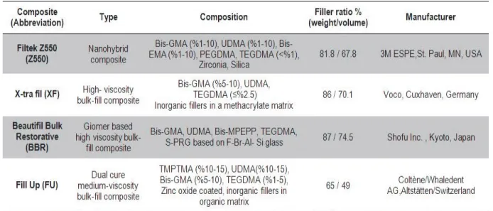
The following composite resins were used for this study: a bulk-fill composite [X-tra fil (VOCO, Cuxhaven, Germany)], a dual cure bulk-fill composite [Fill Up (Coltène/Whaledent AG, Altstätten, Switzerland)], a giomer-based bulk-fill composite [Beautifil Bulk Restorative (Shofu Dental Corporation, San Marcos CA, USA)] and a nanohybrid composite [Filtek Z550 (3M ESPE, St. Paul, MN, USA)]. Detailed information on the composites tested is provided in Table 1 including their composition and manufacturers. As for the light source, a QTH device [Hilux 250 (Benlioğlu Dental, Ankara, Turkey)], a second-generation LED device [Elipar DeepCure-S (3M ESPE, Seefeld, Germany)] and a third-generation LED device [VALO – (Ultradent Products Inc., South Jordan, UT, USA)] device with two different modes (standard and ultra mode) were used. Table 2 shows detailed information on the light devices tested. 2.1. Sample preparation
Twenty-four samples were prepared from each composite. Then, the composite samples were divided into 4 subgroups (n=6 each) according to the light device and light curing mode. Stainless steel molds with a diameter of 5 mm and a thickness of 4 mm were used to prepare the composite samples. Samples of bulk-fill composites were applied in a single bulk increment of 4 mm thickness into the mold and the nanohybrid composite (Z550) was applied in two increments,
each of 2 mm thickness. The procedure and conditions of polymerization are shown in Table 3. After filling the molds with the composites, a matrix strip was placed on top of the composites to prevent formation of an oxygen-inhibited layer and then, each sample was polymerized with different light curing units. The intensity of the curing light was monitored using a radiometer (Peng Lim Enterprise Co, LTD, Taiwan) throughout the measurements. Subsequently, a total of 96 samples were taken out of the molds and each sample was immediately transferred into amber vials containing 1.5 mL of 75% ethanol solution for extraction. The samples were diluted with the solution in the vials in a 1:20 ratio approximately and totally immersed in the solution. The samples were then stored at 37ºC for 24 hours and 1 month and 1 mL of ethanol solution was drawn from the vials at these timepoints for HPLC analysis.
2.2. HPLC analysis
An HPLC device (UFLC-XR, Shimadzu, Japan) and C18 reversed-phase HPLC columns (250 × 4.6 mm CS-2546-C185, Higgins, USA) were used for analysis of residual monomer content of the sample solutions obtained at the end of aforementioned storage periods. For calibration of the HPLC device, standard solutions of pure monomers [HEMA, BİS-GMA, TEGDMA and UDMA (Sigma Aldrich, St Louis, MO, USA)] were initially prepared at a final concentration of 0.1, 1, 10, 100, 500 and 1000 ppm and a total of 24 standard solutions (6 for each monomer) were injected into the HPLC system. At this stage, a mixture of 80% acetonitrile (MERCK, Darmstadt, Germany) and 20% water was used as the mobile phase. Then, retention times, linear calibration curves and peak areas for each monomer were obtained. Data for these monomers are presented in Table 4. A total of 96 solutions obtained from all samples after calibration were stored at 4 ºC until the time of analysis. 1 mL solution was obtained from each solution using Eppendorf pipettes and transferred into 2 mL amber glass vials and then injected into the HPLC device. The detector was set at 254 nm. Since the longest retention time was 5 min, each HPLC run was performed at an average pressure of 6.9 MPa for 7 min and chromatograms were obtained. From the chromatograms, measurements were done on the peaks predetermined for each monomer. In this study, the amount of monomer eluted was determined in ppm using peak areas.
Table 1. Composite resins used in this study and their composition.
Table 2. Light curing units used in this study.
Table 3. Polymerization procedure and conditions according to groups.
2.3. Statistical analysis
The study data were analyzed using the SPSS Version 22.0 (IBM, NY, USA). Since assumptions for parametric testing were met (Kolmogorov-Smirnov), One Way ANOVA test was used for data analysis and post-hoc Tukey test for comparisons among groups. The margin of error was set α= 0.05.
3. Results and discussion
3.1. HEMA
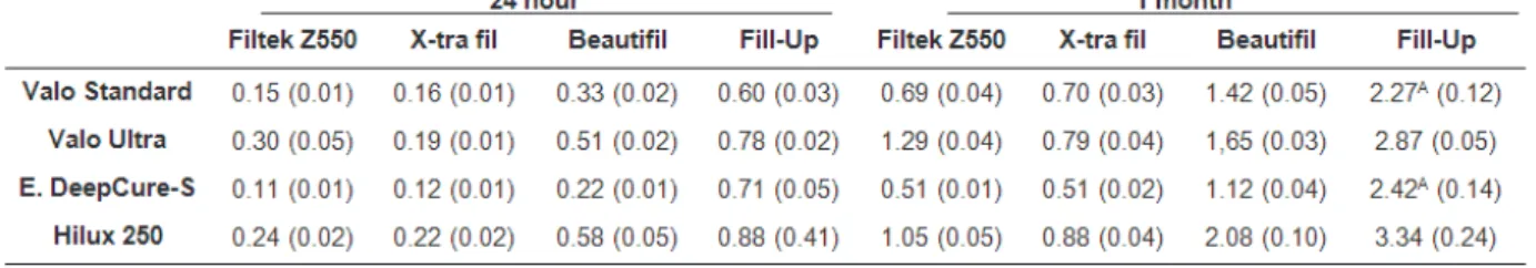
The amount of HEMA monomer (ppm) released from the composite resins polymerized with different light curing units after 24 h and 30 days is shown in Table 5. In all groups, the light curing units used for curing showed a statistically significant effect on the amount of HEMA leached from the composites. The least HEMA release was observed in the groups polymerized with Elipar DeepCure-S except for the FU composite resin. FU groups showed the least HEMA release with the use of standard mode of the Valo light source. The greatest HEMA release was seen in the groups exposed to halogen light source except the nanohybrid group. Among the resin composites, the highest HEMA elution was detected in the FU group and the lowest HEMA elution in the Z550 group. in all groups, the amount of residual monomers leached from the composites was significantly increased after 1 month compared to day 1. HEMA was the least eluted monomer from all tested materials.
3.2. Bis-GMA
The amount of Bis-GMA monomer (ppm) leached from the composite resins polymerized with different light curing units after 24 h and 30 days is shown in Table 6. In all groups, the light curing units showed a statistically significant effect on the amount of Bis-GMA eluted from the composites and none of the groups showed similarity. The least Bis-GMA release was observed in the groups polymerized with Elipar DeepCure-S except for the FU composite resin. The greatest Bis-GMA elution was detected in the groups cured with halogen light source except the nanohybrid group. Among the composites, BBR showed the highest Bis-GMA elution which was increased substantially after 1 month. The amount of residual monomers leached from the composites was significantly increased after 1 month compared to day 1 in all groups. Bis-GMA was identified as the residual monomer type with the greatest elution in the XF group.
3.3. TEGDMA
The quantity of TEGDMA monomer (ppm) released from the composite resins cured with different light curing units after 24 h and 30 days is shown in Table 7. In all groups, the light curing units showed a statistically significant effect on the amount of TEGDMA eluted from the composites. The least TEGDMA elution was observed in the groups polymerized with Elipar DeepCure-S except for the FU composite resin. Except for the nanohybrid group, greatest TEGDMA release was detected in the groups cured with halogen light source. As with HEMA, the greatest TEGDMA release was observed in the FU group and the least in the Z550 group. The amount of residual monomers eluted from the composites was significantly increased after 1 month compared to day 1 in all groups. In the XF group, the levels of TEGDMA eluted from the groups cured with Valo ultra mode and halogen light source after 30 days were comparable.
3.4. UDMA
The amount of UDMA monomer (ppm) eluted from the composite resins polymerized with different light curing units after 24 h and 30 days is shown in Table 8. In all groups, the light curing units showed a statistically significant effect on the amount of UDMA released from the composites and no similarity was observed among the groups. The least UDMA release was observed in the groups polymerized with Elipar DeepCure-S except for the FU composite resin. The greatest UDMA elution was detected in the groups cured with halogen light source except the nanohybrid group. Among the composites, BBR showed the highest UDMA elution and the residual monomer content was increased significantly after 1 month versus day 1. UDMA was the type of residual monomer released at the greatest level in all composites, except the XF composite group. Considering the total amount of residual monomers released from the composites, the greatest monomer elution was detected in the giomer-based bulk-fill composite.
Successful clinical applications of composite restoratives and practical advances have been achieved through improvement of physical and chemical characteristics of resin-based composite materials, increasing their resistance to pressure during mastication, reducing polymerization shrinkage and introduction of a wide array of different products for direct or indirect implementation [33]. Major criteria for success of the materials used in composite resins include good
biocompatibility and no harmful effects on the teeth and surrounding tissues [34,35]. Despite advances in the field of resin composites, leaching of unpolymerized monomers or release of residual monomers after polymerization cause several
problems in a living organism [34,36]. Such problems associated with composite materials sparked a debate on their safety.
In the present study, residual monomer elution from three bulk-fill composites and one nanohybrid
Table 5. Mean (standard deviation) concentrations (ppm) of HEMA eluted from composite resins polymerized with different light curing units.
Table 6. Mean (standard deviation) concentrations (ppm) of Bis-GMA eluted from composite resins polymerized with different light curing units.
Table 7. Mean (standard deviation) concentrations (ppm) of TEGDMA eluted from composite resins polymerized with different light curing units.
Table 8. Mean (standard deviation) concentrations (ppm) of UDMA eluted from composite resins polymerized with different light curing units.
composite following polymerization with different light curing units was analyzed using high performance liquid chromatography (HPLC) technique. There are several studies in the literature reporting significant concerns about the deleterious biological effects of residual monomers leached from resin-based dental materials. All concentrations of compounds eluted from resin-based dental materials have been reported to be associated with biological hazard [37]. While residual monomers leached from the composite materials cause minor effects in the oral cavity, over time, they may diffuse through dentin tubules into the pulpa and affect pulpal cells [4].
Studies on resin-based dental materials and release of their degradation products, namely residual monomers have also focused on the relationship between the type of light curing units used for polymerization and residual monomer leach [38,39]. However, few studies are available on the effect of different light curing units on the amount of residual monomers released from bulk-fill composites in the context of availability of a wide range of products and rapid developments in the dental materials. While light devices differ with respect to their characteristics such as the light intensity, irradiation time and wavelength range, they share common goals including increasing monomer conversion, reducing curing time as much as possible, and minimizing polymerization shrinkage and associated stress [40,41]. The efficiency of the light source used for polymerization is directly related to its polymerization characteristics exhibitied in the composite material [42].
One study reported that LED light devices were associated with higher polymerization efficiency in curing camphorquinone-based dental composites in comparison to halogen light devices [43]. Contrastingly, Öztürk et al. found no significant differences between QTH, LED and plasma arc light-curing units with respect to their effect on the degree of polymerization [44]. Karadas et al. looked at the effect of LED and QTH light curing units on the amount of residual monomers leached from bulk-fill composites and reported less monomer elution with the LED light source in all groups studied and this is consistent with our findings [38]. Conflicting results have been reported by previous studies on the amount of residual monomer released from composites polymerized with LED and QTH light curing units [45-47].
The light curing units used in the current study
showed an effect on the quantity of the residual monomers eluted from the composites with the greatest monomer release observed with the halogen light source and the least with the LED light source. Therefore, the null hypothesis was rejected. Except the FU composite, the second-generation LED light device was associated with less monomer release when compared with the third-generation light device in all groups. The lowest monomer elution detected with Elipar Deep Cure light-curing unit may be explained by its ability to produce a more homogeneous and deeper polymerization through its special fiber transmission system. Additionally, while second-generation light devices have a single-profile wavelength, third-generation light devices produce higher light output with different wavelengths and therefore, longer wavelength photons with less energy might have not been absorbed by the photoinitiator (camphoroquinone) in the composite and thus scattered [48,49].
The lowest level of monomer elution observed in the FU group alone with the Valo light device at standard mode may be related to the different initiator system (camphorquinone-amine) and organic compounds contained in this composite and operation of the Valo device with a multiwavelength LED, producing high-intensity light. Greater monomer release detected with the use of QTH light source may be explained by its lower light intensity compared to other light devices. After halogen light source, the second greatest monomer release values were observed in all groups with the ultra mode of the Valo light device and this might be due to insufficient curing time used for polymerization [22].
Previous studies have reported that although monomer release may last for weeks or months, maximum leaching occurs within the first days after polymerization [50,51]. Polydorou et al. [52] showed that Bis-GMA and TEGDMA monomers eluted from different composites at the highest level within the first 24 hours which decreased on days 7 and 28. In contrast to these studies, a significant increase was detected in residual monomer elution after 1 month versus day 1 in the present study. This may be due to the fact that no interim solutions were obtained for extraction between 24 hours and 1 month. Consistent with our findings, Alshali et al. [12] analyzed storage solutions of samples at 24 h and after 1 and 3 months and showed the highest monomer release within the first days after polymerization, with much lower monomer amounts after 1 month.
In one study, Van Landuyt et al. [4] ranked elution of the most common monomers in the following order: HEMA > TEGDMA > Bis-GMA > UDMA. However, in other studies investigating residual monomer amounts leached from composites containing Bis-GMA, UDMA and TEGDMA, greater elution of UDMA was observed in comparison to the other two monomers, which is consistent with our findings [12,30]. In the current study, while UDMA showed the highest elution among all groups, higher Bis-GMA elution was detected only in the XF group. In contrast to Van Landuyt et al.’s findings [4], residual monomers were ranked as UDMA > Bis-GMA > TEGDMA > HEMA based on their elution. This result might have resulted from differences in materials and methodology used as well as greater content of UDMA and Bis-GMA in the composite compared to TEGDMA. The primary monomer in the XF composite is Bis-GMA, which can explain greater release of this monomer. The least amount of HEMA monomer release observed in all groups and HEMA elution even when it was absent in the composites may be related to its occurrence in trace amounts as a degradation product of UDMA monomer [53].
Among all composites tested, the least residual monomer release was detected in the nanohybrid composite (Z550) in all groups. This may be due to the use of the layering technique for polymerization of this composite, unlike bulk-fill composites. Although bulk-fill composites are applied as a 4 mm-thick single-layer by design, all bulk-fill composites tested in the present study eluted more residual monomers than the nanohybrid composite. Among the tested bulk-fill composites, overall the greatest monomer leach was detected in the groups in which giomer-based bulk-fill (BBR) was used. While the amounts of monomer released from BBR after 1 day was comparable to those of other composites, significantly greater elution of UDMA and Bis-GMA monomers was observed in the giomer-based bulk-fill (BBR) after 1 month versus other composites. The lowest degree of conversion was reported with BBR in two studies analyzing the degree of conversion of giomer-based bulk-fill (BBR) composite in comparison to other bulk-fill composites, as in our study [54,55]. Additionally, BBR composites contain S-PRG (Surface Pre-Reacted Glass) filler particles which release ions over time, possibly affecting monomer leach from the material. Less elution of Bis-GMA and UDMA from the dual-cure bulk-fill composite (FU)
compared to other composites may be due to the dual-cure feature of this composite.
In our study, both the amount of individual monomers and the total amount of monomers released from the composites were at levels that would produce cytotoxic effect on pulpal fibroblast cells [56]. Possible negative effects of residual monomers on patients’ health may be reduced to a minimum by having a good knowledge of light devices used for application of bulk-fill composites, continuous monitoring of the light intensity throughout the procedure, always keeping fiber optic tip clean, preferential use of materials with higher polymerization efficiency, strictly following manufacturers’ instructions on polymerization, positioning the light source as close as possible to the material, using rubber dam and asking the patient not to swallow their saliva for a while.
4. Conclusion
In the current study, the light curing units affected the amount of residual monomers released from the composites in all groups. LED light-curing devices showed less residual monomer elution in comparison to the QTH light device. Bulk-fill composites exhibited greater monomer release versus nanohybrid composite. it was concluded that the light curing units and their modes and the type of composite used in clinical practice may have an effect on the amount of monomers released from the composites.
Acknowledgements
This study is supported by the Scientific Research Project Fund of Sivas Cumhuriyet University under the project number Diş-198. Thanks to Yılmaz Uğur for his efforts for HPLC analysis of samples.
References
1. U. Ortengren, H.Wellendorf, S. Karlsson, and I. E. Ruyter, J. Oral Rehabil., 28 (2001) 1106.
2. J. L. Ferracane, J. Oral Rehabil., 21 (1994) 441.
3. J. Durner, W. Spahl, J. Zaspel, H. Schweikl, R. Hickel, and F. X. Reichl, Dent. Mater., 26 (2010)
91.
4. K. L. Van Landuyt, T. Nawrot, B. Geebelen, J. De Munck, J. Snauwaert, K. Yoshihara, H. Scheers, L. Godderis, P. Hoet, and B. Van Meerbeek, Dent.
Mater., 27 (2011) 723.
5. N. Sasaki, K. Okuda, T. Kato, H. Kakishima, H. Okuma, K. Abe, H. Tachino, K. Tuchida, and K. Kubono, J. Mater. Sci. Mater. Med., 16 (2005)
297.
6. T. M. Gerzina and W. R. Hume, J. Dent. Res., 74
(1995) 369.
7. E. Sandberg, G. Bergenholtz, C. Eklund, and U. I. Dahlgren, J. Dent. Res., 81 (2002) 633.
8. M. Seiss., S. Nitz, N. Kleinsasser, J. T. Buters, H. Behrendt, R. Hickel, and F. X. Reichl, Dent.
Mater., 23 (2007) 9.
9. J. G. Leprince, W. M. Palin, M. A. Hadis, J. Devaux, and G. Leloup, Dent. Mater., 29 (2013)
139.
10. A. K. Luhrs, P. Pongprueksa, J. W. De Munck, B. Geurtsen, and B. Van Meerbeek, Dent. Mater.,
30 (2014) 281.
11. E. M. da Silva, G. S. Almeida, L. T. Poskus, and J. G. Guimaraes, J. Appl. Oral Sci., 16 (2008)
161.
12. R. Z. Alshali, N. A. Salim, R. Sung, J. D. Satterthwaite, and N. Silikas, Dent. Mater., 31
(2015) 1587.
13. R. B. Price, D. G. Murphy, and T. Derand,
Quintessence Int., 31 (2000) 659.
14. J. Park, J. Chang, J. Ferracane, and I. B. Lee,
Dent. Mater., 24 (2008) 1501.
15. A. C. Obici, M. A. C. Sinhoreti, E. Frollini, L. Correr-Sobrinho, M. F. De Goes, and G. E. P. Henriques, Polym. Test., 25 (2006) 282.
16. D. C. Sarrett, Dent. Mater, 21 (2005) 9.
17. A. Alrahlah, N. Silikas, and D. C. Watts, Dent
Mater., 30, (2014) 149.
18. S. Bucuta and N. Ilie, Clin. Oral Investig., 18
(2014) 1991.
19. B. M. Fronza, F. A. Rueggeberg, R. R. Braga, B. Mogilevych, L. E. Soares, A. A. Martin, G. Ambrosano, and M. Giannini, Dent. Mater., 31
(2015) 1542.
20. H. M. Kopperud, G. F. Johnsen, S. Lamolle, I. S. Kleven, H. Wellendorf, and H. J. Haugen, Dent.
Mater., 29 (2013) 824.
21. A. Rencz, R. Hickel, and N. Ilie, Clin. Oral
Investig., 16 (2012) 173.
22. N. Ilie, R. Hickel, and D. C. Watts, Dent. Mater.,
25, (2009) 411.
23. E. K. Viljanen, S. Langer, M. Skrifvars, and P. K. Vallittu, Dent. Mater., 22 (2006) 845.
24. E. B. Tuna, O. Aktoren, Y. Oshida, and K. Gencay, Eur. J. Paediatr. Dent., 11 (2010) 110.
25. S. Y. Lee, H. M. Huang, C. Y. Lin, and Y. H. Shih,
J. Oral Rehabil., 25 (1998) 575.
26. S. A. Mazzaoui, M. F. Burrow, M. J. Tyas, F. R. Rooney, and R. J. Capon, J. Biomed. Mater. Res.,
63 (2002) 299.
27. S. Y. Lee, E. H. Greener, and D. L. Menis, Dent.
Mater., 11 (1995) 348.
28. O. Polydorou, C. Huberty, M. Wolkewitz, R. Bolek, E. Hellwig, and K. Kummerer, J. Biomed.
Mater. Res. B, Appl. Biomater., 100 (2012) 68.
29. I. D. Sideridou, D. S. Achilias, and M. M. Karabela, J. Biomed. Mater. Res. B, Appl.
Biomater., 81 (2007) 207.
30. E. Lempel, Z. Czibulya, B. Kovacs, J. Szalma, A. Toth, S. Kunsagi-Mate, Z. Varga, and K. Boddi, Int. J. Mol. Sci., 17 (2016)732.
31. R. Lagocka, K. Jakubowska, D. Chlubek, and J. Buczkowska-Radlinska, Biomed. Res. Int., 2016
(2016) 3481723.
32. L. Rothmund, F. X. Reichl, R. Hickel, P. Styllou, M. Styllou, K. Kehe, Y. Yang, and C. Hogg,
Dent. Mater., 33 (2017) 54.
33. N. B. Cramer, J. W. Stansbury, and C. N. Bowman, J. Dent. Res., 90 (2011) 402.
34. J. S. Bertram, Mol. Aspects. Med., 21 (2000) 167.
35. H. Darmani, A. S. Al-Hiyasat, and M. M. Milhem, Quintessence Int., 38 (2007) 789.
36. K. Kehe, F. X. Reichl, J. Durner, U. Walther, R. Hickel, and W. Forth, Biomaterials, 22 (2001)
317.
37. G. Schmalz, Eur. J. Oral Sci., 106 (1998) 696.
38. M. Karadas, O. Hatipoglu, H. Er, and E. Akyüz Turumtay, J. Adhes. Sci. Technol., 32 (2018)
2631.
39. D. Manojlovic, M. Radisic, T. Vasiljevic, S. Zivkovic, M. Lausevic, and V. Miletic, Dent.
Mater., 27 (2011) 371.
40. A. U. Yap, Oper. Dent., 25 (2000) 113.
41. A. Peutzfeldt, A. Sahafi, and E. Asmussen, Dent.
Mater., 16 (2000) 330.
42. J. L. Ferracane, J. C. Mitchem, J. R. Condon, and R. Todd, J. Dent. Res., 76 (1997) 1508.
43. J. Leprince, J. Devaux, T. Mullier, J. Vreven, and G. Leloup, Oper. Dent., 35 (2010) 220.
44. B. Ozturk, N. Cobanoglu, A. R. Cetin, and B. Gunduz, Eur. J. Dent., 7 (2013) 102.
45. M. A. Cebe, F. Cebe, M. F. Cengiz, A. R. Cetin, O. F. Arpag, and B. Ozturk, Dent. Mater., 31,
(2015) e141.
46. A. Carvalho Fde, R. C. Almeida, M. A. Almeida, L. H. Cevidanes, and M. C. Leite, Am. J. Orthod.
Dentofacial Orthop., 138 (2010) 617.
47. A. U. Yap, V. T. Han, M. S. Soh, and K. S. Siow,
Oper. Dent., 29 (2004) 448.
48. M. G. Rocha, D. de Oliveira, I. C. Correa, L. Correr-Sobrinho, M. Sinhoreti, J. L. Ferracane, and A. B. Correr, Oper. Dent., 42 (2017) 418.
49. C. Shimokawa, B. Sullivan, M. Turbino, C. Soares, and R. Price, Oper. Dent., 42 (2017) 537.
50. I. D. Sideridou, and D. S. Achilias, J. Biomed.
Mater. Res. B, Appl. Biomater., 74 (2005) 617.
51. O. Polydorou, R. Trittler, E. Hellwig, and K. Kummerer, Dent. Mater., 23 (2007) 1535.
52. O. Polydorou, M. Hammad, A. Konig, E. Hellwig, and K. Kummerer, Dent. Mater., 25
(2009) 1090.
53. V. B. Michelsen, G. Moe, R. Skalevik, E. Jensen, and H. Lygre, J. Chromatogr. B, Analyt. Technol.
Biomed. Life Sci., 850 (2007) 83.
54. P. Yu, A. Yap, and X. Y. Wang, Oper. Dent., 42
(2017) 82.
55. K. Al-Ahdal, N. Ilie, N. Silikas, and D. C Watts,
Dent. Mater., 31 (2015) 1207.
56. W. Geurtsen, F. Lehmann, W. Spahl, and G. Leyhausen, J. Biomed. Mater. Res., 41 (1998)