25
1 Assistant Professor, Department of Endodontics, Faculty of Dentistry, Istanbul Medipol University, Istanbul, Turkey. 2 Professor, Department of Oral and Maxillofacial Surgery, Faculty of Dentistry, Selcuk University, Konya, Turkey. 3 Professor, Department of Endodontics, Faculty of Dentistry, Selcuk University, Konya, Turkey.
PREVALENCE OF VERTICAL ROOT FRACTURE IN EXTRACTED ENDODONTICALLY TREATED TEETH: A PROSPECTIVE STUDY
Endodontik Tedavi Görmüş Çekim Endikasyonu Olan Dişlerde Dikey Kök Kırığı Görülme Sıklığı: İleriye Dönük Bir Çalışma
Keziban OLCAY1,Hanife ATAOĞLU2, Sema BELLI3 Makale Kodu/Article code : 77968
Makale Gönderilme tarihi : 15.02.2016
Kabul Tarihi : 07.12.2016
ABSTRACT
Objectives: Vertical root fracture (VRF) is one of the complications of endodontic treatment which results extraction of the related tooth. The purpose of this prospective study was to evaluate prevalence of VRF in extracted endodontically treated teeth. Materials and Methods: During a 1 year period 241 teeth were observed. All of the cases were from extracted teeth after endodontic treatment. Clinical signs, symptoms, the informations about patient and related tooth were recorded.
Results: A total of 17 (7.02%) cases of VRF occurring among 241 cases were observed. The VRF occurred most frequently in the mesial roots of the mandibular first molars (23.5%) and they were the most frequently extracted teeth (35.2%). The VRF prevalence of maxillary first molars was 23.5%, maxillary first and second premolars with a same frequency (11.7%) were the following most fractured teeth. The fractures were predominantly buccolingual and were more frequent in female patients (58.8%). The mean patient age was 36 years and the mean time to VRF was 45 months. Most of the teeth which undergo VRF had a composite restoration (41.1%). 23.5% of the teeth had a post restoration and from among these posted teeth 75% were fully crowned. VRF was found similar frequency in mandibular teeth and in maxillary teeth (p>0.05).
Conclusions: After average of 45 months from endodontic treatment, mandibular first molar teeth were found most frequently prone to extraction due to VRF. Further investigation is necessary to determine the possible causes and evidence of fracture development.
Key Words: Endodontically treated teeth, vertical root fracture, tooth extraction, failure.
ÖZ
Amaç: Dikey kök kırığı (VRF), ilgili dişin çekimini gerektiren, endodontik tedavinin komplikasyonlarından biridir. Bu ileriye dönük çalışmanın amacı, çekim endikasyonu bulunan endodontik tedavi görmüş dişler arasında VRF görülme sıklığını değerlendirmektir.
Gereç ve Yöntem: 1 yıllık bir süre boyunca 241 diş gözlendi. Tüm vakalar çekim endikasyonu bulunan endodontik tedavi görmüş dişler arasından seçildi. Hasta ve ilgili diş ile ilgili klinik belirtiler, işaretler ve bilgiler kaydedildi.
Bulgular: 241 vaka arasında toplam 17 (%7,02) VRF vakası gözlendi. VRF, en sık alt çene birinci büyük azı dişlerin mesial köklerinde (% 23,5) meydana gelmiş ve bu dişlerin en sık çekilen dişler olduğu bulunmuştur (%35,2). Üst çene birinci büyük azı dişlerde VRF görülme sıklığı % 23,5; takiben, üst çene birinci ve ikinci küçük azı dişler aynı oranla (% 11,7), en sık kırılan dişler olmuştur. Kırıklar ağırlıklı olarak bukko-lingual idi ve kadın hastalarda daha sıktı (% 58,8). Ortalama hasta yaşı 36 idi ve VRF oluşana dek geçen ortalama sure 45 ay idi. VRF görülen dişlerin çoğunda kompozit onarım (% 41,1) vardı. Dişlerin % 23,5'inde post restorasyon vardı ve bu postlu dişlerin % 75'inde tam kron vardı. VRF alt çenedeki dişlerde ve üst çenedeki dişlerde benzer frekansta bulundu (p>0,05).
Sonuç: Endodontik tedaviden ortalama 45 ay sonra, alt çene birinci büyük azı dişler VRF nedeniyle sıklıkla çekime daha yatkın bulundu. Kırık gelişiminin olası nedenlerini ve kanıtlarını belirlemek için daha ileri araştırmalar gereklidir. Anahtar Kelimeler: Endodontik olarak tedavi edilmiş dişler, dikey kök kırığı, diş ekstraksiyonu, başarısızlık.
26
INTRODUCTIONVRF is a complication that may occur after endodontic treatment.1,2 It has unfavourable
prognosis3 and results in extraction of the tooth
or root amputation.2,4,5 Reasons for VRF
include:
a) overzealous widening of the root canal for biomechanical preparation or post placement6, especially when the root canal
has been widened by 40% or more;7
b) particularly when step back techniques were used;8
c) compromised tooth integrity as a result of large carious lesions or trauma;9
d) excessive force during compaction of root filling material, particularly when lateral or lateral-vertical forces are applied10
and
e) lack of sufficient periodontal support, the presence of internal resorption, or both11 can
also lead VRF.
A local deep pocket, dual sinus tracts, and a halo type of lateral radiolucency are the symptoms and radiographic features of VRF 12.
Clinical signs, radiographic features, and symptoms observed in VRF are similar to a failed root canal treatment and periodontal disease. This similarity makes accurate diagnosis difficult.3,5,8,13,14
The prevalence of vertical root fracture was reported as 2-5% in previous studies.8,15
Song et al.16 reported the percentage of
prevalence of VRF in endodontically treated teeth as 1.2% and Zadik et al.17, Fuss et al.18
and Toure et al.19 reported levels of 8.8%,
10.9% and 13.4%, respectively.
The purpose of this study was to investigate the prevalence of VRF in extracted endodontically treated teeth prospectively. MATERIALS AND METHODS
A total of 241 endodontically treated teeth were extracted between January to December 2011. The patients were referred to the Department of
Endodontics, Faculty of Dentistry, and Selcuk University, Turkey by different dental clinics. All of the patients had complained about their endodontically treated teeth. The decisions for extraction was made by specialists (K.O. and S.B.) from totally 1000 failed endodontically treated teeth and 241 teeth were decided for extraction. From these teeth, only 17 were suspected of VRF and referred to an oral surgeon for extraction (H.A.). The questionnaires were filled out via question and answer at the time of extraction for all patients (K.O.); data included information about the patient (age, gender, level of education). Examination of the extracted teeth took into consideration tooth type, status of coronal restoration, root canal filling, radiographic findings, presence of periodontal defects, signs, and the time elapsed from root canal treatment to VRF. After extraction, each tooth was cleaned of external remnants and washed for VRF verification. The diagnosis of VRF was made based on the findings from the extracted tooth. Other extracted teeth were classified by reason for extraction, which included according to prosthetic reasons, periodontal reasons, endodontic failure, nonrestorable caries, nonrestorable cusp/tooth fracture, perforation/ stripping, and teeth extracted because of patient request.
The data were analyzed by using SPSS 17.0 (SPSS, Chicago, IL). The associations between patients’ gender and levels of education were examined using a chi-square test. The differences about age of patients were analyzed with Mann Whitney U test. p0.05 was accepted as the level of significance. RESULTS
VRF was observed in all of the 17 teeth that were referred to an oral surgeon on suspicion of VRF. When these 17 teeth were evaluated, it was observed that 35.2% were mandibular first molars, 23.5% were maxillary first molars, and 11.7% were maxillary first and second premolars (Figure 1). Mesial roots of the
27
mandibular molars were the most frequently extracted because of VRF. The fractures were predominantly buccolingual and were more frequent in female patients (58.8%). The mean patient age was 36 years, and the mean time to vertical root fracture was 45 months. The elapsed time between root canal treatments to VRF is shown in (Figure 2).Figure 1. Distribution of extracted endodontically treated teeth due to vertically root fracture
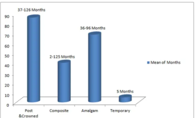
Figure 2. Restorative materials used in 17 teeth with VRF and the mean of elapsed time between root canal treatments to VRF
Most of the teeth that undergo vertical root fracture had a restorative adhesive techniques (41.1%). 23.5% of the teeth had a post restoration, and among these posted teeth, 75% were fully crowned. There is a significant difference among the coronal restorations of teeth (p=0.000) and distrubution of percentages is shown in (Figure 3). None of the posts ended at the coronal third, but all ended at the middle third of the root. All the posts used were prefabricated posts. The frequency of VRF in mandibular teeth and in maxillary teeth was found to be similar (p=0.417).
Figure 3. Distribution of coronal restoration of extracted endodontically treated teeth due to vertically root fracture
The most prevalent clinical observations in this study were the presence of periodontal pocket (17.6%) and painful and/or draining fistula (23.5%) on the buccal side of the tooth. Radiographically, a halo type of lateral radiolucency was observed in 3 of the 17 cases. DISCUSSION
The diagnosis of VRF is problematic and easily misdiagnosed as periodontal disease or endodontic failure.5,8,13 In the present study, the
prevalence of VRF was 7.02%, which was close to the 8.8% found by Zadik et al.17 and 10.9% by
Fuss et al.18 Some clinical retrospective articles
had previously reported lower percentages such as 4%20, 3.7%21, and 1.2% Song et al.16 The lower
percentages of VRF in these studies might be because of the difficulties in the clinical diagnosis of VRF. A prospective study by Toure et al.19
reported the percentage of prevalence of VRF in endodontically treated teeth as 13.4%. A study by Sjögren et al.22 reported the highest percentage of
the VRF in endodontically treated at 30.8%. The differences among these studies might have occurred because of the study periods.
In our study, the ages of patients ranged from 16 to 61. Most of the VRF in endodontically treated teeth were recorded in patients 30 to 50 years of age as in some previous reports.8,12,15,18 Based on this finding,
we can speculate that after endodontic treatment, teeth become more prone to fracture in younger patients.
Fuss et al.5, Testori et al.15 and Tamse et
al.3 reported that premolars have more
frequency of VRF in endodontically treated teeth; however, our results, like those of Chan et al.12 and Llena-Puy et al.4, showed that the
most extracted teeth were mandibular molars. Also, we found that the mesial roots of mandibular molars are more prone to fracture than distal roots, as reported by Fuss et al.5,
Tamse et al.3, and Chan et al.12. The higher
percentage of VRF observed in mesial roots of mandibular molars can be explained by the thin or flat structure of mesial roots, especially after root canal treatment and/or heavier masticatory force associated with first molars.
In Llena-Puy et al.5 study; the mean time
from root canal therapy to vertical root fracture was reported as 54 months. According to our results, it is 45 months. For most teeth, using of a restorative adhesive restoration did not appear to prolong the time between restoration and VRF. In contrast the study of Llena-Puy et al.4, in the
present study teeth that had a post restoration and full crown provided the longest average time elapsed to VRF. Based on this finding, the posted and fully crowned teeth may extend the time elapsed to VRF in endodontically treated teeth.
In the present study, painful and/or draining fistula appearing in the attached gingiva was the most frequent clinical sign. As opposed to the cases of failed endodontic therapies, it was located <4 mm from the gingival margin. Unlike our study (23.5%), this type of fistula was found by Tamse et al.3 in 35% and by Testori et al.15 in
42%. Following the painful and/or draining fistula, periodontal pocket was the second clinical sign observed in our study. Tamse et al.3 reported
that 13% of cases of endodontically treated teeth failed because of VRF; there was no periapical or lateral radiolucency. But in our study, 52.9% of teeth had no periapical or lateral radiolucency, and a halo type of lateral radiolucency was observed only in 3 cases.
Limitations of this study include the fact that, the patients investigated in the present study
were treated at different clinics, not our clinic. Because of this, we do not know what sealers or other materials were used during treatment. A preoperative evaluation of the frequency of a VRF in endodontically treated teeth was investigated in the present study. Further investigations need to investigate frequency of a VRF in a larger sample of patients.
CONCLUSIONS
The results of this study showed that the use of posts and fully crowns for reconstruction of endodontically treated teeth does extend the elapsed time to VRF, but use of adhesive restorative techniques does not shorten the elapsed time to VRF.
ACKNOWLEDGMENTS
This study was performed in Endodontics and Oral & Maxillofacial Surgery Departments of Faculty of Dentistry, Selcuk University. The authors thank Dr. Hilal Erdoğan and Dr. Alperen Bozkurt from Department of Endodontics, Faculty of Dentistry, and Selcuk University for referring the most appropriate clinical cases during this study. The authors also thank Dr.
Serhan Akman from Department of
Prosthodontics, Faculty of Dentistry, and Selcuk University for the statistical analysis of the study. REFERENCES
1. Walton RE, Michelich RJ, Smith GN. The histopathogenesis of vertical root fractures. J Endod 1984;10:48–56.
2. Tsesis I, Rosen E, Tamse A, Taschieri S, Kfir A. Diagnosis of vertical root fractures in endodontically treated teeth based on clinical and radiographic indices: a systematic review. J Endod 2010;36:1455-8.
3. Tamse A, Fuss Z, Lustig J, Kaplavi J. An evaluation of endodontically treated vertically fractured teeth. J Endod 1999;25:506–8.
4. Llena-Puy MC, Forner-Navarro L, Barbero-Navarro I. Vertical root fracture in endodontically treated teeth: a review of 25
29
cases. Oral Surg Oral Med Oral Pathol Oral Radiol Endod 2001;92:553-5.5. Fuss Z, Lustig J, Katz A, Tamse A. An evaluation of endodontically treated vertical root fractured teeth: impact of operative procedures. J Endod 2001;27:46-8.
6. Guzy GE, Nicholls JI. In vitro comparison of intact endodontically treated teeth with and without endo-post reinforcement. J Prosthet Dent 1979;42:39-44.
7. Wilcox LR, Roskelley C, Sutton T. The relationship of root canal enlargement to finger-spreader–induced vertical root fracture. J Endod 1997;23:533-4.
8. Meister F, Lommel TJ, Gerstein H. Diagnosis and possible causes of vertical root fracture. Oral Surg Oral Med Oral Pathol 1980;49:243–53.
9. Sedgley CM, Messer HH. Are
endodontically treated teeth more brittle? J Endod 1992;18:332–5.
10. Joyce AP, Loushine RJ, West LA, Runyan DA, Cameron SM. Photoelastic comparison of stress induced by using stainless steel versus nickel-titanium spreaders in vitro. J Endod 1998;24:714-5.
11. Telli C, Gulkan P, Raab W. Additional studies on the distribution of stresses during vertical compaction of gutta-percha in the root canal. Braz Dent J 1999;187:32-7.
12. Chan CP, Lin CP, Tseng SC, Jeng JH. Vertical root fracture in endodontically versus nonendodontically treated teeth: a survey of 315 cases in Chinese patients. Oral Surg Oral Med Oral Pathol Oral Radiol Endod 1999;87:504-7.
13. Hassan B, Metska ME, Ozok AR, van der Stelt P, Wesselink R. Detection of vertical root fractures in endodontically treated teeth by a cone beam computed tomography scan. J Endod 2009;35:719–22.
14. Ozer SY. Detection of vertical root fractures of different thicknesses in endodontically enlarged teeth by cone beam computed tomography versus digital radiography. J Endod 2010;36:1245-9.
15. Testori T, Badino M, Castagnola M. Vertical root fractures in endodontically treated teeth: a clinical survey of 36 cases. J Endod 1993;19:87-91.
16. Song M, Kim HC, Lee W, Kim E. Analysis of the cause of failure in nonsurgical endodontic treatment by microscopic inspection during endodontic microsurgery. J Endod 2011;37:1516-9.
17. Zadik Y, Sandler V, Bechor R, Salehrabi R. Analysis of factors related to extraction of endodontically treated teeth. Oral Surg Oral Med Oral Pathol Oral Radiol Endod 2008;106:31-5.
18. Fuss Z, Lustig J, Tamse A. Prevalence of vertical root fractures in extracted endodontically treated teeth. Int Endod J 1999;32:283-6.
19. Touré B, Faye B, Kane AW, Lo CM, Niang B, Boucher Y. Analysis of reasons for extraction of endodontically treated teeth: a prospective study. J Endod 2011;37:1512-5. 20. Hansen EK, Asmussen E, Christiansen N. In vivo fractures of endodontically treated posterior teeth restored with amalgam. Endod Dent Traumatol 1990;6:49–55.
21. Morfis AS. Vertical root fractures. Oral Surg Oral Med, Oral Pathol 1990;69:631-5. 22. SjoÈgren U, HoÈgglund B, Sundqvist G, Wing K. Factors affecting long term results of endodontic treatment. J Endod 1990;16:498-504.
Corresponding Author Keziban OLCAY Atatürk Bulvarı, No:27
Department of Endodontics Faculty of Dentistry
Istanbul Medipol University Unkapanı, Fatih, Istanbul. E-mail: [email protected] Phone: +90 0212 453 4800-4958 Fax: +90 0332 223 006