DOI: 10.5455/annalsmedres.2018.12.308 2019;26(4):588-92
Determination of normal lung volume using computed
tomography in children and adolescents
Emine Caliskan1, Mehmet Ozturk2
1Seyhan State Hospital, Clinic of Radiology Adana, Turkey
2Selcuk University Faculty of Medicine Department of Radiology, Konya, Turkey
Copyright © 2019 by authors and Annals of Medical Research Publishing Inc.
Abstract
Aim: To determine the normal reference values for both lung inspiratory volumes in children and adolescents with routine computed
tomography (CT) and compare relationships with gender and age.
Material and Methods: This retrospective study evaluated 100 cases (53 males, 47 females) with normal chest CT (age range: 4-17
years). The cases were divided into three subgroups based on age as 4-7 years (preschool; n=31), 8-12 years (school-age; n=30) and 13-17 years (adolescent; n=39). Demographic data like sex and age were recorded. Right, left and total lung volumes were calculated with automatic volumetric software. Volume comparisons were made for lungs, gender and age groups. Statistical analysis used the Wilcoxon signed-ranks test, Mann-Whitney U test and Tamhane test.
Results: For all cases the right lung volume was greater than the left lung (p<0.001). There was no significant difference identified for
the total lung volumes between the genders (p=0.421). The right, left and total lung median volume values were identified as 379 cm3,
310 cm3 and 689 cm3 in the preschool group, 840 cm 3.710 cm3 and 1550 cm3 in the school-age group and 1670 cm3, 1391 cm3 and
3061 cm3 in the adolescent group, respectively. In the transitions from preschool to school-age and from school-age to adolescent
periods, the right, left and total lung volumes significantly increased.
Conclusion: Quantitative reference values for normal lung inspiratory volume were revealed for children and adolescents using CT.
As age group increased, there were significant increases in lung volume. Gender did not have a significant effect on lung volume. Keywords: Adolescent; Children; Computed Tomography; Lung Volume.
Received: 26.12.2018 Accepted: 22.01.2019 Available online: 28.01.2019
Corresponding Author: Emine Caliskan, Seyhan State Hospital, Clinic of Radiology Adana, Turkey E-mail: [email protected]
INTRODUCTION
Computed tomography (CT) is a routine imaging method commonly used in clinical practice. CT imaging was shown to be superior to radiography (X-ray) and magnetic resonance imaging (MRI) for evaluation of lung diseases (1). The latest-technology product of post-processing work stations allow the automatic measurement of lung volume from CT data sets (2). These measurements are interesting for assessment of pulmonary diseases progressing with lung volume changes. Volume measurements may be indicative in the pediatric period as they are for adults. For examples, emphysema, allergic asthma and foreign body aspiration may cause an increase in lung volume, or chronic cystic fibrosis and interstitial lung disease may reduce lung volume. Lung volume in these diseases may be an appropriate determinant of disease activity, progression or response to treatment (3). As a result, it is important to know the normal lung volume in the healthy population.
There are a variety of pulmonary function tests to measure total lung volume. These include spirometry test and body plethysmography. They are used to monitor progression of disease and response to treatment (3). Additionally, though not used routinely, imaging techniques can be used for lung volume measurements. These techniques include methods from chest radiography to CT or MRI (4). There is good correlation shown between CT and body plethysmography results (5,6). A variety of approaches are used for volumetric measurements on CT. The manual method defines contours for volume calculations and was the first method used with this aim. With developing technology, CT work stations may automatically calculate lung volumes. Thereare studies in the literature determining the normal lung volume of adults, both healthy and with diseases, using CT (5,7,8). However, to the best of our knowledge, there is no basal data related to the normal lung volumes of the pediatric population.
normal reference values for inspiratory volumes of lungs in children and adolescents with routine CT and assess association with potential factors like age and gender.
MATERIAL and METHODS
Subjects
This study included 100 children and adolescents aged from 4-17 years with chest CT taken from January 2015 to July 2018 for a variety of reasons. The study was conducted with ethics approval from the Ethics Committee. “Informed consent” was not obtained from parents as the study was retrospective. Cases included in the study did not provide personal information and were presented anonymously. Cases were divided into groups as 4-7 years (preschool), 8-12 years (school-age) and 13-17 years (adolescent). Comparisons were made between both lung, girl-boy gender and age groups.
CT protocol
CT examinations were performed with or without contrast by a multi-slice CT scanner using helical mode in the supine position (SOMATOM Definition Flash CT 64-slice scanner, Siemens Medical Solutions, Erlangen, Germany). The imaging data for enhanced CT were acquired during an intravenous injection of 1.5 to 2 mL/kg of the contrast agent at a rate of 1 to 3 mL/s. Saline solution of 5 to 20 mL followed the contrast material. The scanning parameters for both enhanced and non-enhanced CT were 25-30 mA, 80-90 kV, depending on child age and weight, section thickness of 1 mm and reconstruction interval of 0.5 mm. The scan revolution time was 0.4 seconds. Axial and sagittal-coronal reformat images were analyzed for each case. CT data was transferred from the archive to a workstation (Syngo, CT Pulmo 3D, 2012B, VA44 version).
CT image analysis
CT images of cases were identified in the hospital archive system (PACS). Chest CTs with pathologic findings like lung masses, atelectasis, emphysema, pneumothorax, pleural fluid and infiltration were excluded. Normal routine chest CTs with no known chronic diseases, sufficient quality and taken during sufficient inspirium were investigated. These images were downloaded to a work station from the internet database. Then right, left and total lung volumes were calculated. Lung volume calculations were based on the principle of the computer determining threshold values for structures containing air density with Hounsfield units (HU) and using a mathematical algorithm to automatically give the volume in cm3 (Figures 1. a and b). Checking of the automatically measured data was performed visually by a pediatric radiologist with more than 10 years of chest CT and 3 years of using automatic volumetric software experiences. If necessary, the procedure was repeated until equivalent to lung boundaries and optimal measures accepted as accurate were obtained.
Statistical Analysis
SPSS 22.0 was used for statistical analysis.The compliance of the data with normal distribution was
Wallis test.Comparisons between both lungs, gender and age groups used the Wilcoxon Signed-ranks test, Mann-Whitney U test and Tamhane test due to non-normal distribution.Quantitative variables are shown as median range (maximum-minimum) in the tables. Variables were investigated at the 95% confidence interval with p values below 0.05 accepted as significant.
Figures 1. a-b. A 15-year old male subject showing right
(2181 cm3), left (2222 cm3) and total (4403 cm3) lung volumes
calculated with CT using automatic volumetry software. Based on axial and coronal reformatted images, right lung is shown in green and left lung is shown in red (a). Upper shows right, left
RESULTS
One hundred children and adolescents with the median age of 11.3 (4-17) years were included in the study. Fifty-three of them were males and 47 of them were females. The preschool group included 17 males and 14 females for a total of 31 subjects (median age: 5.1 [4-7] years); the school-age group included 14 males and 16 females for a total of 30 subjects (median age: 10.8 [8-12] years); and the adolescent age group included 20 males and 19 females for a total of 39 subjects (median age: 15.0 [13-17] years).
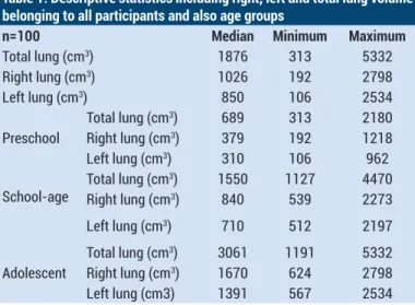
Descriptive statistics of the demographic data including right, left and total lung volume belonging to all participants and also age groups are given in Table 1.
Table 1. Descriptive statistics including right, left and total lung volume belonging to all participants and also age groups
n=100 Median Minimum Maximum
Total lung (cm3) 1876 313 5332
Right lung (cm3) 1026 192 2798
Left lung (cm3) 850 106 2534
Preschool Total lung (cm
3) 689 313 2180
Right lung (cm3) 379 192 1218
Left lung (cm3) 310 106 962
School-age Total lung (cm
3) 1550 1127 4470
Right lung (cm3) 840 539 2273
Left lung (cm3) 710 512 2197
Adolescent Total lung (cm
3) 3061 1191 5332
Right lung (cm3) 1670 624 2798
Left lung (cm3) 1391 567 2534
For all cases, there was a statistically significant difference between right and left lung volumes and right lung volume was greater than left lung volume (p<0.001). There was no significant difference identified for total lung volumes between the genders (p=0.421) (Table 2).
There were significant differences between the right, left and total lung volumes in the preschool, school-age and adolescent groups (p<0.001) (Table 3).
The right, left and total lung volumes significantly increased from preschool to school age (median volume: 379 cm3 vs 840 cm3, 310 cm3 vs 710 cm3, 689 cm3 vs 1550 cm3, respectively).
From school-age to adolescent period, the right, left and total lung volumes significantly increased (median volume: 840 cm3 vs 1670 cm3, 710 cm3 vs 1391 cm3, 1550 cm3 vs 3061 cm3, respectively) (Figure 2).
Table 2. Volume comparisons between right and left lung, and genders
n=100 pvalue
Right-Left lungvolume* <0.001
Male-Female total lung volume** 0.421 *Wilcoxon Signed-ranks test,
**Mann-Whitney U Test
Bold value depicts significant difference
Table 3. Lung volume comparisons between three different age groups
Preschool School-age School-age Adolescent Preschool Adolescent pvalue pvalue pvalue
Right lung volume <0.001 <0.001 <0.001
Left lung volume <0.001 <0.001 <0.001
Total lung volume <0.001 <0.001 <0.001
Tamhane test cm: centimeter, f: female, m: male, n: number of subjects
DISCUSSION
CT of the chest is commonly performed because it combines rapid image taking with detailed anatomic description for a variety of clinical applications. It is a standard diagnostic tool for both oncologic imaging and general assessment of pulmonary pathologies. Compared to other imaging methods such as MRI, X-ray or ultrasound, CT offers an accurate image quality with high resolution (9). Though it is more frequently used in adults, it has now become a frequently chosen imaging method in the pediatric period. However, the benefits of chest CT must be balanced by the risk of life-long malignancy associated with ionizing radiation (10). When imaging a young patient, one should always take into account the ALARA concept, to balance an appropriate and low-dose technique with imaging quality and to reduce the amount of ionizing radiation exposure (11). Screening protocols specific to pediatrics reduce the radiation dose associated with CT.
This study presents basal data related to normal inspiratory lung volume values in the pediatric population. Important results were reached which contribute to the literature. In this study, as age group increased lung volume was found to increase. Similarly, Jill M. Stein et al. published that normal lung parenchymal X-ray CT attenuation decreases with increasing lung volume and age; lung attenuation decreases rapidly in the first 2 years of age and more slowly thereafter (12). Wen Y et al. in a study of adolescent idiopathic scoliosis patients compared lung volume determined from CT scans with pulmonary function tests (13). They found that total lung volume was positively correlated with vital capacity, forced vital capacity, and total lung capacity of adolescent idiopathic scoliosis patients, which were less than those of age-matched controls.
This study found the right lung volume was higher than the left. This situation is easily explained by classic anatomic knowledge. There is some form of segmental symmetry between the right and left lungs, even though the left lung is smaller and only contains two lobes. In general, each lung has 10 segments: the upper lobes contain 3 segments, the middle lobe / lingual 2 and the lower lobes 5. Bilaterally, the upper lobes have apical, posterior and anterior segments and the lower lobes superior (apical) and 4 basal segments (anterior, medial, posterior and lateral). Additionally, this study found no significant difference in lung volume between males and females. This means that in routine practice standard lung volumetry can be used in the same way for both genders. There are a range of clinical scenarios for the interest in CT lung volumetry in adults. For example, inspiratory vital capacity is defined as an important marker of chronic obstructive lung disease with possible contribution to clinical studies (14,15). Another study showed CT analysis of lung surface area in emphysema can be used to assess
Similarly, the results of our study may provide valuable basal data for acute or chronic diseases involving lung volume changes in children. Among these diseases, cystic fibrosis, allergic asthma, foreign body aspiration, bronchiectasis, bronchiolitis, bronchiolitis obliterans, interstitial diseases and Swyer James syndrome may be listed. Additionally, future studies may determine the cut-off values for each disease.
CT scoring systems are a tool for describing semi-quantitative abnormalities. In spite of many scoring systems existing, they have been modified in recent years (17,18). Chest CT scores are sensitive for early detection, regional disease, and are more sensitive to progression of disease than traditional spirometry (3). The important abnormalities included in most scoring systems are bronchiectasis, mucous plugging, airway wall thickening, parenchymal opacity, small nodules, mosaic attenuation, sacculations, overinflation and air trapping. In the progression of cystic fibrosis, air trapping and overinflation are thought to reflect early occurrence of small airway disease. As a result, some studies have investigated expiratory images (19). Differently more current studies have found for young children with cystic fibrosis radiologic assessment of early bronchiectasis with chest CT scan depends on lung volume; thus, expiratory scans may not be appropriate for evaluating bronchiectasis in this population. Lung volume during CT image acquisition should be standardized to evaluate airway dimensions in young children (20). Early and late period findings may create volume changes in both lungs. As a result, we think our study may be used for a new, updated and current scoring system.
Our study has five major limitations. The first is the low number of patients, the second is not including children younger than 4 years of age. The third is that it is a retrospective study and did not include body mass index (BMI) data. BMI and possible lung volume changes were not investigated. The fourth limitation is that clinical and laboratory data were not used to support the fact that the subjects were healthy while determining normal lung volumes. The fifth limitation is that only inspiratory normal lung volumes were assessed. As the study was retrospective and required repeat CT imaging, expiratory data reflecting residual volume were not investigated.
CONCLUSION
In conclusion, this study provided quantitative basal data for normal pediatric inspiratory lung volume. Gender had not significant effect on total lung volume. As age group increased, there was a significant increase in lung volume. Studies of pathologic pediatric lung disease will provide important contribution to the literature in terms of indicating future paths.
Acknowledgements
We would acknowledge Dr. Burak Polat in assisting to review the publications and for his assistance in the online
Competing interests: The authors declare that they have no competing interest.
Financial Disclosure: There are no financial supports
Ethical approval: The study was conducted with ethics approval from the Ethics Committee. “Informed consent” was not obtained from parents as the study was retrospective
Emine Caliskan ORCID: 0000-0001-9869-1396 Mehmet Ozturk ORCID: 0000-0001-5585-1476
REFERENCES
1. Porcel JM, Leung CC, Restrepo MI, et al. Year in review 2012: lung cancer, respiratory infections, tuberculosis, pleural diseases, bronchoscopic intervention and imaging. Respirology 2013;18:573-83.
2. Garfield JL, Marchetti N, Gaughan JP, et al. Total lung capacity by plethysmography and high-resolution computed tomography in COPD. Int J Chron Obstruct Pulmon Dis 2012;7:119-26.
3. de Jong PA, Lindblad A, Rubin L, et al. Progression of lung disease on computed tomography and pulmonary function tests in children and adults with cystic fibrosis. Thorax 2006;61:80-5.
4. Chen F, Kubo T, Shoji T, et al. Comparison of pulmonary function test and computed tomography volumetry in living lung donors. J Heart Lung Transplant 2011;30:572-5. 5. Kauczor HU, Heussel CP, Fischer B, et al. Assessment of
lung volumes using helical CT at inspiration and expiration: comparison with pulmonary function tests. AJR Am J Roentgenol 1998;171:1091-5.
6. Kendrick AH. Comparison of methods of measuring static lung volumes. Monaldi Arch Chest Dis 1996;51:431-9. 7. Haas M, Hamm B, Niehues SM. Automated lung volumetry
from routine thoracic CT scans: how reliable is the result? Acad Radiol 2014;21:633-8.
8. Shin JM, Kim TH, Haam S, et al. The repeatability of computed tomography lung volume measurements:Comparisons in healthy subjects, patients with obstructive lung disease, and patients with restrictive lung disease. PLoS One 2017;12:e0182849.
9. Gruber-Rouh T, Thalhammer A, Klingebiel T, et al. Computed tomography-guided biopsies in children: accuracy, efficiency and dose usage. Ital J Pediatr 2017;43:4.
10. Gilchrist FJ, Buka R, Jones M, et al. Clinical indications and scanning protocols for chest CT in children with cystic fibrosis: a survey of UK tertiary centres. BMJ Paediatr Open 2018;2:e000367.
11. Larson DB, Molvin LZ, Wang J, et al. Pediatric CT quality management and improvement program. Pediatr Radiol 2014;44:519-24.
12. Stein JM, Walkup LL, Brody AS, et al. Quantitative CT characterization of pediatric lung development using routine clinical imaging. Pediatr Radiol 2016;46:1804-12.
13. Wen Y, Kai S, Yong-Gang Zet al. Relationship between Lung Volume and Pulmonary Function in Patients With Adolescent Idiopathic Scoliosis: Computed Tomographic-based 3-Dimensional Volumetric Reconstruction of Lung Parenchyma. Clin Spine Surg 2016;29:396-400.
14. Molho M, Shulimzon T, Benzaray S, et al. Importance of inspiratory load in the assessment of severity of airways obstruction and its correlation with CO2 retention in chronic obstructive pulmonary disease. Am Rev Respir Dis 1993;147:45-9.
15. Taube C, Kanniess F, Grönke L, et al. Reproducibility of forced inspiratory and expiratory volumes after bronchodilation in patients with COPD or asthma. Respir Med 2003;97:568-77. 16. Coxson HO, Rogers RM, Whittall KP, et al. A quantification
of the lung surface area in emphysema using computed tomography. Am J Respir Crit Care Med 1999;159:851-6. 17. Brody AS, Molina PL, Klein JS, et al. High-resolution
computed tomography of the chest in children with cystic fibrosis: support for use as an outcome surrogate. Pediatr Radiol 1999;29:731-5.
18. Robinson TE, Leung AN, Northway WH, et al. Spirometer-triggered high-resolution computed tomography and pulmonary function measurements during an acute exacerbation in patients with cystic fibrosis. J Pediatr 2001;138:553-9.
19. Bonnel AS, Song SM, Kesavarju K, et al. Quantitative air-trapping analysis in children with mild cystic fibrosis lung disease. Pediatr Pulmonol 2004;38:396-405.
20. Mott LS, Graniel KG, Park J, et al. Assessment of early bronchiectasis in young children with cystic fibrosis is dependent on lung volume. Chest 2013;144:1193-8.