R E S E A R C H
Open Access
Different accelerated corneal collagen
cross-linking treatment modalities in
progressive keratoconus
Ahmet Kirgiz
1,3*, Mustafa Eliacik
2and Yusuf Yildirim
3Abstract
Background: To compare the outcomes of two different protocols of accelerated corneal crosslinking (CXL) on visual, corneal high order aberrations (HOA) and topographic parameters in patients with progressive keratoconus.
Methods: In this prospective comparative study, sixty-six eyes of 66 patients with progressive keratoconus were divided into two groups; 37 eyes in Group 1 received 18 mW/cm2 for five minutes, and 29 eyes in Group 2 were treated with 9 mW/cm2 for 10 min. The uncorrected distant visual acuity (UCVA), best-corrected distant visual acuity (BCVA), corneal HOAs and topography parameters were measured preoperatively and postoperatively at the end of 12 months. The data for the two groups were compared statistically.
Results: The mean UCVA and BCVA were significantly improved at the postoperative 12 months compared with the preoperative values in both groups (P < 0.05 for all). A significant improvement in corneal HOAs was observed in both groups (P < 0.05 for all). The change in corneal coma value was significantly higher in Group 2 (P < 0.05). The change in keratometric values K1, K2, AvgK and maximum keratometry (AKf) were significantly higher in Group 2 (P < 0.05 for all). The regression model showed that the most important factor predicting the change in AKf was the type of CXL (β = − 0.482, P = 0.005).
Conclusions: Accelerated CXL using 10 min of UVA irradiance at 9 mW/cm2showed better topographic improvements and coma values than five minutes of UVA irradiance at 18 mW/cm2independent of keratoconus severity.
Keywords: Accelerated crosslinking, Corneal collagen CXL, High order aberrations, Keratoconus, Topography Background
Keratoconus is a bilateral ectatic disease characterized by progressive thinning of the cornea, paracentral steepening and irregular astigmatism with onset at puberty in most cases. Although spectacle correction and rigid gas-perme-able contact lenses are the main treatment for mild cases, surgery becomes necessary because of the progressive na-ture of the disease.
In 2003, an encouraging study demonstrating a suc-cessful parasurgical method for halting keratoconus pro-gression was published by Wollensak et al. [1]. The basic principle of this method is the chemical interaction of
ultraviolet (UV) A and riboflavin to induce covalent bond formation between collagen fibers of the cornea. In this way, the stiffness and rigidity of the cornea are pro-vided. In this study, a conventional crosslinking (CXL)
method was described as a 3 mW/cm2 irradiance with
370 nm UVA light with a total dose of 5.4 J/cm2applied to the cornea after a 30 min installation of riboflavin.
The conventional method of CXL has become an import-ant treatment of keratoconus since it was introduced [2]. However, the length of the standard treatment time has led to the development of accelerated crosslinking methods [3]. The effectiveness of accelerated CXL protocols began to be
compared with conventional methods over time [4–8].
Despite these comparative studies with standard protocol, the studies comparing different accelerated CXL protocols with each other are limited [9–11].
© The Author(s). 2019 Open Access This article is distributed under the terms of the Creative Commons Attribution 4.0 International License (http://creativecommons.org/licenses/by/4.0/), which permits unrestricted use, distribution, and reproduction in any medium, provided you give appropriate credit to the original author(s) and the source, provide a link to the Creative Commons license, and indicate if changes were made. The Creative Commons Public Domain Dedication waiver (http://creativecommons.org/publicdomain/zero/1.0/) applies to the data made available in this article, unless otherwise stated. * Correspondence:[email protected]
1
Department of Ophthalmology, University of Health Sciences, Bagcilar Training and Research Hospital, Istanbul, Turkey
3University of Health Sciences, Beyoglu Eye Training and Research Hospital,
Istanbul, Turkey
In this study, we compared the visual, corneal high order aberrations (HOA) and topographic parameters of acceler-ated CXL protocols using five minutes of UVA irradiance
at 18 mW/cm2 and 10 min of UVA irradiance at 9 mW/
cm2. This is the first study that verifies if preoperative data could be a predictive factor of success of two different ac-celerated CXL methods on progressive keratoconus. Methods
In this prospective comparative study, patients with pro-gressive keratoconus who underwent two different ir-radiance doses of accelerated CXL in Bagcilar Education and Research Hospital, Istanbul, Turkey, were included. Between January 2016 and March 2017, 80 consecutive patients who were diagnosed with progressive keratoco-nus were randomly given accelerated CXL with an
ir-radiance of 18 mW/cm2for five minutes (Group 1) or an
irradiance of 9 mW/cm2 for 10 min (Group 2). The
pa-tients were equally placed into the groups for
randomization with the help of a computer-generated random number table and those who were followed regularly for one year were included in this study (37 eyes for Group 1 and 29 eyes for Group 2). The study was carried out in accordance with the principles stated in the Helsinki Declaration and approved by the Clinical Research Ethics Committee of Bagcilar Education and Research Hospital (Project # 2015/233). Written in-formed consent was obtained from all patients before participation in the study.
The keratoconus progression was evaluated as follows: an increase of 1.00 diopter (D) or more in the steepest keratometry (K) measurement, manifest cylinder or mani-fest refraction spherical equivalent or the loss of at least two lines of best-corrected distance visual acuity (BCVA) in the past 12 months. Patients were excluded if they were older than 40 years of age and had a corneal thickness less than 400μm at the thinnest point, apical corneal scarring, hydrops or severe dry eye, a history of corneal surgery, pregnant or lactating women throughout the study. Wearers of hard contact lenses discontinued their use three weeks before any assessment and surgery.
All the patients had a complete ophthalmic examination including the uncorrected distant visual acuity (UCVA) and BCVA using a Snellen chart, manifest refraction, slit-lamp biomicroscopy and dilated fundoscopy. Topo-graphical measurements and corneal HOAs were obtained using a placido disk topography with Sheimpflug tomog-raphy of the anterior segment (Sirius, Costruzione Stru-menti Oftalmici, Italy) according to the manufacturer’s guideline by the same trained examiner. Corneal topo-graphic parameters as well as the corneal total HOAs, spherical aberrations, coma and trefoil values (anterior cornea at 6 mm zone) were recorded from the topography. All patients underwent these investigations in the
preoperative period and at one, three, six, and twelve months postoperatively. Visual acuity based on the Snellen chart was converted to LogMAR units for statistical ana-lysis. Patients were divided into two subgroups: those with a maximum keratometry (Kmax) value of < 58.0 D defined
as mild to moderate keratoconus and Kmax of≥ 58.0 D as
advanced keratoconus according to the baseline Kmax in both groups [12].
The demarcation line depth was measured by using
anterior segment optical coherence tomography
(AS-OCT) (Visante, Carl Zeiss, Dublin, CA) at 1 month postoperatively to allow for an accurate comparison. The measurements were taken from the center of the cornea where the reflection of the demarcation line was visible. It was manually marked with the help of a caliper tool provided by the software of the device.
Surgical technique
Accelerated CXL has been performed on all patients. Ini-tially, a topical anesthetic agent, proparacaine 0.5%, (Alcaine; Alcon Laboratories, Inc.) was administered and 8 mm central corneal epithelium was removed with a blunt spatula. Riboflavin with dextran (0.1% riboflavin in 20% dextran, Medicross, Germany) solution was then ad-ministered topically every two minutes for 20 min in both groups. After the installation of the riboflavin as a photo-sensitizer, the cornea was exposed to a UV-A 365 nm light for five minutes at an irradiance of 18 mW/cm2in Group
1 and 10 min at an irradiance of 9 mW/cm2in Group 2
(Peschke Meditrade, GmbH, Switzerland). Riboflavin was instilled every minute during the UV-A exposure in Group 1 and every two minutes in Group 2. After treat-ment, the eye surface was washed with 20 mL of a bal-anced salt solution, medicated with antibiotic eye drops (moxifloxacin 0.5%, Vigamoxª; Alcon Co., Inc.) and dressed with a bandage soft contact lens until the closure of epithelial defect. Topical moxifloxacin eye drops (four times per day for one week; Vigamox; Alcon Co., Inc.), artificial tears (four times per day for one month) and lote-prednol etabonate ophthalmic suspension 0.5% (four times per day two weeks after epithelial healing; Lotemax; Bausch & Lomb) were administered postoperatively. Statistical analysis
Statistical package for the social sciences, version 22 (SPSS, Chicago, IL, USA), was used to analyze the data. The Kolmogorov-Smirnov test was performed to test dis-tribution of the sample means. The categorical variables were compared between the groups using theχ2test. The Student’s t-test was applied between the groups and paired t-test was used for independent variables. Spearman’s cor-relational and regression analysis was applied to predict change in Kmax as independent variables (age, sex,
treatment type and preoperative measures). AP value less than 0.05 was considered significant.
Results
In total, 66 eyes of 66 patients with a mean age of 24.5 ± 6.36 years (range: 14 to 38 years) were evaluated in this study. Thirty-seven eyes (Group 1) of 37 patients (21 males, 16 females) were treated with 18 mW/cm2accelerated CXL for five minutes and 29 eyes (Group 2) of 29 patients (13 males, 16 females) were treated with 9 mW/cm2accelerated CXL for 10 min. The mean ages of Groups 1 and 2 were 23.59 ± 6.85 and 25.66 ± 5.57 years, respectively. There were no significant differences between the groups with respect to age and sex (P = 0.194 and P = 0.336, respectively).
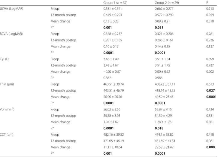
The preoperative values and postoperative alterations after 12 months in visual acuities, corneal HOAs and topographic findings are summarized in Tables1,2, and3. There was no significant difference between the groups in terms of all preoperative values (P > 0.05). The UCVA and
BCVA were significantly improved at 12 months
postoperative compared with preoperative values in both groups (P < 0.05 for all). Corneal total HOAs, spherical aberrations, coma and trefoil values were significantly im-proved at the postoperative 12 months compared with the preoperative values in both groups (P < 0.05 for all). K1, K2 and AvgK values did not differ significantly in Group 1 (P > 0.05), but the difference in K2 and AvgK was signifi-cant in Group 2 (P = 0.0001). There was a signifisignifi-cant de-crease in the maximum keratometry (AKf or Kmax) in both groups (P = 0.005 and P = 0.001, respectively). The thinnest point of the cornea (Thin), corneal volume and the central corneal thickness (CCT) decreased signifi-cantly at 12 months in both groups (P < 0.05 for all).
The mean changes for each parameter from baseline and postoperative 12 months were compared between
the two groups (Tables 1, 2, and 3). The changes in
UCVA and BCVA did not differ significantly in a statis-tical way between the two groups (P = 0.510 and P = 0.137, respectively). The changes in corneal total HOA, spherical aberrations and trefoil values did not differ Table 1 Comparison of visual acuities and corneal topographic findings between the two groups
Group 1 (n = 37) Group 2 (n = 29) P
UCVA (LogMAR) Preop 0.581 ± 0.341 0.662 ± 0.277 0.213
12-month postop 0.449 ± 0.293 0.572 ± 0.299 0.059
Mean change 0.13 ± 0.22 0.09 ± 0.21 0.510
P* 0.001 0.031
BCVA (LogMAR) Preop 0.378 ± 0.237 0.421 ± 0.206 0.281
12-month postop 0.281 ± 0.185 0.283 ± 0.161 0.936 Mean change 0.10 ± 0.13 0.14 ± 0.15 0.137 P* 0.0001 0.0001 Cyl (D) Preop 3.46 ± 1.49 3.51 ± 1.54 0.899 12-month postop 3.48 ± 1.67 3.51 ± 1.75 0.937 Mean change −0.02 ± 0.57 0.00 ± 0.62 0.902 P* 0.862 0.986 Thin (μm) Preop 463.51 ± 38.74 458.72 ± 37.11 0.613 12-month postop 443.51 ± 46.79 418.14 ± 43.35 0.027 Mean change 20.00 ± 20.76 40.59 ± 25.45 0.0001 P* 0.0001 0.0001 Vol (mm3) Preop 56.62 ± 3.56 55.87 ± 4.15 0.434 12-month postop 55.58 ± 3.93 54.59 ± 4.29 0.331 Mean change 1.03 ± 1.62 1.28 ± ± .75 0.561 P* 0.0001 0.018 CCT (μm) Preop 482.16 ± 39.52 474.1 ± 38.82 0.410 12-month postop 471.05 ± 46.19 451.59 ± 41.84 0.081 Mean change 11.11 ± 18.64 22.52 ± 21.42 0.008 P* 0.001 0.0001
UCVA = uncorrected visual acuity; BCVA = best-corrected visual acuity; Cyl = topographic cylindrical value; Thin = thinnest point of cornea; Vol = corneal volume; CCT = central corneal thickness; D = diopters
significantly in a statistical way between the two groups (P = 0.08, P = 0.81 and P = 0.22, respectively). Statisti-cally, the change in corneal coma value was significantly higher in Group 2 (P < 0.05). The change in kerato-metric values K1, K2, AvgK and AKf were significantly higher in Group 2 (P < 0.05 for all). The change in sym-metry index back (SIb) did not differ between the two groups (P = 0.99), but the change in symmetry index front (SIf ) was significantly higher in Group 2 (P = 0.008). The change in Thin and CCT was also higher in Group 2 (P = 0.0001 and P = 0.008, respectively).
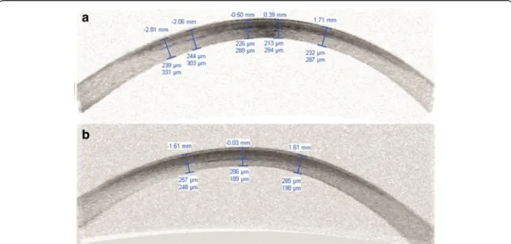
The mean depth of demarcation line was 228.03 ±
36.74μm (min 169 μm to max 305 μm) in Group 1 and
279.86 ± 31.95μm (min 208 μm to max 355 μm) in
Group 2. The difference between the two groups was
significant (P = 0.001). The fig. 1 shows examples of de-marcation lines from both groups.
Subgroup analysis showed no difference in terms of the mean change in UCVA and BCVA between the mild to moderate and advanced keratoconus in both groups (P > 0.05 for all). However, both subgroups showed a sig-nificant decrease in the mean changes in K2 and AvgK in Group 2, with K1 in advanced cases (P < 0.05 for all). AKf also showed an evident but not significant decrease in both subgroups in Group 2 (P > 0.05) (Table4).
Both groups showed no correlation between preopera-tive measurements (visual acuity, thinnest and central cor-neal thickness, corcor-neal volume and AKf ) and the change in AKf at 12 months (P > 0.05 for all) (Table5). Regression analysis showed that the strongest relationship with the Table 2 Comparison of keratometry readings and symmetry indices between the two groups
Group 1 (n = 37) Group 2 (n = 29) P K1 (D) Preop 45.64 ± 2.8 45.88 ± 2.3 0.708 12-month postop 45.71 ± 2.91 45.30 ± 3.02 0.580 Mean change −0.07 ± 0.72 0.58 ± 2.03 0.001 P* 0.559 0.135 K2 (D) Preop 49.10 ± 3.56 49.39 ± 3.06 0.727 12-month postop 49.19 ± 3.63 48.47 ± 3.27 0.408 Mean change −0.09 ± 0.79 0.92 ± 1.22 0.0001 P* 0.506 0.0001 AvgK (D) Preop 47.3 ± 3.09 47.56 ± 2.56 0.715 12-month postop 47.37 ± 3.15 46.63 ± 2.76 0.322 Mean change −0.08 ± 0.69 0.93 ± 1.14 0.0001 P* 0.513 0.0001 AKf (D) Preop 56.38 ± 5.26 56.29 ± 5.34 0.949 12-month postop 55.59 ± 4.98 54.44 ± 5.12 0.364 Mean change 0.79 ± 1.59 1.85 ± 1.58 0.003 P* 0.005 0.001 AKb (D) Preop 80.9 ± 10.76 80.95 ± 12.09 0.985 12-month postop 84.61 ± 11.94 85.9 ± 12.64 0.672 Mean change −3.71 ± 6.06 −4.95 ± 4.35 0.108 P* 0.001 0.0001 SIf (D) Preop 6.48 ± 3.74 6.6 ± 3.63 0.900 12-month postop 6.14 ± 3.57 5.72 ± 3.44 0.631 Mean Change 0.34 ± 0.93 0.88 ± 0.83 0.008 P* 0.031 0.0001 SIb (D) preop 1.67 ± 0.84 1.63 ± 0.82 0.845 12-month postop 1.75 ± 0.79 1.7 ± 0.85 0.796 Mean change −0.08 ± 0.26 −0.06 ± 0.29 0.999 P* 0.091 0.254
K1 = flat keratometry; K2 = steep keratometry; AvgK = average keratometry; AKf = apical keratoscopy front (Kmax); AKb = apical keratoscopy back; SIf = symmetry index front; SIb = symmetry index back; D = Diopters
change in AKf was the type of treatment protocol (β = − 0.482,P = 0.005) (Table6).
Discussion
Keratoconus progression, especially at younger ages, is aggressive and may not stop on its own. Improving the biomechanical strength of the cornea could be a greater benefit than waiting for patients to undergo corneal transplantation. CXL provides the effect of stopping the progression of keratoconus by increasing the degree of interfibrillar linkages through photopolymerization of riboflavin. The long-term results of patients undergoing standard CXL have proven the positive effect of this treatment on stabilizing the keratometric parameters [13–18]. However, this protocol is time-consuming and with their time- and cost-saving benefits, accelerated CXL protocols have been replaced in recent years in ophthalmology practice [3–7,19].
The first article comparing the standard and acceler-ated techniques was published by Tomita et al. [7]. They reported no significant differences in postoperative changes in UCVA and BCVA, in the manifest refraction spherical equivalent or in the postoperative changes in the keratometric readings and the corneal biomechanical responses between the two procedures. According to their findings, a similar demarcation line was formed by
three minutes 30 mW/cm2accelerated CXL and
conven-tional CXL. This first result encouraged ophthalmolo-gists in the use of accelerated technique as a valid alternative for the standard protocol.
Table 3 Preoperative and 12-month postoperative corneal high order aberrations HOAs 6-mm Group 1 n = 37 Group 2n = 29 P Total (RMS,μm) Preoperative 4.60 ± 2.0 4.66 ± 2.06 0.82 12 months postoperative 4.12 ± 1.78 4.08 ± 2.01 0.68 Mean difference 0.48 ± 0.26 0.58 ± 0.30 0.08 P* 0.04 0.04 Spherical (RMS,μm) Preoperative 0.56 ± 0.38 0.58 ± 0.46 0.72 12 months postoperative 0.48 ± 0.41 0.49 ± 0.37 0.80 Mean difference 0.08 ± 0.06 0.08 ± 0.05 0.81 P* 0.02 0.01 Coma (RMS,μm) Preoperative 4.28 ± 2.06 4.44 ± 1.92 0.16 12 months postoperative 3.82 ± 1.95 3.76 ± 1.81 0.32 Mean difference 0.46 ± 0.18 0.68 ± 0.21 0.04 P* 0.02 < 0.01 Trefoil (RMS,μm) Preoperative 0.56 ± 0.36 0.59 ± 0.42 0.60 12 months postoperative 0.42 ± 0.38 0.44 ± 0.40 0.42 Mean difference 0.14 ± 0.09 0.15 ± 0.10 0.22 P* 0.03 0.02
HOAs = higher order aberrations; RMS = root mean square
P: Paired t test; P*: Student’s t-test; Values in bold are significant (P < 0.05)
Fig. 1 AS-OCT shows the demarcation lines of two patients treated with accelerated CXL ((a) 5 min 18 mW/cm2; 213μm depth, (b) 10 min 9 mW/
Since 2014, many published clinical trials, with at least one year of follow-up, were conducted to investigate the therapeutic effect of different types of accelerated CXL
techniques (9 mW/cm2, 18 mW/cm2, 30 mW/cm2 and
45 mW/cm2), comparing them with the Dresden
proto-col [6–8, 20–23]. Even though most of these studies demonstrated successful clinical results, they could not achieve a different perspective. One of the reports that compared the two different types of protocols for
accel-erated CXL (30 mW/cm2for four minutes and 18 mW/
cm2for five minutes) with a one-year follow-up in a lar-ger cohort was recently published and showed no
signifi-cant changes in spherical equivalent, visual and
topographic results [9]. According to their findings, the authors concluded that both modalities of accelerated CXL (total doses of 7.2 J/cm2 and 5.4 J/cm2) exhibited comparable efficacy and applying higher energy for lon-ger periods of time could reach satisfactory results in the stabilization of keratoconus progression following a mean of 12 months. In contrast to these findings, Choi
et al. [22] reported that increasing the UV intensity (30 mW/cm2) and decreasing the irradiation time (to three minutes and 40 s) exhibited less topographical flattening than that of the conventional Dresden protocol. Another study published by Peyman et al. [24] supported Choi’s
conclusion by investigating the stromal demarcation line depth in pulsed and continuous four minutes of acceler-ated CXL protocols. Peyman and associates reported that total fluence of 7.2 J/cm2could not induce a deeper demarcation line in contrast to previous studies.
Although the results of all these studies are chaotic di-versity, most of them indicate that the lower threshold of human corneas for irradiance is the main reason for Bunsen-Roscoe reciprocity law becoming invalid. Our observation corroborates with previous literature re-ports. Our study showed a strong positive relationship between CXL duration and topographic flattening. Al-though improvement in visual acuity was similar in both groups, the change in keratometric values K1, K2, AvgK,
and AKf were significantly higher in 9 mW/cm2for the
Table 4 Changes in parameters from baseline after accelerated crosslinking with 18 mW/cm2(Group 1) and 9 mW/cm2(Group 2) in mild to moderate and advanced keratoconus at 12 months
Mild to Moderate Keratoconus Advanced Keratoconus Group 1 (n = 23) (Mean ± SD) Group 2 (n = 17) (Mean ± SD) P Group 1 (n = 14) (Mean ± SD) Group 2 (n = 12) (Mean ± SD) P UCVA (LogMAR) −0.12 ± 0.18 − 0.07 ± 0.17 0.354 −0.14 ± 0.26 −0.11 ± 0.25 0.803 BCVA (LogMAR) −0.07 ± 0.11 −0.10 ± 0.13 0.515 −0.13 ± 0.14 −0.19 ± 0.16 0.37 K1 (D) −0.01 ± 0.54 −0.22 ± 2.43 0.688 0.20 ± 0.95 − 1.08 ± 1.16 0.005 K2 (D) 0.05 ± 0.61 − 0.82 ± 1.13 0.003 0.14 ± 1.03 − 1.06 ± 1.37 0.019 AvgK (D) 0.01 ± 0.53 − 0.82 ± 1.10 0.003 0.16 ± 0.90 − 1.06 ± 1.21 0.007 Cycl (D) 0.06 ± 0.41 − 0.01 ± 0.60 0.619 − 0.06 ± 0.77 0.02 ± 0.67 0.757 AKf (D) − 0.69 ± 1.36 −1.54 ± 1.47 0.067 − 0.94 ± 1.95 −2.27 ± 1.68 0.078 AKb (D) 3.89 ± 5.69 4.97 ± 4.80 0.531 3.40 ± 6.82 4.91 ± 3.82 0.504 Slf (D) −0.28 ± 0.66 −0.64 ± 0.90 0.153 − 0.44 ± 1.27 −1.21 ± 0.56 0.065 Slb (D) 0.13 ± 0.25 0.12 ± 0.22 0.827 − 0.02 ± 0.25 −0.02 ± 0.36 0.94 Thin (μm) −21.08 ± 23.60 −42.00 ± 28.61 0.016 − 18.21 ± 15.65 −38.58 ± 21.21 0.01 Vol (mm3) −0.93 ± 1.46 −1.13 ± 2.83 0.777 − 1.18 ± 1.9 − 1.48 ± 2.73 0.747 CCT (μm) − 11.82 ± 18.94 −21.05 ± 20.15 0.146 − 9.92 ± 18.77 −24.58 ± 23.86 0.092
UCVA = uncorrected visual acuity; BCVA = best-corrected visual acuity; K1 = flat keratometry; K2 = steep keratometry; AvgK = average keratometry; Cyl = topographic cylindrical value; AKf = apical keratoscopy front (Kmax); AKb = apical keratoscopy back; SIf = symmetry index front; SIb = symmetry index back; Thin = thinnest point of cornea; Vol = corneal volume; CCT = central corneal thickness; D = diopters
P: Paired t test; Values in bold are significant (P < 0.05)
Table 5 Correlation between AKf change at 12-month and preoperative assessment variables
Ch_AKf Preop UCVA Preop BCVA Preop Thin Preop Vol PreopCCT Preop AKf
Group 1 R 0.004 0.041 0.188 −0.088 0.244 −0.320
P 0.982 0.810 0.266 0.606 0.146 0.053
Group 2 R 0.031 0.059 −0.057 −0.136 −0.081 − 0.287
P 0.872 0.762 0.768 0.481 0.678 0.131
UCVA = uncorrected visual acuity; BCVA = best-corrected visual acuity; Thin = thinnest point of cornea; Vol = corneal volume; CCT = central corneal thickness, AKf = apical keratometry front (Kmax); Ch_AKf = change in AKf at 12 months
10 min group (P < 0.05 for all). Also, the change in SIf was significantly higher in the 9 mW/cm2protocol.
Shetty et al. [10] compared the three accelerated CXL
protocols (9 mW/cm2 for 10 min, 18 mW/cm2 for five
minutes, and 30 mW/cm2 for three minutes) with the
conventional CXL. In their prospective randomized interventional study, they reported that conventional CXL and 10 min accelerated CXL provided similar topo-graphic improvement, while the effect of three minutes accelerated CXL was lower compared with the other groups at the end of 12 months. Furthermore, they highlighted the relationship between the flattening effect and energy of radiation, concluding that the efficiency decreases as the amount of energy increases. Although the authors provided a valuable contribution to the treatment capacity of accelerated CXL types, a few points in the study have overshadowed the results. Fol-lowing CXL, improvements in topographical and
aberro-metry parameters indicate CXL’s functional success,
even if its long term biomechanical impact in stabilizing progressive corneal ectasia is limited. Epstein et al. [25] reported that the Kmax value was the most important criterion in the follow-up of the progression of keratoco-nus. Many long-term studies that support this conclu-sion have stated that Kmax is the most commonly used
parameter to determine keratoconus progression [16,
26]. The use of Kmax as an indicator of CXL success is
the most important difference that distinguishes our work from Shetty and colleagues. We also verified
Shetty’s preliminary results by changing methods and
using Sirius topography. Since Sirius combines Placido disk topography with Sheimpflug tomography of the an-terior segment, we could measure the large number of topographic parameters more precisely in a short period of time. Previous studies have found that these two
devices differed significantly; Sirius showed good to ex-cellent repeatability for all measured parameters, espe-cially in healthy corneas [27–29]. In another study, Shetty and colleagues analyzed the effect of post collagen CXL haze on the measurement and repeatability of pachymetry and mean keratometry of four corneal to-pographers [30]. They reported that postoperative mean keratometry values measured with the Pentacam were affected by post-CXL haze. As a result of all these stud-ies, we believe that the Sirius device gives more reliable results than the Pentacam for the tests performed after crosslinking.
In another retrospective case series supporting our findings, Toker and colleagues examined a series of 134 crosslinked eyes. They reported that although the
stand-ard and accelerated CXL (10 min, 9 mW/cm2; 4 min, 30
mW/cm2) results were similar in terms of keratometric
stabilization, the four-minute method showed less topo-graphic improvement. In this study, the demarcation line depth was similar between the standard and 10-min groups (266 mμ, and 273 mμ, respectively), and it was
found 173 mμ in the four-minute group [11]. In our
study, despite no correlation emerging between demar-cation line depth and AKf, the change in corneal coma value was significantly higher in Group 2 (P < 0.05) where the demarcation line was deeper and closer to conventional epithelium-off CXL. Another consideration
was that the concept of “efficacy” after CXL should be
based mainly on its biomechanical impact because it is not a refractive procedure. The shallower demarcation
line in the 18 mW/cm2group may be due to the limited
CXL activity because of insufficient oxygen in the envir-onment due to increased oxygen consumption with high
UVA intensity. Therefore, Mazzotta et al. [31] showed
that the demarcation line reached an average of 280 mμ Table 6 The results of regression analysis of model
Beta T P 95% CI Lower Upper (Constant) 1.290 0.202 −3.248 14.985 Gender 0.007 0.059 0.953 −0.766 0.813 Treatment Type* − 0.482 −2.922 0.005 −2.699 − 0.503 Age − 0.023 −0.183 0.855 −0.072 0.060
Preop AKf level (moderate or advanced) −0.357 −1.976 0.053 −0.227 0.002
Pre BCVA 0.294 1.783 0.080 −0.269 4.637
Preop thinnest point −0.022 −0.043 0.966 −0.045 0.043
Preop corneal volume −0.225 −0.999 0.322 −0.294 0.098
Preop central corneal thickness 0.251 0.580 0.564 −0.026 0.047
Demarcation line 0.206 1.247 0.217 −0.004 0.016
a. Dependent Variable: Change in AKf at 12 months
AKf = apical keratoscopy front (Kmax); BCVA = best-corrected visual acuity; CI = confidence interval of difference * Significant
when they used 15 mW/cm2 UV-A power with pulsed light applied for 6 min.
In keratoconus, increased corneal HOAs further worsen optical quality and visual acuity [32]. By inter-preting the changes in HOAs after CXL, it is possible to establish the benefit of CXL on optical and refractive functions. Studies have shown that in progressive kerato-conus, there is an apparent reduction in HOAs after CXL [33]. However, to the best of our knowledge, there is no study in the literature comparing the corneal HOAs changes after accelerated CXL.
Alió and Shabayek [34] suggested using anterior cor-neal aberrations to evaluate keratoconus and reported that in eyes with keratoconus, coma-like aberrations were found to be significantly higher compared with normal eyes. In our study, we evaluated anterior corneal HOAs and compared their changes in the 6 mm zone. All anterior corneal aberrations demonstrated a signifi-cant reduction in both groups, in accordance with
previ-ous studies presenting improvement in HOAs [33, 35].
In their study with 96 eyes and a 12-month follow-up time, Greenstein et al. [33] reported a significant reduc-tion in total anterior corneal HOAs, total coma, three order coma, and vertical coma. Caporossi et al. [35] also found a significant reduction in the total cornea HOAs and coma aberration from immediately after treatment up to 24 months in 44 eyes. In our study, further im-provement was observed in coma values in the 9 mW/
cm2group (Group 2) at 12 months.
Another point to consider is the real association be-tween the success rate of accelerated CXL types and pre-operative keratoconus severity. Comparisons between the two subgroups revealed that there was no significant difference in terms of the mean change in UCVA and BCVA between the mild to moderate and advanced ker-atoconus at any examination. However, both subgroups showed a significant decrease in the mean changes in K2
and AvgK for the 9 mW/cm2 protocol, with K1 in
ad-vanced cases. Even though the differences did not reach a significant level, the 9 mW/cm2for 10 min application had a greater flattening effect in the maximum kerato-metry in both subgroups.
Some limitations of this study are that there was no comparison with conventional CXL, this study had a short follow-up period, and the sample size was relatively small.
Conclusions
Based on a mean follow-up time of 12 months,
acceler-ated CXL using 10 min of UVA irradiance at 9 mW/cm2
showed better topographic improvements than five
mi-nutes of UVA irradiance at 18 mW/cm2, independent of
keratoconus severity. Significant decreases were
ob-served in HOAs in both groups, but in the 9 mW/cm2
group with the deeper demarcation line depth, coma ab-errations improved further. Although shortening the duration of the procedure is a practical approach in terms of patient and physician comfort, optimum treat-ment protocol should be preferred to slow the progression.
Acknowledgements None.
Funding None.
Availability of data and materials
The datasets used and analyzed for the present study are available from the corresponding author.
Authors’ contributions
AK and YY collected the data. ME interpreted the data and performed statistical analysis. AK, ME and YY were the major contributors in writing the manuscript. All authors read and approved the final manuscript.
Ethics approval and consent to participate
This research followed the tenets of the Declaration of Helsinki. The study was approved by the Clinical Research Ethics Committee of Bagcilar Education and Research Hospital (Project # 2015/233). A written informed consent was obtained from all patients.
Consent for publication
All study subjects gave their informed consent.
Competing interests
The authors declare that they have no competing interests.
Author details
1Department of Ophthalmology, University of Health Sciences, Bagcilar
Training and Research Hospital, Istanbul, Turkey.2Department of
Ophthalmology, Istanbul Medipol University, Istanbul, Turkey.3University of
Health Sciences, Beyoglu Eye Training and Research Hospital, Istanbul, Turkey.
Received: 14 January 2019 Accepted: 12 May 2019
References
1. Wollensak G, Spoerl E, Seiler T. Riboflavin/ultraviolet-a induced collagen crosslinking for the treatment of keratoconus. Am J Ophthalmol. 2003; 135(5):620–7.
2. Subasinghe SK, Ogbuehi KC, Dias GJ. Current perspectives on corneal collagen crosslinking (CXL). Graefes Arch Clin Exp Ophthalmol. 2018; 256(8):1363–84.
3. Medeiros CS, Giacomin NT, Bueno RL, Ghanem RC, Moraes HV Jr, Santhiago MR. Accelerated corneal collagen crosslinking: technique, efficacy, safety, and applications. J Cataract Refract Surg. 2016;42(12):1826–35.
4. Aldahlawi NH, Hayes S, O'Brart DP, Meek KM. Standard versus accelerated riboflavin-ultraviolet corneal collagen crosslinking: resistance against enzymatic digestion. J Cataract Refract Surg. 2015;41(9):1989–96.
5. Hashemi H, Fotouhi A, Miraftab M, Bahrmandy H, Seyedian MA, Amanzadeh K, et al. Short-term comparison of accelerated and standard methods of corneal collagen crosslinking. J Cataract Refract Surg. 2015;41(3):533–40. 6. Elbaz U, Shen C, Lichtinger A, Zauberman NA, Goldich Y, Ziai S, Rootman
DS. Accelerated versus standard corneal collagen crosslinking combined with same day phototherapeutic keratectomy and single intrastromal ring segment implantation for keratoconus. Br J Ophthalmol. 2015;99(2):155–9. 7. Tomita M, Mita M, Huseynova T. Accelerated versus conventional corneal
collagen crosslinking. J Cataract Refract Surg. 2014;40(6):1013–20. 8. Sadoughi MM, Einollahi B, Baradaran-Rafii A, Roshandel D, Hasani H, Nazeri
M. Accelerated versus conventional corneal collagen cross-linking in patients with keratoconus: an intrapatient comparative study. Int Ophthalmol. 2018;38(1):67–74.
9. Yildirim Y, Olcucu O, Gunaydin ZK, Ağca A, Ozgurhan EB, Alagoz C, et al. Comparison of accelerated corneal collagen cross-linking types for treating keratoconus. Curr Eye Res. 2017;42(7):971–5.
10. Shetty R, Pahuja NK, Nuijts RM, Ajani A, Jayadev C, Sharma C, et al. Current protocols of corneal collagen cross-linking: visual, refractive, and tomographic outcomes. Am J Ophthalmol. 2015;160(2):243–9.
11. Toker E, Çerman E, Özcan DÖ, Seferoğlu ÖB. Efficacy of different accelerated corneal crosslinking protocols for progressive keratoconus. J Cataract Refract Surg. 2017;43(8):1089–99.
12. Chan TC, Chow VW, Jhanji V, Wong VW. Different topographic response between mild to moderate and advanced keratoconus after accelerated collagen cross-linking. Cornea. 2015;34(8):922–7.
13. Henriquez MA, Villegas S, Rincon M, Maldonado C, Izquierdo L Jr. Long-term efficacy and safety after corneal collagen crosslinking in pediatric patients: three-year follow-up. Eur J Ophthalmol. 2018;28(4):415–8.
14. Galvis V, Tello A, Ortiz AI. Corneal collagen crosslinking with riboflavin and ultraviolet for keratoconus: long-term follow-up. J Cataract Refract Surg. 2015;41(6):1336–7.
15. De Bernardo M, Capasso L, Lanza M, Tortori A, Iaccarino S, Cennamo M, et al. Long-term results of corneal collagen crosslinking for progressive keratoconus. J Optom. 2015;8(3):180–6.
16. Raiskup F, Theuring A, Pillunat LE, Spoerl E. Corneal collagen crosslinking with riboflavin and ultraviolet-a light in progressive keratoconus: ten-year results. J Cataract Refract Surg. 2015;41(1):41–6.
17. Yildirim A, Cakir H, Kara N, Uslu H, Gurler B, Ozgurhan EB, et al. Corneal collagen crosslinking for ectasia after laser in situ keratomileusis: long-term results. J Cataract Refract Surg. 2014;40(10):1591–6.
18. Kymionis GD, Grentzelos MA, Kankariya VP, Liakopoulos DA, Karavitaki AE, Portaliou DM, et al. Long-term results of combined transepithelial phototherapeutic keratectomy and corneal collagen crosslinking for keratoconus: Cretan protocol. J Cataract Refract Surg. 2014;40(9):1439–45. 19. Elbaz U, Shen C, Lichtinger A, Zauberman NA, Goldich Y, Chan CC, et al.
Accelerated (9-mW/cm2) corneal collagen crosslinking for keratoconus-a 1-year follow-up. Cornea. 2014;33(8):769–73.
20. Woo JH, Iyer JV, Lim L, Hla MH, Mehta JS, Chan CM, et al. Conventional versus accelerated collagen cross-linking for keratoconus: a comparison of visual, refractive, topographic and biomechanical outcomes. Open Ophthalmol J. 2017;11:262–72.
21. Kymionis GD, Kontadakis GA, Hashemi KK. Accelerated versus conventional corneal crosslinking for refractive instability: an update. Curr Opin Ophthalmol. 2017;28(4):343–7.
22. Choi M, Kim J, Kim EK, Seo KY, Kim TI. Comparison of the conventional Dresden protocol and accelerated protocol with higher ultraviolet intensity in corneal collagen cross-linking for keratoconus. Cornea. 2017;36(5):523–9. 23. Ng AL, Chan TC, Lai JS, Cheng AC. Comparison of the central and
peripheral corneal stromal demarcation line depth in conventional versus accelerated collagen cross-linking. Cornea. 2015;34(11):1432–6. 24. Peyman A, Nouralishahi A, Hafezi F, Kling S, Peyman M. Stromal
demarcation line in pulsed versus continuous light accelerated corneal cross-linking for keratoconus. J Refract Surg. 2016;32(3):206–8. 25. Epstein RL, Chiu YL, Epstein GL. Pentacam HR criteria for curvature
change in keratoconus and postoperative LASIK ectasia. J Refract Surg. 2012;28(12):890–4.
26. O'Brart DP, Patel P, Lascaratos G, Wagh VK, Tam C, Lee J, et al. Corneal cross-linking to halt the progression of keratoconus and corneal ectasia: seven-year follow-up. Am J Ophthalmol. 2015;160(6):1154–63.
27. Finis D, Ralla B, Karbe M, Borrelli M, Schrader S, Geerling G. Comparison of two different scheimpflug devices in the detection of keratoconus, regular astigmatism, and healthy corneas. J Ophthalmol. 2015;2015:315281. 28. Lanza M, Paolillo E, Gironi Carnevale UA, Lanza A, Irregolare C, Mele L, et al.
Central corneal thickness evaluation in healthy eyes with three different optical devices. Cont Lens Anterior Eye. 2015;38(6):409–13.
29. Kumar M, Shetty R, Jayadev C, Rao HL, Dutta D. Repeatability and agreement of five imaging systems for measuring anterior segment parameters in healthy eyes. Indian J Ophthalmol. 2017;65(4):288–94. 30. Shetty R, Agrawal A, Deshmukh R, Kaweri L, Rao HL, Nagaraja H, et al. Effect
of post crosslinking haze on the repeatability of Scheimpflug-based and slit-scanning imaging devices. Indian J Ophthalmol. 2017;65(4):305–10. 31. Mazzotta C, Baiocchi S, Bagaglia SA, Fruschelli M, Meduri A, Rechichi M.
Accelerated 15 mW pulsed-light crosslinking to treat progressive keratoconus: two-year clinical results. J Cataract Refract Surg. 2017;43(8):1081–8.
32. Schlegel Z, Lteif Y, Bains HS, Gatinel D. Total, corneal, and internal ocular optical aberrations in patients with keratoconus. J Refract Surg. 2009;25(10 Suppl):S951–7.
33. Greenstein SA, Fry KL, Hersh MJ, Hersh PS. Higher-order aberrations after corneal collagen crosslinking for keratoconus and corneal ectasia. J Cataract Refract Surg. 2012;38(2):292–302.
34. Alió JL, Shabayek MH. Corneal higher order aberrations: a method to grade keratoconus. J Refract Surg. 2006;22(6):539–45.
35. Caporossi A, Mazzotta C, Baiocchi S, Caporossi T. Long-term results of riboflavin ultraviolet a corneal collagen cross-linking for keratoconus in Italy: the Siena eye cross study. Am J Ophthalmol. 2010;149(4):585–93.