Introduction
Orthodontists are among the health care professionals who can undertake functional orthopaedics. They can produce functional or/and orthopaedic changes in three dimensions within the maxillofacial region in subjects with a range of malocclusions. However, one of the most difficult aspects is the diagnosis and treatment of subjects with a Class III malocclusion. Children with a Class III malocclusion may have an underdeveloped maxilla, an overdeveloped mandible, or a combination of both. These skeletal problems may be combined with dental abnormalities, such as a protrusive maxillary and retrusive mandibular dentition (Guyer et al., 1986; Ngan, 2001).
According to Fränkel (1970), the development of a Class
III malocclusion may be a consequence of an imbalance in
the related craniofacial structures. Although Fränkel (1970)
and Fränkel and Fränkel (1989) stated that neuromuscular aberration was not a primary causative factor in the development of the malocclusion, they believed that a functional aberration, including an incorrect habitual position and abnormal activity of the oral and facial muscles, aggravated the deformation by involving adaptive functional activity, which may impede antero-posterior development of the maxilla. Thus, it could be expected that the functional regulator III (FR-3) appliance may work by counteracting the forces of the surrounding muscles that restrict forward maxillary skeletal development and retrude maxillary tooth position, and that the appliance may stimulate forward
Effects of the functional regulator III on profile changes in
subjects with maxillary deficiency
Nihat Kilic*, Mevlüt Celikoglu* and Hüsamettin Oktay**
Department of Orthodontics, Faculty of Dentistry, *Atatürk University, Erzurum and **Istanbul Medipol University, Istanbul, Turkey
Correspondence to: Dr Husamettin Oktay, Istanbul Medipol Üniversitesi Unkapanı, Atatürk Bulvarı No: 27 34083 Fatih-İstanbul, Turkey. E-mail: [email protected]
SUMMARY The aim of this study was to determine the hard and soft tissue profile changes in Class III malocclusion subjects following functional regulator III (FR-3) treatment. The material comprised the cephalometric films of 15 patients (11 males and four females; mean ages 10.22 and 10.44 years, respectively) with a Class III malocclusion and a concave profile treated with the FR-3, and a control group of 15 subjects (11 males and four females; mean ages 10.39 and 10.27 years, respectively) with a Class I malocclusion matched for chronological age and observation period with the study group. Fourteen linear and seven angular measurements were measured on the cephalometric films taken before (T1) and after (T2) treatment/observation.
The results of the Student’s t-test showed that the treatment group had a concave facial profile when compared with the controls. At the end of treatment, the maxilla and surrounding soft tissues showed significant anterior movement (P < 0.001 and P < 0.01, respectively), whereas mandibular growth was restricted. The vertical dimensions increased, the upper incisors proclined, and the lower incisors retroclined significantly (P < 0.001). The FR-3 appliance produced significant improvements in the hard and soft tissues of Class III subjects with a concave profile.
growth of the maxilla resolving the muscle-blocking effects and allowing stretching of the periosteum. In addition, this appliance may produce a restriction of mandibular
development (Fränkel, 1970; McNamara and Huge, 1985;
Fränkel and Fränkel, 1989; Wahl, 2006).
There have been limited studies on the effects of FR-3 therapy on the dentoskeletal structures, although the introduction of this appliance to clinical orthodontics dates back to the 1970s. Almost all authors using the FR-3 appliance are in agreement about the redirection of mandibular growth in a backward and downward direction (Robertson, 1983; Loh and Kerr, 1985; Kerr and TenHave, 1988; Kerr et al., 1989; Ulgen and Firatli, 1994; Baik et al., 2004; Kalavritinos et al., 2005). However, there is considerable controversy in relation to the maxillary skeletal
effects. Fränkel (1970), McNamara and Huge (1985),
Miethke et al. (2003), Falck and Zimmermann-Menzel (2008), and Levin et al. (2008) observed substantial effects due to FR-3 therapy on the maxillary basal bone. On the
other hand, many clinicians (Robertson, 1983; Loh and
Kerr, 1985; Kerr and TenHave, 1988; Kerr et al., 1989; Biren and Erverdi, 1993; Ulgen and Firatli, 1994; Baik et al., 2004; Kalavritinos et al., 2005) do not support Fränkel’s supposition that FR-3 therapy has a favourable effect on the maxillary basal bone. According to those authors, correction of a Class III malocclusion is produced mainly by the downward and backward rotation of the mandible and/or alterations in upper and lower incisor inclinations.
Figure 1 Functional regulatorIII appliance.
As previously stated, little interest has been shown in investigating the changes in dentoskeletal structures induced by the FR-3 appliance. Surprisingly, hard tissue and concomitant soft tissue effects of the FR-3 appliance
were often neglected in the published literature (McNamara
and Huge, 1985; Kerr and TenHave, 1987; Biren and Erverdi, 1993; Kalavritinos et al., 2005).
Soft tissue profile effects of FR-3 therapy were first
mentioned by McNamara and Huge (1985). Kerr and
TenHave (1987) found considerable changes in the soft tissue profile of patients treated by FR-3 appliances when compared
with a control sample. Biren and Erverdi (1993) compared
soft tissue changes in 25 patients treated with the FR-3 with those of 20 untreated subjects with a Class III malocclusion and noted the beneficial effects of FR-3 therapy on the soft
tissue profile. Recently, Kalavritinos et al. (2005) in a study
of 14 subjects who had undergone FR-3 therapy over a period of 2.4 years reported favourable outcomes such as a significant decrease in SNB angle and lower lip convexity and an increase in ANB angle, overjet, facial convexity, nose prominence, lower soft tissue face height, and upper lip thickness.
These studies investigating the soft tissue profile have some deficiencies that can be summarized as follows: (1)
case report (McNamara and Huge, 1985), (2) inadequate
evaluation of soft tissue profile (McNamara and Huge,
1985; Biren and Erverdi, 1993), (3) no control group (Kalavritinos et al., 2005), and (4) a control group with
various malocclusion (Kerr and TenHave, 1987).
The primary aim of treatment in Class III subjects must be focused on soft tissue profile and appearance rather than occlusion, as opposed to other malocclusion groups, since the facial profile of skeletal Class III deformities is usually the main concern of these patients (Kerr and TenHave, 1987). This presents a considerable challenge to orthodontists and it is essential when developing a treatment plan to estimate facial changes along with occlusal improvements.
This study aimed to investigate hard tissue and concomitant soft tissue profile changes induced by FR-3 appliance therapy.
Subjects and methods
Ethical approval from the local ethics committee and informed consent from the parents of the children were obtained.
Fifteen patients (11 males and four females) treated using the original FR-3 (Figure 1) and 15 subjects (11 males and four females) with a normal occlusion (control group) were included in the present study. The chronological ages of the subjects in both groups are shown in Table 1. The normal occlusion group was chosen from the longitudinal archive of the Department of Orthodontics, Ataturk University. Selection of the control subjects was based on normal growth and development, a balanced skeletal
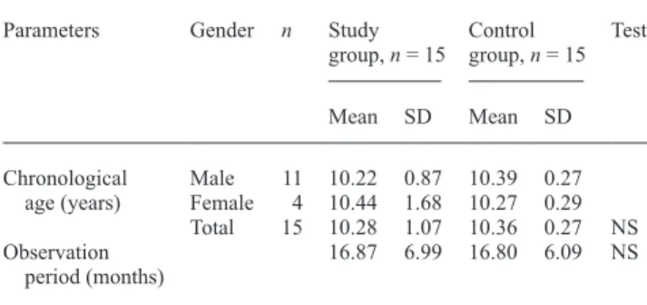
Table 1 Initial chronological ages and observation periods of the
groups and their comparisons. Parameters Gender n Study
group, n = 15 Control group, n = 15 Test Mean SD Mean SD Chronological
age (years) Male 11 10.22 0.87 10.39 0.27 NS Female 4 10.44 1.68 10.27 0.29 Total 15 10.28 1.07 10.36 0.27 Observation period (months) 16.87 6.99 16.80 6.09 NS NS, not significant.
profile, a Class I occlusion, and absence of anterior and/or posterior crossbites. The patients in the treatment group had maxillary retrusion or a combination of maxillary retrusion and mandibular protrusion (skeletal Class III relationship with anterior crossbite and retrusive nasomaxillary area). Subjects with a history of orthodontic treatment were not included.
During the first 2 weeks, the patients wore their appliances 4–5 hours during the day for slow adaptation. If, after this time, the mucosa looked healthy, the patients were instructed to wear the appliance all day but not at nights. After the patients had adapted to wearing the FR-3 during the day, they wore their appliances at all times except while eating or brushing their teeth. The appliance and treatment progress were checked at 4 weekly intervals. Active functional treatment was terminated when the negative overjet and concave profile were corrected.
Cephalometric radiographs were obtained from all subjects at the beginning and after treatment and at the control periods. The films were taken in a standard position by one operator using the same X-ray machine and cephalostat (Siemens Nanodor 2; Siemens AG,
Figure 2 Landmarks used in the study: S, sella; N, nasion; Or, orbitale; Ss, sulcus superior; Ls, labiale superior; Li, labiale inferior; Si, sulcus inferior; Pog′, soft tissue pogonion; A, Point A; B, Point B; Pog, pogonion; Me, menton; Go, gonion; Po, porion; Se, intersection of the greater wing of the sphenoid bone with the floor of the anterior cranial fossa; ptm, the most inferior and posterior point on the anterior outline of the pterygomaxillary fissure; Prn, pronasale.
Figure 3 Lines and linear and angular measurements used in the study: y-axis (PMV plane): a line passing through Se and Ptm points. E line: Ricketts’ aesthetic line extending between the nose tip (Prn) and soft tissue pogonion (Pog′). Mandibular plane: a plane passing through gonion and menton. Frankfort horizontal plane: horizontal plane passing through porion and orbitale. (1) A–y, (2) Ss–y, (3) Ls–y, (4) Li–y, (5) Si–y, (6) B–y, (7) Pog′–y, (8) Pog–y, (9) Ss–E, (10) Ls–E, (11) Li–E, (12) Si–E, (13) U1–SN, (14) Z angle, (15) nasolabial angle, (16) labiomental angle, (17) IMPA, (18) FMA (Frankfort-mandibular plane angle), (19) SN–GoMe. Munich, Germany). In order to determine hard and soft tissue profile changes, 14 linear and seven angular measurements were used. These measurements were
adopted from the studies of Varlik et al. (2008) and
Nanda et al. (1990) (Figures 2 and 3).
Statistical analysis
To determine the errors associated with digitizing and measurements, 15 radiographs were randomly selected. All procedures such as landmark identification, tracing, and measurement were repeated after a period of 2 weeks by the same author (MC). Intraclass correlation coefficients were performed to assess the reliability of the measurements as
described by Houston (1983).
Comparisons between the groups were carried out by means of a Student’s t-test. All statistical analyses were performed using the Statistical Package for Social Sciences (Windows 98, version 10.0; SPSS Inc., Chicago, Illinois, USA).
Results
The coefficients of reliability of the measurements were above 0.90 for all parameters. Initial chronological ages and observation periods of the groups and their comparisons are shown in Table 1. There was no statistically significant difference between the groups in relation to these parameters.
The results of the Student’s t-test comparing initial values for the groups are shown in Table 2. The distances from point A, sulcus superior, and lip superior to PMV line (y-line); sulcus inferior to E line measurement; U1–SN angle; IMPA; overjet; and overbite were significantly smaller in the treatment group, whereas the distances from sulcus superior and lip superior to E line; point B to PMV line measurement; and Z, nasolabial, labiomental, SN– GoMe, and FMA angles were significantly larger than those of the control group.
Changes occurring during the treatment and observation periods are shown in Table 3. The changes in all measurements, except Ls–E and Ls–y distances and labiomental angle, showed statistically significant differences between the groups, i.e. the maxilla (A–y) and superior sulcus (Ss–y and Ss–E) showed significantly more anterior movement, while anterior movements of the mandible (B–y, Pog–y, and Pog′–y) and lower lip (Li–y and Si–y) were inhibited (Table 3). Lip inferior and sulcus inferior to E line measurements increased significantly (P < 0.01). The upper incisors proclined and the lower incisors retroclined significantly in the FR-3 group (P < 0.001). The Z angle decreased and the nasolabial angle increased with treatment (P < 0.001). The vertical dimensions of the face also significantly increased (P < 0.001) in the FR-3 group. Overjet increased (P < 0.001) and overbite decreased (P < 0.01).
Discussion
The soft tissue facial profile has been considered as an important objective of orthodontic/orthopaedic therapy in patients with
Class III malocclusions (Kilicoglu and Kirlic, 1998). The
evaluation of the treatment effects. The present study intended to evaluate the profile changes induced by FR-3 therapy.
Since ethical considerations did not allow postponing treatment of Class III subjects for scientific purposes, the control group was formed of subjects with an acceptable occlusion and a skeletal Class I relationship.
In the present study, FR-3 therapy was undertaken in Class III subjects aged from 8 to 12 years, mean age 10.28 years. All subjects were prepubertal and in the mixed dentition stages at the beginning of treatment. There is a consensus that treatment with the FR-3 should be
commenced during the prepubertal stage (Fränkel, 1970;
Loh and Kerr, 1985; Ulgen and Firatli, 1994; Baik et al., 2004; Falck and Zimmermann-Menzel, 2008; Levin et al., 2008). Loh and Kerr (1985), Kerr and TenHave (1987), Kerr et al. (1989), Kalavritinos et al. (2005), and Ulgen and Firatli (1994) applied the FR-3 to subjects aged between 9.50 and 11.10 years. However, some clinicians (Fränkel, 1970; Biren and Erverdi, 1993; Baik et al., 2004; Falck and Zimmermann-Menzel, 2008; Levin et al., 2008) started FR-3 therapy at a younger age.
Table 2 shows that the FR-3 group had a retrusive maxilla and upper lip, protrusive mandible and lower lip, retroclined lower incisors, and reduced overjet and overbite. These pre-treatment findings are coincident with those of similar
relevant studies (Loh and Kerr, 1985; Kerr and TenHave,
1987; Kerr et al., 1989; Ulgen and Firatli, 1994). Skeletal changes
There are conflicting opinions regarding the treatment effects of the FR-3 appliance on maxillary growth. Some
reports (Fränkel, 1970; McNamara and Huge, 1985;
Kohmura et al., 1986; Aytan et al., 1989; Isiksal and Seckin, 1990; Miethke et al., 2003; Falck and Zimmermann-Menzel, 2008; Levin et al., 2008) support the idea that FR-3 therapy produces a favourable growth effect on the maxilla, while Robertson (1983), Loh and Kerr (1985), Kerr and TenHave (1987, 1988), Ulgen and Firatli (1994), and Kalavritinos et al. (2005) did not find similar results. Fränkel (1970) investigated the effects of FR-3 therapy on maxillary development and observed significant increases in maxillary growth. This favourable effect on the basal maxilla was also
supported by the studies of McNamara and Huge (1985),
Kohmura et al. (1986), Aytan et al. (1989), Isiksal and Seckin (1990), Miethke et al. (2003), and Falck and Zimmermann-Menzel (2008). Recently, Levin et al. (2008) showed that the FR-3 appliance produced considerable improvements in maxillary size and position. They found that SNA increased 1.3 degrees and effective midfacial
length was 1.3 mm more than in the control group. Baik et al.
(2004) found excellent results in most of their treated cases, although no statistically significant difference were observed between the treatment and control group regarding the basal maxilla. The findings of the present study indicate that the
Table 2 Results of the Student’s t-test comparing the initial
cephalometric values between the groups.
Parameters Study group Control group P value
Mean SD Mean SD A–y 48.00 2.68 51.47 3.08 0.000 Ss–y 63.30 3.07 66.22 3.89 0.002 Ls–y 67.17 3.94 70.52 4.72 0.004 Li–y 70.12 5.62 70.00 5.33 0.935 Si–y 65.92 5.00 63.52 5.71 0.089 B–y 55.27 4.97 52.38 5.39 0.036 Pog–y 57.98 5.72 55.43 6.32 0.107 Pog′–y 69.43 6.09 67.13 6.86 0.175 Ss–E −9.07 1.70 −7.67 1.40 0.001 Ls–E −5.03 2.40 −2.00 2.07 0.000 Li–E −1.23 2.78 −0.57 2.52 0.335 Si–E −4.32 1.37 −5.57 1.90 0.005 U1–SN 98.47 5.97 102.40 4.70 0.006 Z angle 77.88 5.83 75.23 3.37 0.035 Nasolabial angle 116.57 9.33 107.65 9.87 0.001 Labiomental angle 136.90 7.29 122.33 11.20 0.000 IMPA 88.95 4.79 98.30 3.00 0.000 SN–GoMe 37.18 5.93 32.90 4.26 0.002 FMA 29.52 4.41 26.47 3.71 0.002 Overjet −1.23 1.06 1.47 1.06 0.000 Overbite 1.13 1.73 2.43 1.06 0.001
Table 3 Results of the Student’s t-test comparing the mean changes
between groups.
Parameters Study group Control group P value
Mean SD Mean SD A–y 2.45 1.08 1.32 0.72 0.000 Ss–y 2.85 1.46 2.07 1.05 0.020 Ls–y 2.65 1.30 2.55 1.52 0.785 Li–y 0.73 1.78 2.47 1.58 0.000 Si–y 0.48 1.70 1.75 1.34 0.002 B–y 0.40 1.70 1.88 1.10 0.000 Pog–y 0.98 2.01 2.17 1.33 0.010 Pog′–y 1.07 1.81 2.43 1.56 0.003 Ss–E 0.27 0.93 −0.28 0.83 0.038 Ls–E 0.27 1.69 0.03 1.46 0.570 Li–E −0.97 1.54 0.01 1.85 0.027 Si–E −1.22 0.99 −0.60 0.67 0.007 U1–SN 2.50 2.42 0.12 1.03 0.000 Z angle −2.22 1.85 −0.47 1.76 0.000 Nasolabial angle 2.90 3.24 −0.22 2.07 0.000 Labiomental angle −0.43 3.67 −1.00 2.35 0.479 IMPA −4.05 2.14 0.35 1.50 0.000 SN–GoMe 2.28 0.86 0.15 1.27 0.000 FMA 1.80 0.77 0.22 1.28 0.000 Overjet 2.00 0.96 0.47 0.75 0.000 Overbite −0.33 1.58 0.37 0.94 0.042
improvement of facial aesthetics has been one of the major concerns for seeking treatment. Soft tissue changes following orthodontic treatment are sometimes regarded as secondary to the
underlying hard tissue alterations (Kerr and TenHave, 1987).
However, soft tissue changes play an important role in the
response of the craniofacial complex to the FR-3 appliance therapy included favourable changes in the basal maxilla.
As mentioned, some studies (Robertson, 1983; Loh and
Kerr, 1985; Kerr and TenHave, 1987, 1988; Kerr et al., 1989; Biren and Erverdi, 1993; Ulgen and Firatli, 1994; Kalavritinos et al., 2005) failed to find significant
improvements in the maxilla. Some (Robertson, 1983; Loh
and Kerr, 1985) used no control group, while others (Kerr and TenHave, 1987, 1988; Kerr et al., 1989) selected their control groups from subjects with various malocclusions. Kalavritinos et al. (2005) compared their results with those of an untreated control sample from a published study. Ulgen and Firatli (1994) used FR-3 therapy in a group of subjects with a functional Class III malocclusion.
In relation to the mandibular changes, the present findings showed that FR-3 appliance therapy restricted mandibular growth (B–y and Pog–y) and increased the vertical
dimensions (SN–GoMe and FMA angles). Baik et al. (2004)
observed that the mandibular plane angle was increased, but mandibular growth was not inhibited with FR-3 treatment. In
the short term, Levin et al. (2008) found that SNB angle
decreased 0.3 degrees in the treatment group and increased 0.4 degrees in the control, while mandibular length (Go–Gn) increased by 5.4 mm in the study group and 6.5 mm in the control group. These values were not significant between the groups. They also found that the position of pogonion relative to nasion-perpendicular showed a significant difference between the groups (−0.7 mm in the treatment
group and 1 mm in the control group). McNamara and Huge
(1985) showed that FR-3 therapy directed mandibular growth in a vertical direction. The vertical changes observed in the present research are in agreement with studies that found the FR-3 appliance to stimulate mandibular growth in a vertical
direction (Loh and Kerr, 1985; Kerr et al., 1989; Ulgen and
Firatli, 1994; Baik et al., 2004; Kalavritinos et al., 2005) Dentoalveolar changes
After FR-3 appliance therapy, the molar relationship and anterior crossbite were corrected, and the changes in overjet and overbite measurements were statistically significant between the groups (Table 3). Proclination of the upper incisors and retroclination of the lower incisors contributed to the improvements in overjet and overbite. It has been generally accepted that the lower incisors retrocline
significantly after FR-3 therapy (Loh and Kerr, 1985; Kerr
et al., 1989; Isiksal and Seckin, 1990; Ulgen and Firatli, 1994; Baik et al., 2004; Levin et al., 2008), while the findings regarding upper incisor inclination differ
somewhat. Kerr et al. (1989) and Levin et al. (2008)
observed a significant increase in 1–SN angle, but contrary
to the finding of Ulgen and Firatli (1994) and Baik et al.
(2004). Compatible with the present findings, many authors (Kerr et al., 1989; Ulgen and Firatli, 1994; Baik et al., 2004; Levin et al., 2008) found that the overjet increased
significantly after FR-3 therapy. The present results are
coincident with those of Kerr et al. (1989) and Ulgen and
Firatli (1994), who found a statistically significant reduction in overbite when compared with the controls. In contrast, Baik et al. (2004) and Levin et al. (2008) found insignificant changes in overbite measurements (−0.3 and 0.2 mm and 0.9 and 0.5 mm in the treatment and the control group, respectively).
Soft tissue changes
Significant increases in A–y, Ss–y, and Ss–E measurements showed that the FR-3 appliance caused considerable changes in the basal maxilla and surrounding soft tissue structures, although anterior movement of the upper lip did not reach significance. The Z angle decreased, and the nasolabial angle increased in the treated subjects. These changes could be explained by anterior movement of the upper lip.
The effects of FR-3 therapy on the soft tissue profile of the
face have been reported (McNamara and Huge, 1985; Kerr
and TenHave, 1987; Kerr et al., 1989; Biren and Erverdi, 1993). McNamara and Huge (1985) observed mild-to-moderate profile improvements in three treated patients, and Biren and Erverdi (1993) noted the beneficial effects of the
FR-3 on the soft tissue facial profile. According to Kerr and
TenHave (1987), the main treatment response of the FR-3 was in the lower facial area, and this effect caused a reduction in the concavity of the Class III soft tissue profile.
A balanced soft tissue profile is a desired treatment
objective in orthodontics (Kerr and TenHave, 1987). The
findings of the present study indicate that significant improvements in soft tissue profile occur after FR-3 therapy. These small significant changes were similar to those found by other authors. The appliance tended to reduce the concavity of the hard and soft tissue profile by allowing growth of the maxilla and surrounding soft tissues and inhibiting mandibular growth.
Conclusions
1. Initial cephalometric measurements showed the group treated with the FR-3 appliance to have concave profiles relative to the normal subjects.
2. The maxilla and surrounding soft tissues showed significant anterior movement, whereas mandibular and surrounding soft tissue growth were inhibited, and the vertical dimensions were increased
3. The Class III occlusion and overjet were improved by means of skeletal changes in conjunction with upper incisor proclination and lower incisor retroclination.
References
Aytan S, Yukay F, Ciger S, Enacar A, Aksoy A, Telli A E 1989 Fränkel III appliance. Turkish Journal of Orthodontics 2: 338–345
Baik H S, Jee S H, Lee K J, Oh T K 2004 Treatment effects of Fränkel functional regulator III in children with Class III malocclusions. American Journal of Orthodontics and Dentofacial Orthopedics 125: 294–301 Biren S, Erverdi N 1993 Cephalometric evaluation of maxillary
retrognathism cases treated with FR-3 appliance. Journal of Marmara University Dental Faculty 1: 354–360
Falck F, Zimmermann-Menzel K 2008 Cephalometric changes in the treatment of Class III using the Fränkel appliance. Journal of Orofacial Orthopedics 69: 99–109
Fränkel R 1970 Maxillary retrusion in Class 3 and treatment with the function corrector 3. Transactions of the European Orthodontic Society, pp. 249–259
Fränkel R, Fränkel C 1989 Orofacial orthopedics with the function regulator. Karger, Basel
Guyer E C, Ellis E E, McNamara Jr J A, Behrents R G 1986 Components of Class III malocclusion in juveniles and adolescents. Angle Orthodontist 56: 7–30
Houston W J B 1983 The analysis of errors in orthodontic measurements. American Journal of Orthodontics 83: 382–390
Isiksal E, Seckin O 1990 The functional regulators (FR III, FR IV). Turkish Journal of Orthodontics 3: 150–156
Kalavritinos M, Papadopoulos M A, Nasiopoulos A 2005 Dental arch and cephalometric changes following treatment for class III malocclusion by means of the function regulator (FR-3) appliance. Journal of Orofacial Orthopedics 66: 135–147
Kerr W J, TenHave T R 1987 Changes in soft tissue profile during the treatment of Class III malocclusion. British Journal of Orthodontics 14: 243–249
Kerr W J, TenHave T R 1988 A comparison of three appliance systems in the treatment of Class III malocclusion. European Journal of Orthodontics 10: 203–214
Kerr W J S, TenHave T R, McNamara J A 1989 A comparison of skeletal and dental changes produced by functional regulators (FR-2 and FR-3). European Journal of Orthodontics 11: 235–242
Kilicoglu H, Kirlic Y 1998 Profile changes in patients with Class III malocclusions after Delaire mask therapy. American Journal of Orthodontics and Dentofacial Orthopedics 113: 453–462
Kohmura T et al. 1986 Effects of therapy using the function regulator (FR III) on the cases with the reversed occlusion. Nippon Kyosei Shika Gakkai Zasshi 45: 693–711
Levin A S, McNamara J A Jr, Franchi L, Baccetti T, Fränkel C 2008 Short-term and long-Short-term treatment outcomes with the FR-3 appliance of Fränkel. American Journal of Orthodontics and Dentofacial Orthopedics 134: 513–524
Loh M K, Kerr W J 1985 The function regulator III: effects and indications for use. British Journal of Orthodontics 12: 153–157
McNamara J A, Huge S A 1985 Functional regulator (FR-3) of Fränkel. American Journal of Orthodontics 88: 409–424
Miethke R R, Lindenau S, Dietrich K 2003 The effect of Fränkel’s function regulator type III on the apical base. European Journal of Orthodontics 25: 311–318
Nanda R S, Meng H, Kapila S, Goorhuis J 1990 Growth changes in the soft tissue facial profile. Angle Orthodontist 60: 177–190
Ngan P 2001 Treatment of Class III malocclusion in the primary and mixed dentitions. In: Bishara S E (ed.). Textbook of orthodontics. W.B. Saunders Company, Philadelphia, pp. 324–374.
Robertson N R E 1983 An examination of treatment changes in children treated with the function regulator of Fränkel. American Journal of Orthodontics 83: 299–310
Ulgen M, Firatli S 1994 The effects of the Fränkel’s function regulator on the Class III malocclusion. American Journal of Orthodontics and Dentofacial Orthopedics 105: 561–567
Varlik S K, Gultan A, Tumer N 2008 Comparison of the effects of Twin Block and activator treatment on the soft tissue profile. European Journal of Orthodontics 30: 128–134
Wahl N 2006 Orthodontics in 3 millennia. Chapter 9: functional appliances to midcentury. American Journal of Orthodontics and Dentofacial Orthopedics 129: 829–833