Therapeutic Nanomaterials, First Edition. Edited by Mustafa O. Guler and Ayse B. Tekinay.
© 2016 John Wiley & Sons, Inc. Published 2016 by John Wiley & Sons, Inc.
SAFETY OF NANOMATERIALS
Nuray Gunduz, Elif Arslan, Mustafa O. Guler,
and Ayse B. Tekinay
Institute of Materials Science and Nanotechnology, National Nanotechnology Research Center (UNAM), Bilkent University, Ankara, Turkey
11.1 INTRODUCTION
Advances in the design and fabrication of nanomaterials have created novel opportunities for the development of effective therapeutic agents against hard‐to‐treat disorders. However, the long‐term biocompatibility of these materials remains an important concern for their use in biomed ical applications. A thorough assessment of potential toxicity is therefore necessary for any material intended for use in biological environments; nonetheless standard methods for the evaluation of nanomaterial toxicity are often lacking. In this chapter, we will overview the attempts in under standing the biocompatibility of nanomaterials and provide an account of how these views changed in light of recent findings, with emphasis on the methodology used in nanotoxicology studies.
Nanoparticles (NPs) have commonly been used in the fields of engi neering, medicine, pharmacology, and biotechnology over the last 20 years (Granqvist et al., 1976). NPs of iron and noble metals are especially popular in biomedical research, as they are largely inert toward biological agents, display optical properties ideal for in vivo imaging, and can be
functionalized with a variety of targeting molecules. These properties render NPs and other engineered nanomaterials (ENMs) attractive for use in clinical studies, and ENM‐based methods have been proposed for appli cations such as targeted gene and drug delivery, computed tomography, Raman spectroscopy, photoacoustic imaging, and photothermal therapy (Choi and Wang, 2011; Neuberger et al., 2005; Prasad, 2009). However, the popularity of ENMs in biomedical and industrial applications has led to questions regarding their safety and especially on their long‐term toxic effects on living organisms. As with any other foreign element, nanomate rials can affect the functioning of the body by disrupting any of a large number of interconnected pathways, and the nanotoxicology of ENMs should be evaluated in a broad and comprehensive manner by taking account of the individual and synergistic aspects of ENM biology. The physicochemical features, exposure routes, uptake mechanisms, cellular localization, and biodistribution, clearance, adverse molecular responses (inflammation and ROS generation), and genotoxicity of ENMs should therefore all be considered to provide a coherent account of their potential toxic effects.
A large number of techniques exist for the evaluation of these features under in vitro or in vivo conditions, and some (but not all) are typically employed to test the safety of ENMs intended for biomedical use. However, despite the considerable volume of research regarding the adverse effects of nanostructures, a comprehensive toxicity assessment of ENMs is yet to be attempted in the literature, and guidelines for the safe use and manufac turing of these materials are likewise wanting. Nonetheless, institutional organizations such as the Environmental Protection Agency (EPA), World Health Organization (WHO), National Institute for Occupational Safety and Health (NIOSH), and the European Commission frequently perform toxicology assessments on nanostructures to better understand the risks involved with this class of materials (Park, 2012).
11.2 CHARACTERIZATION, DESIGN, AND SYNTHESIS OF NANOMATERIALS
11.2.1 Chemical Identity and Physicochemical Properties
NPs are commonly used in various in vitro and in vivo toxicity studies, and computational approaches have also been used for the in silico toxicity assessment of ENMs with minimal cost and no harm to living organisms (Clark et al., 2011; García‐Remesal et al., 2013; Han et al., 2011; Sayes
and Ivanov, 2010). The determination of the chemical identity of ENMs is a vital aspect of their characterization, and potential impurities or batch‐ to‐batch variances may play a major role in the toxicity of NPs. ENMs can be synthesized with various shapes, structures, sizes, and chemical and physical properties. Methods for the synthesis and assembly of ENM are likewise variable and may involve a large number of physical and chemical processes using solid, liquid, or gas precursors (Saravanan et al., 2008). Synthesis methods are broadly classified as either bottom‐up or top‐down approaches (Saravanan et al., 2008): Bottom‐up techniques assemble a larger structure from smaller units or “building blocks” (such as the syn thesis of NPs from a solvent in a sol‐gel method), while top‐down tech niques reduce a bulk material to an array of smaller structures through microfabrication processes such as grinding, polishing, and lithography (Ghosh Chaudhuri and Paria, 2011).
The design approach chosen for ENM synthesis may heavily influence its toxicity; as such, the biocompatibility of an ENM can be considered to be a major design characteristic. Biocompatibility for ENMs is defined as the ability to show an intended biomedical function without any adverse effects (Williams, 2008) and depends heavily on the size, shape, surface area, composition, solubility, surface charge, and purity of the nanomaterial. In addition, it must be noted that ENMs are typically functionalized with a variety of biological moieties to modulate their toxic effects or impart additional properties to the material. These moieties represent another criti cal aspect of ENM fabrication, as most NPs are designed and developed according to their intended biological functionality. It is therefore vital to preserve this functionality through the path of the nanomaterial from its site of introduction to the site of action, and the failure to do so may result in adverse effects due to the nonspecific activity of the nanomaterial.
Surface coating is the most common method for introducing a fun ctional moiety onto an NP. An outer layer of biofunctional material not only provides bioactivity to the NP but also increases its stability in serum or storage. Surface conjugation can be performed for purposes such as cell adhesion or targeting of a specific tissue, organ, or disease (Li et al., 2008; Matsuo et al., 2003; Xie et al., 2005). Targeting moieties also eliminate potential side effects by preventing the NPs from binding to cells or tissues outside their site of interest. As such, another important role of coating is to reduce the toxic or adverse effects of ENMs, and it is known that differ ent surface coatings may modulate the physicochemical properties and cytotoxic effects of NPs (Suresh et al., 2012).
The primary structural features of ENMs are their sizes and surface areas. The size of the ENM not only affects its physicochemical characteristics
but also plays roles in its uptake and interaction with cells, tissues, organs, and, in cases where the NP is not administered but surreptitiously encoun tered as a contaminant, whole organisms. Nanomaterials with smaller sizes have greater surface areas per mass, which allows a better interac tion between the nanomaterial surface and biological materials (Ghosh Chaudhuri and Paria, 2011). For example, the uptake mechanism of ENMs was dramatically influenced by the sizes of the nanomaterials in case of silver nanoparticles (NPs) with different sizes. Smaller NPs were also more active in increasing the generation of ROS and expression of inflam matory markers (Park et al., 2011). Size characterization is therefore critical for ENMs that are intended for use in biomedical applications and can be performed by TEM analysis (visual characterization) and UV/vis spectroscopy (quantified characterization) (Browning et al., 2013). In addition, while ENMs may be stable in water or standard buffers, biological environments are often complex and contain proteins that may trigger the aggregation of NPs. As such, size characterization should also be per formed in a medium that adequately simulates the biological environment at the site of action (Powers et al., 2007).
The biological activity of an ENM is also influenced by its morphology, the importance of which is addressed in several studies. One such paper demonstrates that AuNR−PEG (Au nanorods functionalized with PEG) nanostructures were more cytotoxic to human skin cells than AuNS−MPS (Au nanospheres functionalized with mercaptopropane sulfonate) and suggests that differences in shape and agglomeration responses play a cru cial role in NP toxicity (Schaeublin et al., 2012). The aspect ratio of an ENM is often a good predictor for the potential effects of its morphology, as high‐aspect ratio ENMs (nanotubes and nanowires) and low‐aspect ratio ENMs (nanospheres and similar structures) display distinct behaviors in uptake and cytotoxicity.
Chemical composition is another critical parameter that is frequently characterized for the nanotoxicology assessment of ENMs: Prahalad et al., for example, have shown that material composition, solubility, and acidity are important factors for determining the responses of cells to transition metal‐based ENMs (Prahalad et al., 1999). In addition to the three major parameters of size, shape, and composition, properties such as surface chemistry, surface structure and charge, purity, solubility, crystal structure, catalytic activity, acidity/alkalinity, and agglomeration behavior also affect the toxicity of ENMs (Arora et al., 2012; Donaldson and Poland, 2013; Kroll et al., 2009; Park, 2012). All these parameters may alter cellular responses to ENMs by changing uptake mechanisms (through associations with endo‐ or exocytosis pathways) or facilitating interactions with proteins
and other biological macromolecules (Lynch et al., 2009; Nel et al., 2009). As such, their evaluation is key for predicting the potential toxicity of an NP.
The chemistry, surface charge, and nanotopology of an ENM can change greatly through the process of its synthesis and application, especially due to the moieties used in surface modification and the structural alterations they undergo in biological environments. Therefore, the characterization of the surface chemistry of ENMs is an essential step for nanotoxicology studies (Johnston et al., 2010a). The charge of the ENM is another critical physicochemical aspect and should be characterized to determine its potential interactions with molecules, cells, and especially membranes, which are often charged (Johnston et al., 2010a). The solubility of the ENM is important for its biocompatibility (as biological environments are mostly aqueous) and for the evaluation of biopersistence (Johnston et al., 2010a). Both of these parameters must therefore be investigated in detail to provide an adequate explanation for the toxicity (or the lack thereof) of a material, as well as to design novel ENMs with minimal toxic effects. The reliability and applicability of the analysis method used is also crucial, as experimental protocols may potentially alter the physicochemical features of ENMs, making it difficult to predict or explain their in vivo behavior. 11.2.2 Biological Identity
Biological responses toward ENMs are strongly affected by the interac tions between the ENM and cells, body fluids, proteins, and other macro molecules, which are also important for the distribution of ENMs in the target organism (Nel et al., 2006). The adsorption capacity of an ENM correlates with its size and influences material toxicity. NPs with smaller sizes possess larger surface areas per mass, which increases their adsorp tion capacity and biological interactions. These interactions trigger the formation of a protein coat, called the corona, when NPs are exposed to a biological environment (Kroll et al., 2009). The corona affects the effec tive size, charge, and accumulation/aggregation behaviors of ENMs and is in turn influenced by the surface chemistry of the NP (Kroll et al., 2009; Park, 2012). After the ENM assembles its corona of aggregating proteins, it may also gain the ability to interact with cells and tissues that it would normally fail to recognize (Nel et al., 2006). In addition, these interactions may result in the degradation or denaturation of proteins.
Blood is the first biological medium encountered by ENMs delivered by intravenous injection and contains a large variety of proteins, lipids, and other macromolecules that may interact with NPs (Rahman et al., 2013). These interactions generate two types of coronae: hard coronae and
soft coronae. The hard corona is formed by those proteins that display a greater affinity to NPs than to other proteins. These proteins make strong bonds with the ENM and are not easily detached. The soft corona, in con trast, is formed by proteins with low affinity toward NPs and is a more dynamic layer (Rahman et al., 2013). It is also suggested that soft corona proteins interact or associate with hard corona proteins rather than the ENM itself.
The structure and composition of the protein corona are determined by the physicochemical characteristics of NPs, such as size, shape, curvature, surface charge, surface modification, and solubility. In addition, the type of biological environment, route of administration, and exposure time also affect the corona formation process (Rahman et al., 2013; Walkey and Chan, 2012). As such, both the inherent properties of NPs and the influence of the biological environment must be characterized to perform an adequate toxicology assessment of NPs.
One of the most important features of the protein corona is its thickness, which is affected by protein concentration, and ENM’s size and surface properties. For instance, blood proteins generate a hydrodynamic diameter of approximately 3–15 nm, which triggers the formation of a thick, multi layer protein corona (Rahman et al., 2013). The surface charge of the ENM has been indicated as a critical parameter for protein corona formation, as higher surface charges are generally associated with greater protein adsorption (Park, 2012). Surface modifications on ENMs also alter the adsorption of proteins and thus the formation of the corona, which can be used to engineer the corona composition by changing the surface chemistry of an ENM.
Cell culture media used in in vitro studies also possess protein compo nents that can form coronae around material surfaces, which should be considered for the nanotoxicological assessment of ENMs. It has been demonstrated that different media compositions have different impacts on both the formation of the protein corona and the cellular responses given to the NPs (Rahman et al., 2013). Nonetheless, culture media are not fully adequate substitutes for the tissue microenvironment, and corona compositions and cell responses may differ significantly under in vitro and in vivo conditions. In addition, the composition of the protein corona is also altered when the ENM is transported from one tissue microen vironment to another, which in turn triggers a different set of cellular res ponses (García‐Remesal et al., 2013). As such, a standardized toxicology assessment of ENMs should ideally be performed through the integration of both in vitro and in vivo results in various media that the ENM is expected to encounter.
11.3 INTERACTIONS AT THE CELL–MATERIAL INTERFACE
The high costs and ethical concerns associated with large‐scale animal research have necessitated the development of reliable, high‐throughput in vitro screening assays and the use of computational models in toxicology studies (Gangwal et al., 2011; Krewski et al., 2010). Although various in vitro assays have been developed for the toxicity analysis of ENMs, these methods are not widely employed and may yield results inconsistent with in vivo studies (Demokritou et al., 2013; Han et al., 2012; Oberdorster, 2012). This is hardly surprising, as in vivo systems are directed through a complex multicellular network that connects the actions of cells, tissues, and organs, while these interactions are largely lacking in in vitro models (DeLoid et al., 2014). However, the toxicity of ENMs is dependent on the physicochemical pro perties of nanomaterials, including size (Dykman and Khlebtsov, 2014), morphology (Agarwal et al., 2013; Barua et al., 2013; Gitrowski et al., 2014), surface chemistry (Yang et al., 2014), agglomeration state (Grassian et al., 2007) and solubility (Xia et al., 2008), biological milieu (Zhu et al., 2012), and cell type (dos Santos et al., 2011; Kroll et al., 2011; Kuhn et al., 2014), which are important for both in vivo and in vitro systems.
Investigations on the toxicity of ENMs often fail to consider that the cell itself is a bionano cosmos assembled from a great variety of nanoscale machines. For instance, the ribosome is an impressive example of a nano material, with a diameter around approximately 20 nm. Another example of a nanoscale biological machine is the adenovirus capsid (~30 nm), which selectively transports its nucleic acid cargo to specific target organs and has been utilized for gene therapy due to this property. The targeted delivery mechanism of adenoviruses is in turn stopped by the immunological detec tion and capture methods employed by macrophages in the liver. As such, the human body is already subject to a large number of nanoscale interac tions, and a better understanding of these interactions will assist greatly in the understanding of the effects of ENMs (Feliu and Fadeel, 2010).
The biological milieu plays an important role during cell–nanomaterial interactions. Lipids and proteins cover the surfaces of ENMs immedi ately after their introduction into biological systems (Nel et al., 2009), and this effect, in tandem with other physicochemical properties, affects the entry mechanisms of ENMs in all cell types. The localization and intracellular fate of ENMs depend on their internalization pathways, which in turn are affected by surface chemistry (Yang et al., 2014), size (Dykman and Khlebtsov, 2014), and shape (Agarwal et al., 2013; Barua et al., 2013; Gitrowski et al., 2014) of ENMs, in addition to biological
milieu (Zhu et al., 2012), cell type (dos Santos et al., 2011; Kuhn et al., 2014), and other factors. Internalization pathways and interactions at the cell–ENM interface are dependent on the biomolecular corona, which partially blocks the bare material surface from interacting with the cell membrane and therefore reduces the surface free energy of ENMs, result ing in a decrease in their cellular uptake (Lesniak et al., 2013). Other studies have also reported that the thickness of the biocorona, as directed by changing serum concentrations, may affect the cellular uptake of ENMs (Zhu et al., 2012).
11.3.1 Intracellular Activity
After cellular entry, ENMs are transported within the cell and may concentrate in various organelles. However, owing to the lack of infor mation regarding the intracellular transport mechanisms used by ENMs, there is no uniform consensus regarding the precise series of events that occur following internalization. Thus, the intracellular transport of ENMs cannot be generalized, especially in view of the structural and biochemical variability of the ENMs themselves (Wu et al., 2014). Once inside the cell, ENMs may interact with cellular compartments such as the cytosol, endo some, mitochondria, lysosome, nucleus, and Golgi apparatus (Fig. 11.1). The cytoskeleton is another common target for cytotoxic ENMs and plays significant roles in cellular events such as intracellular transport, cell divi sion, and motility, all of which can be disrupted by NP activity. It has been reported that the aggregation state of AuNPs strongly influences their toxicity against human dermal fibroblast cells: NP clusters were fourfold more toxic than their nonaggregated counterparts and triggered the disrup tion of F‐actin fibers. In addition, the aggregation states of these NPs were closely related to their surface chemistry and the conditions under which the NPs were introduced to the biological milieu (Yang et al., 2014). Despite their lack of acute cytotoxicity, single‐walled carbon nanotubes (SWCNTs) were also shown to greatly reduce the proliferation of HeLa cells, which was attributed to actin‐related cell division defects (Holt et al., 2010). Similar effects were observed in neuronal progenitor cells and endothelial cells treated with iron oxide NPs, resulting in concentration‐ dependent alterations in cellular physiology. High levels of iron oxide NPs affected the actin cytoskeleton through the formation and maturation of focal adhesion complexes (FACs) and resulted in a reduction in cell prolif eration (Soenen et al., 2010). ENMs might also localize in other organelles after escaping from endosomes: Rhodamine‐loaded poly(lactic‐co‐glycolic acid) (PLGA) NPs, for example, were reported to localize in various
intracellular compartments such as the Golgi apparatus, cytosol, and endoplasmic reticulum (Cartiera et al., 2009).
Mitochondria are another potential endpoint for ENM transport, as the high‐membrane potential (~ −200 mV) of mitochondria would strongly attract positively charged NPs (Szabo et al., 2012). The inner membrane of the mitochondrion might be especially suitable for the accumulation of ENMs, as a result of its hydrophobic, anionic, and high‐membrane potential characteristics. Mitochondriotropic moieties have been devel oped to exploit this property for mitochondrial gene therapy and chemo therapy applications. The accumulation or localization of ENMs inside mitochondria can be accomplished through surface functionalization via
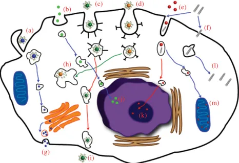
(a) (b) (c) (d) (e) (f) (l) (j) (k) (h) (g) (i) (m)
FIGURE 11.1 Uptake mechanisms and the intracellular transport of ENMs.
Uptake may occur through micropinocytosis (a), pinocytosis (b), caveolin‐mediated endocytosis (c), clathrin‐mediated endocytosis (d), unknown endocytic pathways (e), or diffusion/penetration (f). After cellular entry, ENMs may localize into dif ferent organelles and cellular compartments. ENMs that enter to the Golgi apparatus can cause organelle damage and/or leave the cell via vesicles involved in the regular secretion system (g). ENMs remaining in early endosomes move slowly along cyto skeletal elements, fuse with late endosomes, and finally accumulate in the lysosome (h), nucleus (j), nucleolus (k), and mitochondria (m). In addition to these possible endpoints, ENMs can also accumulate in the cytoplasm (l) after escaping from late endosomes or lysosomes. Ultimately, ENMs can be excreted from the cell by transcytosis (i).
triphenylphosphonium (TTP), which is a mitochondriotropic moiety. The localization of TPP‐conjugated ENMs inside the mitochondria has also been confirmed by experimental reports. It has been demonstrated that polystyrene NPs functionalized with both TPP and poly‐l‐lysine (PLL) localize in mitochondria due to the hydrophobic properties of TPP (Wang et al., 2013). Doxorubicin‐loaded, TPP‐functionalized poly ethylenimine hyperbranched polymer (PEI) NPs likewise triggered rapid and severe cytotoxicity within a few hours of incubation at low concentrations, in contrast to free doxorubicin (Theodossiou et al., 2013). Another study detailed the design of a multifunctional envelope‐coated nanocarrier system, which consists of mesoporous silica nanoparti cles (MSNs) functionalized with TPP, the antineoplastic drug topotecan (TPT), and a charge‐conversional shielding layer. This system was shown to target tumor cells and initiate apoptosis through mitochondrial damage (Luo et al., 2014). Mitochondrion‐mediated apoptotic cell death was also achieved in drug‐resistant breast cancer cells via NP‐directed efficient subcellular localization of doxorubicin into the mitochondria (Zeng et al., 2014). The surface chemistry and size of ENMs are impor tant parameters for mitochondrial targeting and localization: While carboxylated Polystyrene NPs with 20 nm diameters could be located in the mitochondria of different hepatocyte cell lines, the mitochondrial uptake and localizations of 200 nm diameter Polystyrene NPs were limited (Johnston et al., 2010b).
Lysosomes can also be targeted depending on the functionalization moiety of NPs. Glycol chitosan NPs decorated with hydrophobic moi eties have been reported to eventually localize into the lysosome (Nam et al., 2009). The intracellular fate of AuNPs has also been reported in different studies: Protein‐coated AuNPs are generally entrapped in endo somes but can be directed to several cellular compartments by the addition of targeting moieties such as TAT cell‐penetrating peptides or a liposome coating (Krpetic et al., 2011; Nativo et al., 2008). The uptake and local ization of ENMs allow these molecules to interfere with intracellular mechanisms, potentially resulting in organelle damage, membrane leak age, or the production of reactive oxygen species (ROS). These effects may be medically beneficial if the ENMs are designed to specifically target cancer cells or other diseased cell types. SPIONs covalently coated with the lysosomal protein marker LAMP1 (LAMP1‐SPION), for example, were used to apply mechanical stress to the lysosomes of rat insulinoma tumor cells. The activation of the SPIONs was triggered using a dynamic field generator, decreasing the sizes and numbers of the lysosomes and resulting in a corresponding increase in cellular apoptosis. The remote
control of apoptosis was therefore achieved through the activation of magnetic ENMs (Zhang et al., 2014).
The excretion and clearance of ENMs will be discussed in the in vivo part of this chapter, but here it should be noted that cells themselves respond to ENMs by activating their exocytosis mechanisms, thus extruding or degrading ENMs at the subcellular level. The exocytosis of ENMs has been reported for a variety of materials. For example, Bartczak et al. characterized the exocytosis trends of AuNPs functionalized with angiogenesis‐inhibiting (“inhibitor”) or similar but noninhibitory (“mutant”) peptides and made the interesting observation that mutant peptides were first exocytosed and subsequently reuptaken and reexocytosed from the cell, while inhibitors were progressively removed with no reuptake (Bartczak et al., 2012).
The intracellular localization of ENMs determines whether they can be exocytosed through the activity of lysosomes or the Golgi apparatus. The exocytosis of phosphate‐modified mesoporous nanoparticles (P‐MSNs) showed a good correlation with the rates of lysosomal exocytosis in dif ferent cell lines, which has significant implications for the development of efficient drug delivery agents (Yanes et al., 2013). The degradation of ENMs is a big concern for the delivery of biological cargos. Sahay et al. reported that siRNA‐conjugated cationic lipid NPs are eventually degraded in the lysosome (Sahay et al., 2013), which is a common drawback of biodegradable ENMs utilized for gene therapy, cancer treatment, or drug delivery.
Various methods exist for the evaluation of the intracellular activity of ENMs: (i) Deactivation of cellular uptake mechanisms (phagocytosis, macropinocytosis, clathrin‐mediated endocytosis, caveolae‐mediated endocytosis, and transcytosis); (ii) assays for cell viability/proliferation (MTT (3‐(4,5‐dimethylthiazol‐2‐yl)‐2,5‐diphenyltetrazolium bromide), alamar blue, XTT, lactade dehydrogenase (LDH), calcein AM/ethidium homodimer‐2 staining, 5‐ethynyl‐20‐deoxyuridine (EdU), and 5‐bromo‐2′‐ deoxyuridine (BrdU)); (iii) mechanistic assays (detection of ROS by dichlorofluorescein (DCF) and GSH level assays, apoptosis (caspase activity assays), necrosis); (iv) microscopic evaluation of intracellular localization (transmission electron microscopy (TEM) or scanning elec tron microscopy (SEM), confocal laser scanning microscopy (CLSM), atomic force microscopy, and magnetic resonance imaging); (v) quanti fication of ENM uptake and accumulation (inductively coupled plasma spectroscopy techniques: ICP‐MS, ICP‐OES, and flow cytometry); (vi) genotoxicity assays (DNA damage, micronuclei formation, DNA fragmentation).
TABLE 11.1 Inhibitors Used to Study the Uptake Mechanisms of ENMs
Inhibitor Mechanism of Action Affected Uptake Mechanism Nystatin Disrupts caveolar function Caveolae‐mediated
endocytosis (Greulich et al., 2011) Amyloride hydrochloride Blocker of epithelial Na+ channels Macropinocytosis (Gitrowski et al., 2014; Weissleder et al., 2014) Cytochalasin A/B/D
Polymerization of actin Caveolae‐mediated endocytosis (Gao et al., 2013)
Chlorpromazine Reversible translocation of clathrin from cell membrane
Clathrin‐mediated endocytosis (Gao et al., 2013)
Genistein Tyrosine kinase inhibitor Caveolae‐mediated endocytosis Lovastatin Cholesterol synthesis Macropinocytosis and
clathrin‐ and caveolae‐ mediated endocytosis Nocodazole Polymerization of microtubules Clathrin‐mediated endocytosis (Gao et al., 2013) Sodium azide Energy Gao et al. (2013) Sucrose Blocking agents of
clathrin‐coated pit formation
Clathrin‐mediated endocytosis (Gao et al., 2013)
Filipin Inhibitor of caveolae‐ associated endocytosis
Caveolae‐mediated endocytosis (Gao et al., 2013)
BFA Disruption of Golgi apparatus
Intracellular transportation of ENMs (Gao et al., 2013)
Monensin Lysosome inhibitor Intracellular cargo transport and disposition (Gao et al., 2013)
Phenylarsine oxide
Endocytosis Gao et al. (2013) Wortmannin Inhibits phosphoinositide‐3
kinases (PI3Ks) through covalent interactions
Macropinocytosis (Sokolova et al., 2013)
11.3.2 Cellular Uptake Mechanisms
Imaging and spectroscopy techniques are widely used in monitoring the cellular uptake and intracellular localization of ENMs. Selective targeting is an important design concern for ENMs, and the ability to engineer the localization of a nanostructure within the cell significantly improves its capacity for selective delivery. Some applications require the retention of ENMs in specific organelles or necessitate their uptake by specific cell types, which can be achieved through the activation of different uptake mechanisms. As such, pathway‐specific inhibitors are commonly used to determine the methods of uptake used by ENMs. Some of these inhibitors are listed in Table 11.1.
11.4 ASSAYS FOR CELL VIABILITY/PROLIFERATION
The evaluation of the short‐ and long‐term effects of ENMs is commonly conducted through cell viability and proliferation assays. ENM toxicity is mainly quantified by metabolic activity assays (e.g., MTT, XTT, alamar blue, LDH) and direct cell death assays (calcein AM/ethidium homodimer‐2 staining). Cytotoxicity screening of an ENM is often a preliminary step that determines whether the material is suitable for further testing. As such, comparative assays have been developed for this process. One par ticularly stringent example is provided by Kroll et al., who have developed a matrix‐based test system to evaluate the cytotoxic effects of 23 ENMs against a set of sensitive cell lines and suggest that such a system can be used to predict in vivo nanostructure behavior, minimizing the number of animals used in toxicity assessments by preselecting against cytotoxic ENMs (Kroll et al., 2011). The matrix‐based, high‐throughput cytotoxicity screening of ENMs is of great importance for the development standard ized cytotoxicity assessments and modeling studies.
Cell proliferation assays are also performed frequently in ENM research, often to investigate the short‐term detrimental effects of ENMs or to determine whether the material can trigger cell cycle arrest in cancer cells. Although ENMs may not necessarily cause short‐term cytotoxicity in cells, they may nonetheless interfere with DNA synthesis and cell division, which is a desirable property for materials intended for use in anticancer treatments (Kim et al., 2013). EdU and BrdU, synthetic nucleosides that replace the thymidine moiety in DNA during replication, are commonly used to analyze cell proliferation, cell division, DNA synthesis, and cell cycle arrest.
11.4.1 Assays for Oxidative Stress and Apoptosis Mechanisms
The generation of ROS or reactive nitrogen species (RNS) may alter the homeostatic oxidative states of cells and induce cell death through apoptosis, necrosis, or other mechanisms. ENMs may affect the cellular oxidative state in various ways. They typically cause generation of exces sive ROS or RNS at higher concentrations, which is considered a major factor in the severity of ENM cytotoxicity. On the other hand, ENMs with reductive properties can also be fabricated through surface modification methods to act as free radical scavengers and decrease oxidative damage in cells (Karakoti et al., 2010). The most widely used method for the quantification of ROS is the DCF assay, which relies on the oxidation of nonfluorescent DCF to fluorescent DCF by various intracellular oxidants. ROS generation can also be monitored by the evaluation of GSH level, free radical content, mitochondrial membrane potential, and stress‐related gene/protein expression analyses.
The apoptotic state of cells can be analyzed by annexin V staining, cytochrome c release, caspase‐3 activity, and other caspase activity assays. Annexin V staining is the most widely used method for the evaluation of early apoptosis in ENM‐treated cells. Normally, phosphatidylserine is located on the cytoplasmic surface of the cell membrane. However, after the initiation of apoptosis, Polystyrene translocates from the inner surface and is found on the cell exterior, which can be detected by fluorescent dye‐tagged annexin V.
11.4.1.1 Microscopic Evaluation of Intracellular Localization of ENMs CLSM or electron microscopy (SEM/TEM) can also be used for
qualitative, semiquantitative, and quantitative evaluations of the uptake, localization, intracellular fate, accumulation, and exocytosis profiles of ENMs (Bartneck et al., 2010; Havrdova et al., 2014; Krpetic et al., 2011; Lesniak et al., 2013; Ma et al., 2011; Nativo et al., 2008; Rodriguez‐ Lorenzo et al., 2014; Soenen et al., 2012; Ye et al., 2014).
11.4.2 Evaluation of Uptake and Accumulation of ENMs
The evaluation of nanomaterial uptake, accumulation, and exocytosis might also be performed through inductively coupled plasma spectroscopy tech niques, laser scanning microscopy techniques, flow cytometry (in the case of fluorescent nanomaterials), and TEM. Inductively coupled plasma spec trometry (i.e., ICP‐AES (Bartczak et al., 2012; Krpetic et al., 2011), ICP‐MS
(Wang et al., 2011), and ICP‐OES (Ferrati et al., 2010)) are the most widely used methods for quantitative assessment of endocytosis, accumulation, and exocytosis. However, ICP‐based techniques cannot distinguish ENMs and their breakdown products (i.e., metal ions) from naturally existing ions and elements within the cells. As such, this quantification method is applicable only to ENMs composed of materials not normally found in biological systems. In addition, the reproducibility of ICP spectroscopy measurements requires the ENMs to have a monodispersed size distri bution, well‐defined geometry, and well‐characterized crystalline lattice.
Fluorescence‐based techniques (i.e., fluorescence spectroscopy, flow cytometry, and laser scanning microscopy) can be used for the quantitative and qualitative evaluations of internalization, localization, accumulation, and exocytosis of ENMs (Rodriguez‐Lorenzo et al., 2014; Slowing et al., 2011). Although these techniques are very precise, they can only be used with ENMs displaying fluorescent activity, either as an intrinsic property (as in quantum dots) or through surface modification with fluorescent dyes. However, it must be kept in mind that functionalization of an ENM with a fluorescent dye might change its surface properties and biological activities. In addition, weakly conjugated fluorescent dyes may detach from the ENM in biological environments and result in major errors in the quantification of ENM uptake and localization.
11.4.3 Genotoxicity Assays
ENMs are capable of localizing into the nucleus, where they interact with and potentially damage genomic DNA. The genotoxicity of ENMs can be analyzed through techniques such as chromosome aberration test, comet assay, and micronucleus assay. The chromosome aberration test is an indicator of chromosomal alterations induced by ENMs and involves visual evaluation following the isolation and Giemsa staining of metaphasic chromosomes. The comet assay, also known as single‐cell gel electro phoresis (SCGE), is the most widely used assay for evaluating DNA damage at the level of individual cells. The underlying mechanism of comet assay is that damaged DNA migrates faster than undamaged DNA in an agarose matrix. Quantitative analysis of DNA damage is per formed by staining and imaging the comet‐like structures formed follow ing electrophoresis process. However, as the comet assay is low throughput and often faces reproducibility issues, high‐throughput screening tools are necessary to reliably assess ENM‐mediated DNA damage (Watson et al., 2014).
11.5 ANIMAL MODELS AND LONG‐TERM RISK ASSESSMENT
Despite the widespread production and application of ENMs, there is lack of satisfactory information regarding their impact on humans, animals, and the ecosystem. Exposure to ENMs may directly or indirectly cause detrimental effects on the human health. ENMs can be introduced to the human body through inhalation, ingestion, dermal penetration, or injection, either unin tentionally or through the course of an ENM‐based therapy (Oberdorster et al., 2005). As with any foreign material, ENMs may trigger adverse effects following their administration. As such, the biocompatibility, biodistribution, biodegradation, excretion, and acute and long‐term toxicities of ENMs must be carefully assessed before their use in clinical trials. Although the in vitro tests previously outlined in this chapter provide means of predicting the behavior of nanomaterials, it is nonetheless possible that unforeseen in vivo effects may occur. In addition, the behavior of a nanomaterial can change through the spontaneous and rapid formation of protein coronae in biological media, which has a major impact on the interaction of ENMs with cells and organelles. It has also been reported that the immune system can detect NPs in the same way that it senses microbes and other foreign agents (Feliu and Fadeel, 2010; Mahmoudi et al., 2011). It is difficult to see many of these effects under in vitro conditions, which necessitates in vivo experiments.
ENMs that are intended for biomedical purposes are almost universally administered directly into the human body. Examples of such ENM designs include SPIONs and dendrimers for MRI studies; gold NPs for photother mal therapy, imaging, and drug and gene delivery; MSNs for MRI studies, drug delivery, and topical treatment; and carbon nanotubes for drug delivery and tumor detection and treatment. These materials are promising for the development of efficient and innovative solutions for the major challenges faced by the biomedical field, especially with regards to the treatment of common disorders such as cancer, obesity, and neurodegener ative diseases. Nevertheless, as with any material intended for human use, their safety must first and foremost be investigated to sufficient detail, through both in vitro experiments and animal studies, before they can be administered to human patients.
11.5.1 The Blood–Brain Barrier
Diseases of the central nervous system (CNS) can severely affect the health and comfort of patients through deterioration of either the spinal cord (myelopathy) or brain (encephalopathy). Development of ENMs for
monitoring and treatment of CNS‐related diseases is now a topic of great interest, as surface functionalization allows nanomaterials to display prop erties necessary for both diagnosis and therapy (this combined approach is also called theranostics). Although the fundamental mechanisms of certain CNS‐related disorders are still not fully understood, there has been some progress in developing effective diagnosis‐and‐treatment methods. However, many of these efforts are hampered due to the protective role of the blood–brain barrier (BBB), which blocks the transport of drugs and other large molecules into the brain. ENMs may facilitate the passage of their cargo through the BBB, which makes them attractive for improving drug targeting, increasing theranostic efficacy and reducing drug toxicity in CNS‐related disorders (Krol et al., 2013). ENMs can therefore be used for the early diagnosis, imaging, or therapy of CNS‐related disorders or taken as a prophylactic measure against the advancement of potential disease.
The mechanisms ENMs employ to pass through the airway epithelia and BBB into the secondary target organs (STOs: liver, spleen, kidneys, heart, and brain) are not fully understood. Oberdorster and his group per formed an inhalation study in rats with iridium (2–4 nm) and carbon (5–10 nm) NPs, as well as their aggregated forms (mean mobility diame ters between 20 and 80 nm) and found the translocation of iridium NPs into the STOs to be significantly higher than that of carbon NPs. Further more, the translocation and accumulation of 80 nm aggregates of iridium were lower than 20 nm aggregates, and inhaled NPs could also be detected in the skeleton and soft tissues. However, the underlying mechanism for cerebral localization following NP inhalation remains uncertain (Kreyling et al., 2009).
Two types of strategies are used to enhance the transport of drugs into the brain: BBB manipulation and drug manipulation (Krol et al., 2013). The utilization of ENMs is classified as drug manipulation, and its impor tance has increased during the past few years. Surface functionalization of ENMs can increase their ability to cross the BBB (depicted in Fig. 11.2), although the precise mechanisms involved in surface functionalization‐ mediated BBB transport are an issue of contention. The stimulation of endocytosis or low‐density lipoprotein (LDL)‐mediated transcytosis, the weakening of the BBB due to toxicity, and the loosening of tight junctions between endothelial cells are among the mechanisms proposed for this phenomenon (Gidwani and Singh, 2014). Regardless of the mechanism involved, surface modification of ENMs is nonetheless advantageous for drug delivery into the CNS, as nanomaterials provide a high drug‐loading capacity, prevent the degradation of the drug in the biological milieu, enhance the retention of the drug in the bloodstream, avoid efflux pump action
at the brain microvessels, and can be synthesized in the size ranges required for efficient transport across the BBB.
A variety of targeting moieties have been studied for the enhancement of BBB‐crossing efficiency. The targeting molecule is often involved in receptor‐mediated transport pathways; surface functionalization with materials that bind to the cellular receptors for insulin (Ulbrich et al., 2011), LDL (Bertrand et al., 2010; Wagner et al., 2012), and glutathione (Gaillard et al., 2012) has been reported in the literature. In addition, the BBB mani pulation and drug manipulation strategies can be combined to increase the penetration efficiency of ENMs. Surfactants such as dimethylsulfoxide (DMSO), sodium dodecylsulphate (SDS), and polysorbate 80 (PS‐80) are commonly used to loosen the adherent and tight junctions between the endothelial cells of the BBB and can be used in conjunction with ENMs to increase targeting efficiency. PS‐80 is a commonly employed reagent for the coating of NPs intended to target brain parenchyma (Kreuter, 2013).
(a) (b) Blood Junctions Neuron Astrocyte Endothelial cell Microglia Brain (c) (d)
FIGURE 11.2 Schematic representation of the blood–brain barrier (BBB).
Microvascular endothelial cells are connected through tight junctions (TJs) and adherens junctions (AJs) that prevent the leakage of foreign material into the brain. Nanoparticles ordinarily cannot pass through the BBB (a) and must be functionalized with targeting moieties that facilitate BBB transport through endo cytosis (b), receptor‐mediated transcytosis (c), or the loosening of the junctions between endothelial cells (d).
ENMs designed for BBB penetration typically do not display drug activity on their own and are instead loaded with a drug cargo that is released at the site of interest. ENMs have been loaded with well‐known chemotherapeutics such as doxorubicin (Li et al., 2014; Wohlfart et al., 2011), paclitaxel (Dilnawaz et al., 2012; Zhang et al., 2012), and prodrugs like curcumin (Tsai et al., 2011). It has been shown that doxorubicin‐loaded PS‐80 functionalized poly(methacrylic acid) (PMAA) NPs accumulated in brain tumors and brain metastases of breast cancer and reduced the growth of brain tumors to a greater extent than free Dox. Similarly, Dox‐ loaded PS‐PMAA NPs could be detected in tumor‐bearing brain tissue and induced apoptosis in cancer cells while leaving the rest of the brain unharmed, suggesting cancer cell‐specific targeting capacity (Li et al., 2014). Another study with doxorubicin was performed with rats treated intrave nously with doxorubicin‐loaded poly(n‐butyl cyanoacrylate) (PBCA) NPs with and without a 1% polysorbate 80 coating, and PS‐80 coating was shown to significantly improve the transcytosis of the drug into the brain parenchyma compared to free Dox and noncoated NPs (Wohlfart et al., 2011). Paclitaxel‐loaded magnetic NPs were also reported to be trans ported to the STOs and the brain stem, cerebellum, and cortex more compared to native paclitaxel. In addition, Pac‐MNPs displayed effective MRI contrast agent properties and can be used to acquire MRI images of the rat brain and liver (Dilnawaz et al., 2012).
An effective chemotherapy for brain tumors requires a nanoformu lated drug that can both penetrate the BBB and selectively target cancer cells. As such, ENMs have also been functionalized with peptides and proteins that target membrane receptors that are overexpressed in cancer cells. It has been shown that transferrin‐ (Tf) modified, cyclo‐[Arg‐ Gly‐Asp‐d‐Phe‐Lys] (c[RGDfK])‐paclitaxel conjugate loaded micelles (TRPM) accumulate in the brain to a greater extent than free taxol, paclitaxel‐ loaded micelles (PM), and transferrin‐modified, PM. More importantly, the nanoformulated paclitaxel (TRPM) exhibited the strongest anti glioma activity, as measured through an in vivo survival experiment. The mean survival time of mice treated with TRPM (42.8 days) was significantly longer that those treated with TPM (39.5 days), PM (34.8 days), taxol (33.6 days), and saline (34.5 days) (Zhang et al., 2012). Likewise, Tsai et al. showed that curcumin‐loaded PLGA NPs were accumulated in all STOs, except the heart, to a greater extent than free curcumin. In addition, PLGA NPs loaded with curcumin also accumulated in the hippocampus and displayed extended retention times in both the hippo campus (increased by 83%) and the cerebral cortex (increased by 96%) (Tsai et al., 2011).
Although nanomaterials are highly efficient platforms for the treatment of a great variety of disorders, their short‐ and long‐term effects should be investigated in great detail prior to their in vivo application. Recent reports demonstrate that the presence of the protein corona on the surface of ENPs can alter the theranostic behaviors of nanomaterials; for instance, the formation of amyloid β fibrils is inhibited in the case of corona‐coated NPs, whereas the rate of amyloid β fibrillation is accelerated in the presence of uncoated NPs (Ghavami et al., 2013). As such, greater attention should be given to the therapeutic behaviors of ENMs and how these behaviors may be altered in biological environments.
11.6 CONCLUSIONS AND FUTURE PERSPECTIVES
In this chapter, we presented some of the emerging research regarding the considerable impact that ENMs may have on living systems. The potential toxicity of ENMs is a significant concern throughout their design, modification, and functionalization, and the fabrication of ENMs with well‐defined physicochemical properties is critical for efficient tar geting with minimal side effects. The uptake and cellular localization of ENMs are affected by their physicochemical properties and chemical and biological identities. While the former two can be controlled during fabrication, the biological identity of an ENM depends on the biological environments it encounters and may change considerably inside living organisms. Interactions at cell–ENM interface are also dependent on physicochemical properties (e.g., size, shape, surface charge, etc.) and the biological identity of nanomaterials (e.g., protein corona formation) and cell/organism types. The entry and localization of nanomaterials within the cells are also important for their function, as well as for risk assessment studies. Methods listed in this chapter are the most widely used techniques and assays of the investigation of ENM localization and toxicity. In general, in vitro models and computational simulations are used for short‐term toxicity analyses, while in vivo animal studies are preferred for long‐term toxicity and risk assessments. In addition to experimental research, advanced computational simulations and models can facilitate the investigation of the possible effects of nanomaterials, through the simulation of nanomaterial–cell interactions for the predic tion of ENM efficiency and potential toxicity. In addition, the development of reliable and high‐throughput in vitro models may also allow the in vitro evaluation of the long‐term impacts of ENMs. The combination of advanced in vitro, in vivo, and in silico methods will no doubt further our
understanding of nanomaterial function and may allow the design of safer and more effective ENMs for the treatment of a wide range of disorders.
REFERENCES
Agarwal, R., Singh, V., Jurney, P., Shi, L., Sreenivasan, S.V., and Roy, K. (2013). Mammalian cells preferentially internalize hydrogel nanodiscs over nanorods and use shape‐specific uptake mechanisms. Proc Natl Acad Sci U S A 110, 17247–17252.
Arora, S., Rajwade, J.M., and Paknikar, K.M. (2012). Nanotoxicology and in vitro studies: the need of the hour. Toxicol Appl Pharmacol 258, 151–165.
Bartczak, D., Nitti, S., Millar, T.M., and Kanaras, A.G. (2012). Exocytosis of peptide functionalized gold nanoparticles in endothelial cells. Nanoscale 4, 4470–4472.
Bartneck, M., Keul, H.A., Singh, S., Czaja, K., Bornemann, J., Bockstaller, M., Moeller, M., Zwadlo‐Klarwasser, G., and Groll, J. (2010). Rapid uptake of gold nanorods by primary human blood phagocytes and immunomodulatory effects of surface chemistry. ACS Nano 4, 3073–3086.
Barua, S., Yoo, J.W., Kolhar, P., Wakankar, A., Gokarn, Y.R., and Mitragotri, S. (2013). Particle shape enhances specificity of antibody‐displaying nanoparticles. Proc Natl Acad Sci U S A 110, 3270–3275.
Bertrand, Y., Currie, J.C., Demeule, M., Regina, A., Che, C., Abulrob, A., Fatehi, D., Sartelet, H., Gabathuler, R., Castaigne, J.P., et al. (2010). Transport characteristics of a novel peptide platform for CNS therapeutics. J Cell Mol Med 14, 2827–2839.
Browning, L.M., Lee, K.J., Nallathamby, P.D., and Xu, X.‐H.N. (2013). Silver nanoparticles incite size‐ and dose‐dependent developmental phenotypes and nanotoxicity in zebrafish embryos. Chem Res Toxicol 26, 1503–1513.
Cartiera, M.S., Johnson, K.M., Rajendran, V., Caplan, M.J., and Saltzman, W.M. (2009). The uptake and intracellular fate of PLGA nanoparticles in epithelial cells. Biomaterials 30, 2790–2798.
Choi, J. and Wang, N.S. (2011). Nanoparticles in Biomedical Applications and Their Safety Concerns. In R. Fazel (ed.) Biomedical Engineering—From Theory to Applications. InTech, Maastricht.
Clark, K.A., White, R.H., and Silbergeld, E.K. (2011). Predictive models for nanotoxicology: current challenges and future opportunities. Regul Toxicol Pharmacol 59, 361–363.
DeLoid, G., Cohen, J.M., Darrah, T., Derk, R., Rojanasakul, L., Pyrgiotakis, G., Wohlleben, W., and Demokritou, P. (2014). Estimating the effective density of engineered nanomaterials for in vitro dosimetry. Nat Commun 5, 3514.
Demokritou, P., Gass, S., Pyrgiotakis, G., Cohen, J.M., Goldsmith, W., McKinney, W., Frazer, D., Ma, J., Schwegler‐Berry, D., Brain, J., et al. (2013). An in vivo and in vitro toxicological characterisation of realistic nanoscale CeO(2) inhalation exposures. Nanotoxicology 7, 1338–1350.
Dilnawaz, F., Singh, A., Mewar, S., Sharma, U., Jagannathan, N.R., and Sahoo, S.K. (2012). The transport of non‐surfactant based paclitaxel loaded magnetic nanoparticles across the blood brain barrier in a rat model. Biomaterials 33, 2936–2951.
Donaldson, K. and Poland, C.A. (2013). Nanotoxicity: challenging the myth of nano‐specific toxicity. Curr Opin Biotechnol 24, 724–734.
Dykman, L.A. and Khlebtsov, N.G. (2014). Uptake of engineered gold nanopar ticles into mammalian cells. Chem Rev 114, 1258–1288.
Feliu, N. and Fadeel, B. (2010). Nanotoxicology: no small matter. Nanoscale 2, 2514–2520.
Ferrati, S., Mack, A., Chiappini, C., Liu, X., Bean, A.J., Ferrari, M., and Serda, R.E. (2010). Intracellular trafficking of silicon particles and logic‐embedded vectors. Nanoscale 2, 1512–1520.
Gaillard, P.J., Appeldoorn, C.C., Rip, J., Dorland, R., van der Pol, S.M., Kooij, G., de Vries, H.E., and Reijerkerk, A. (2012). Enhanced brain delivery of liposomal methylprednisolone improved therapeutic efficacy in a model of neuroinflam mation. J Control Release 164, 364–369.
Gangwal, S., Brown, J.S., Wang, A., Houck, K.A., Dix, D.J., Kavlock, R.J., and Hubal, E.A. (2011). Informing selection of nanomaterial concentrations for ToxCast in vitro testing based on occupational exposure potential. Environ Health Perspect 119, 1539–1546.
Gao, H., Yang, Z., Zhang, S., Cao, S., Shen, S., Pang, Z., and Jiang, X. (2013). Ligand modified nanoparticles increases cell uptake, alters endocytosis and elevates glioma distribution and internalization. Sci Rep 3, 2534.
García‐Remesal, M., García‐Ruiz, A., Pérez‐Rey, D., de la Iglesia, D., and Maojo, V. (2013). Using nanoinformatics methods for automatically identifying relevant nanotoxicology entities from the literature. BioMed Res Int 2013, 9.
Ghavami, M., Rezaei, M., Ejtehadi, R., Lotfi, M., Shokrgozar, M.A., Abd Emamy, B., Raush, J., and Mahmoudi, M. (2013). Physiological temperature has a crucial role in amyloid beta in the absence and presence of hydrophobic and hydro philic nanoparticles. ACS Chem Neurosci 4, 375–378.
Ghosh Chaudhuri, R. and Paria, S. (2011). Core/shell nanoparticles: classes, properties, synthesis mechanisms, characterization, and applications. Chem Rev 112, 2373–2433.
Gidwani, M. and Singh, A.V. (2014). Nanoparticle enabled drug delivery across the blood brain barrier: in vivo and in vitro models, opportunities and challenges. Curr Pharm Biotechnol 14, 1201–1212.
Gitrowski, C., Al‐Jubory, A.R., and Handy, R.D. (2014). Uptake of different crystal structures of TiO(2) nanoparticles by Caco‐2 intestinal cells. Toxicol Lett 226, 264–276.
Granqvist, C.G., Buhrman, R.A., Wyns, J., and Sievers, A.J. (1976). Far‐infrared absorption in ultrafine Al particles. Phys Rev Lett 37, 625–629.
Grassian, V.H., O’Shaughnessy, P.T., Adamcakova‐Dodd, A., Pettibone, J.M., and Thorne, P.S. (2007). Inhalation exposure study of titanium dioxide nanoparticles with a primary particle size of 2 to 5 nm. Environ Health Perspect 115, 397–402. Greulich, C., Diendorf, J., Simon, T., Eggeler, G., Epple, M., and Koller, M.
(2011). Uptake and intracellular distribution of silver nanoparticles in human mesenchymal stem cells. Acta Biomater 7, 347–354.
Han, Y., Xu, X., Yin, L., Long, L., Liu, R., and Liu, S. (2011). Dynamical sys tems: an effective way in nanotoxicology study. J Algorithms Comput Technol
5, 79–94.
Han, X., Corson, N., Wade‐Mercer, P., Gelein, R., Jiang, J., Sahu, M., Biswas, P., Finkelstein, J.N., Elder, A., and Oberdorster, G. (2012). Assessing the rele vance of in vitro studies in nanotoxicology by examining correlations between in vitro and in vivo data. Toxicology 297, 1–9.
Havrdova, M., Polakova, K., Skopalik, J., Vujtek, M., Mokdad, A., Homolkova, M., Tucek, J., Nebesarova, J., and Zboril, R. (2014). Field emission scanning electron microscopy (FE‐SEM) as an approach for nanoparticle detection inside cells. Micron 67, 149–154.
Holt, B.D., Short, P.A., Rape, A.D., Wang, Y.L., Islam, M.F., and Dahl, K.N. (2010). Carbon nanotubes reorganize actin structures in cells and ex vivo. ACS Nano 4, 4872–4878.
Johnston, H.J., Hutchison, G.R., Christensen, F.M., Aschberger, K., and Stone, V. (2010a). The biological mechanisms and physicochemical characteristics responsible for driving fullerene toxicity. Toxicol Sci 114, 162–182.
Johnston, H.J., Semmler‐Behnke, M., Brown, D.M., Kreyling, W., Tran, L., and Stone, V. (2010b). Evaluating the uptake and intracellular fate of polystyrene nanoparticles by primary and hepatocyte cell lines in vitro. Toxicol Appl Pharmacol 242, 66–78.
Karakoti, A., Singh, S., Dowding, J.M., Seal, S., and Self, W.T. (2010). Redox‐ active radical scavenging nanomaterials. Chem Soc Rev 39, 4422–4432. Kim, J.A., Aberg, C., de Carcer, G., Malumbres, M., Salvati, A., and Dawson,
K.A. (2013). Low dose of amino‐modified nanoparticles induces cell cycle arrest. ACS Nano 7, 7483–7494.
Kreuter, J. (2013). Mechanism of polymeric nanoparticle‐based drug transport across the blood‐brain barrier (BBB). J Microencapsul 30, 49–54.
Krewski, D., Acosta, D., Jr., Andersen, M., Anderson, H., Bailar, J.C., 3rd, Boekelheide, K., Brent, R., Charnley, G., Cheung, V.G., Green, S., Jr.,
et al. (2010). Toxicity testing in the 21st century: a vision and a strategy. J Toxicol Environ Health B Crit Rev 13, 51–138.
Kreyling, W.G., Semmler‐Behnke, M., Seitz, J., Scymczak, W., Wenk, A., Mayer, P., Takenaka, S., and Oberdorster, G. (2009). Size dependence of the translocation of inhaled iridium and carbon nanoparticle aggregates from the lung of rats to the blood and secondary target organs. Inhal Toxicol 21 Suppl 1, 55–60.
Krol, S., Macrez, R., Docagne, F., Defer, G., Laurent, S., Rahman, M., Hajipour, M.J., Kehoe, P.G., and Mahmoudi, M. (2013). Therapeutic benefits from nanoparti cles: the potential significance of nanoscience in diseases with compromise to the blood brain barrier. Chem Rev 113, 1877–1903.
Kroll, A., Pillukat, M.H., Hahn, D., and Schnekenburger, J. (2009). Current in vitro methods in nanoparticle risk assessment: limitations and challenges. Eur J Pharm Biopharm 72, 370–377.
Kroll, A., Dierker, C., Rommel, C., Hahn, D., Wohlleben, W., Schulze‐Isfort, C., Gobbert, C., Voetz, M., Hardinghaus, F., and Schnekenburger, J. (2011). Cytotoxicity screening of 23 engineered nanomaterials using a test matrix of ten cell lines and three different assays. Part Fibre Toxicol 8, 9.
Krpetic, Z., Saleemi, S., Prior, I.A., See, V., Qureshi, R., and Brust, M. (2011). Negotiation of intracellular membrane barriers by TAT‐modified gold nanopar ticles. ACS Nano 5, 5195–5201.
Kuhn, D.A., Vanhecke, D., Michen, B., Blank, F., Gehr, P., Petri‐Fink, A., and Rothen‐Rutishauser, B. (2014). Different endocytotic uptake mechanisms for nanoparticles in epithelial cells and macrophages. Beilstein J Nanotechnol 5, 1625–1636.
Lesniak, A., Salvati, A., Santos‐Martinez, M.J., Radomski, M.W., Dawson, K.A., and Aberg, C. (2013). Nanoparticle adhesion to the cell membrane and its effect on nanoparticle uptake efficiency. J Am Chem Soc 135, 1438–1444. Li, F., Sun, J.‐y., Wang, J.‐y., Du, S.‐l., Lu, W.‐y., Liu, M., Xie, C., and Shi, J.‐y.
(2008). Effect of hepatocyte growth factor encapsulated in targeted liposomes on liver cirrhosis. J Control Release 131, 77–82.
Li, J., Cai, P., Shalviri, A., Henderson, J.T., He, C., Foltz, W.D., Prasad, P., Brodersen, P.M., Chen, Y., DaCosta, R., et al. (2014). A multifunctional poly meric nanotheranostic system delivers doxorubicin and imaging agents across the blood‐brain barrier targeting brain metastases of breast cancer. ACS Nano
8, 9925–9940.
Luo, G.‐F., Chen, W.‐H., Liu, Y., Lei, Q., Zhuo, R.‐X., and Zhang, X.‐Z. (2014). Multifunctional enveloped mesoporous silica nanoparticles for subcellular co‐delivery of drug and therapeutic peptide. Sci Rep 4, 6064.
Lynch, I., Salvati, A., and Dawson, K.A. (2009). Protein‐nanoparticle interactions: what does the cell see? Nat Nanotechnol 4, 546–547.
Ma, X., Wu, Y., Jin, S., Tian, Y., Zhang, X., Zhao, Y., Yu, L., and Liang, X.J. (2011). Gold nanoparticles induce autophagosome accumulation through size‐dependent nanoparticle uptake and lysosome impairment. ACS Nano 5, 8629–8639. Mahmoudi, M., Sahraian, M.A., Shokrgozar, M.A., and Laurent, S. (2011).
Superparamagnetic iron oxide nanoparticles: promises for diagnosis and treatment of multiple sclerosis. ACS Chem Neurosci 2, 118–140.
Matsuo, T., Sugita, T., Kubo, T., Yasunaga, Y., Ochi, M., and Murakami, T. (2003). Injectable magnetic liposomes as a novel carrier of recombinant human BMP‐2 for bone formation in a rat bone‐defect model. J Biomed Mater Res A 66A, 747–754. Nam, H.Y., Kwon, S.M., Chung, H., Lee, S.Y., Kwon, S.H., Jeon, H., Kim, Y.,
Park, J.H., Kim, J., Her, S., et al. (2009). Cellular uptake mechanism and intracellular fate of hydrophobically modified glycol chitosan nanoparticles. J Control Release 135, 259–267.
Nativo, P., Prior, I.A., and Brust, M. (2008). Uptake and intracellular fate of surface‐ modified gold nanoparticles. ACS Nano 2, 1639–1644.
Nel, A., Xia, T., Mädler, L., and Li, N. (2006). Toxic potential of materials at the nanolevel. Science 311, 622–627.
Nel, A.E., Madler, L., Velegol, D., Xia, T., Hoek, E.M., Somasundaran, P., Klaessig, F., Castranova, V., and Thompson, M. (2009). Understanding biophysicochemi cal interactions at the nano‐bio interface. Nat Mater 8, 543–557.
Neuberger, T., Schöpf, B., Hofmann, H., Hofmann, M., and von Rechenberg, B. (2005). Superparamagnetic nanoparticles for biomedical applications: possibilities and limitations of a new drug delivery system. J Magn Magn Mater 293, 483–496. Oberdorster, G. (2012). Nanotoxicology: in vitro‐in vivo dosimetry. Environ
Health Perspect 120, A13; author reply A13.
Oberdorster, G., Oberdorster, E., and Oberdorster, J. (2005). Nanotoxicology: an emerging discipline evolving from studies of ultrafine particles. Environ Health Perspect 113, 823–839.
Park, M.V.D.Z. (2012). Nanotoxicology—an in vitro approach. Proefschrift Maastricht (Maastricht University Maastricht).
Park, M.V.D.Z., Neigh, A.M., Vermeulen, J.P., de la Fonteyne, L.J.J., Verharen, H.W., Briede, J.J., van Loveren, H., and de Jong, W.H. (2011). The effect of particle size on the cytotoxicity, inflammation, developmental toxicity and genotoxicity of silver nanoparticles. Biomaterials 32, 9810–9817.
Powers, K.W., Palazuelos, M., Moudgil, B.M., and Roberts, S.M. (2007). Characterization of the size, shape, and state of dispersion of nanoparticles for toxicological studies. Nanotoxicology 1, 42–51.
Prahalad, A.K., Soukup, J.M., Inmon, J., Willis, R., Ghio, A.J., Becker, S., and Gallagher, J.E. (1999). Ambient air particles: effects on cellular oxidant rad ical generation in relation to particulate elemental chemistry. Toxicol Appl Pharmacol 158, 81–91.
Prasad, G.L. (2009). Biomedical Applications of Nanoparticles. In Safety of Nanoparticles, T.J. Webster, ed. (Springer New York), pp. 89–109.
Rahman, M., Laurent, S., Tawil, N., Yahia, L., and Mahmoudi, M. (2013). Protein‐ Nanoparticle Interactions (Springer Series in Biophysics 15), Springer‐Verlag, Berlin/Heidelberg.
Rodriguez‐Lorenzo, L., Fytianos, K., Blank, F., von Garnier, C., Rothen‐ Rutishauser, B., and Petri‐Fink, A. (2014). Fluorescence‐encoded gold nanoparticles: library design and modulation of cellular uptake into dendritic cells. Small 10, 1341–1350.
Sahay, G., Querbes, W., Alabi, C., Eltoukhy, A., Sarkar, S., Zurenko, C., Karagiannis, E., Love, K., Chen, D., Zoncu, R., et al. (2013). Efficiency of siRNA delivery by lipid nanoparticles is limited by endocytic recycling. Nat Biotechnol 31, 653–658.
dos Santos, T., Varela, J., Lynch, I., Salvati, A., and Dawson, K.A. (2011). Effects of transport inhibitors on the cellular uptake of carboxylated polystyrene nanoparticles in different cell lines. PLoS One 6, e24438.
Saravanan, P., Gopalan, R., and Chandrasekaran, V. (2008). Synthesis and charac terisation of nanomaterials. Def Sci J 58, 504516.
Sayes, C. and Ivanov, I. (2010). Comparative study of predictive computational models for nanoparticle‐induced cytotoxicity. Risk Anal 30, 1723–1734. Schaeublin, N.M., Braydich‐Stolle, L.K., Maurer, E.I., Park, K., MacCuspie, R.I.,
Afrooz, A.R.M.N., Vaia, R.A., Saleh, N.B., and Hussain, S.M. (2012). Does shape matter? bioeffects of gold nanomaterials in a human skin cell model. Langmuir 28, 3248–3258.
Slowing, II, Vivero‐Escoto, J.L., Zhao, Y., Kandel, K., Peeraphatdit, C., Trewyn, B.G., and Lin, V.S. (2011). Exocytosis of mesoporous silica nanoparticles from mammalian cells: from asymmetric cell‐to‐cell transfer to protein harvesting. Small 7, 1526–1532.
Soenen, S.J., Nuytten, N., De Meyer, S.F., De Smedt, S.C., and De Cuyper, M. (2010). High intracellular iron oxide nanoparticle concentrations affect cellular cytoskeleton and focal adhesion kinase‐mediated signaling. Small 6, 832–842. Soenen, S.J., Manshian, B., Montenegro, J.M., Amin, F., Meermann, B., Thiron,
T., Cornelissen, M., Vanhaecke, F., Doak, S., Parak, W.J., et al. (2012). Cytotoxic effects of gold nanoparticles: a multiparametric study. ACS Nano 6, 5767–5783.
Sokolova, V., Kozlova, D., Knuschke, T., Buer, J., Westendorf, A.M., and Epple, M. (2013). Mechanism of the uptake of cationic and anionic calcium phos phate nanoparticles by cells. Acta Biomater 9, 7527–7535.
Suresh, A.K., Pelletier, D.A., Wang, W., Morrell‐Falvey, J.L., Gu, B., and Doktycz, M.J. (2012). Cytotoxicity induced by engineered silver nanocrystal lites is dependent on surface coatings and cell types. Langmuir 28, 2727–2735.
Szabo, I., Leanza, L., Gulbins, E., and Zoratti, M. (2012). Physiology of potassium channels in the inner membrane of mitochondria. Pflugers Arch 463, 231–246. Theodossiou, T.A., Sideratou, Z., Katsarou, M.E., and Tsiourvas, D. (2013).
Mitochondrial delivery of doxorubicin by triphenylphosphonium‐functional ized hyperbranched nanocarriers results in rapid and severe cytotoxicity. Pharm Res 30, 2832–2842.
Tsai, Y.M., Chien, C.F., Lin, L.C., and Tsai, T.H. (2011). Curcumin and its nano‐ formulation: the kinetics of tissue distribution and blood‐brain barrier penetra tion. Int J Pharm 416, 331–338.
Ulbrich, K., Knobloch, T., and Kreuter, J. (2011). Targeting the insulin receptor: nanoparticles for drug delivery across the blood‐brain barrier (BBB). J Drug Target 19, 125–132.
Wagner, S., Zensi, A., Wien, S.L., Tschickardt, S.E., Maier, W., Vogel, T., Worek, F., Pietrzik, C.U., Kreuter, J., and von Briesen, H. (2012). Uptake mechanism of ApoE‐modified nanoparticles on brain capillary endothelial cells as a blood‐ brain barrier model. PLoS One 7, e32568.
Walkey, C.D. and Chan, W.C.W. (2012). Understanding and controlling the inter action of nanomaterials with proteins in a physiological environment. Chem Soc Rev 41, 2780–2799.
Wang, L., Liu, Y., Li, W., Jiang, X., Ji, Y., Wu, X., Xu, L., Qiu, Y., Zhao, K., Wei, T., et al. (2011). Selective targeting of gold nanorods at the mitochondria of cancer cells: implications for cancer therapy. Nano Lett 11, 772–780.
Wang, X.‐H., Peng, H.‐S., Yang, L., You, F.‐T., Teng, F., Tang, A.‐W., Zhang, F.‐J., and Li, X.‐H. (2013). Poly‐l‐lysine assisted synthesis of core‐shell nanoparti cles and conjugation with triphenylphosphonium to target mitochondria. J Mater Chem B 1, 5143–5152.
Watson, C., Ge, J., Cohen, J., Pyrgiotakis, G., Engelward, B.P., and Demokritou, P. (2014). High‐throughput screening platform for engineered nanoparticle‐mediated genotoxicity using CometChip technology. ACS Nano 8, 2118–2133.
Weissleder, R., Nahrendorf, M., and Pittet, M.J. (2014). Imaging macrophages with nanoparticles. Nat Mater 13, 125–138.
Williams, D.F. (2008). On the mechanisms of biocompatibility. Biomaterials 29, 2941–2953.
Wohlfart, S., Khalansky, A.S., Gelperina, S., Begley, D., and Kreuter, J. (2011). Kinetics of transport of doxorubicin bound to nanoparticles across the blood‐ brain barrier. J Control Release 154, 103–107.
Wu, X.A., Choi, C.H., Zhang, C., Hao, L., and Mirkin, C.A. (2014). Intracellular fate of spherical nucleic acid nanoparticle conjugates. J Am Chem Soc 136, 7726–7733.
Xia, T., Kovochich, M., Liong, M., Madler, L., Gilbert, B., Shi, H., Yeh, J.I., Zink, J.I., and Nel, A.E. (2008). Comparison of the mechanism of toxicity of
zinc oxide and cerium oxide nanoparticles based on dissolution and oxidative stress properties. ACS Nano 2, 2121–2134.
Xie, Y., Ye, L., Zhang, X., Cui, W., Lou, J., Nagai, T., and Hou, X. (2005). Transport of nerve growth factor encapsulated into liposomes across the blood‚ Äìbrain barrier: in vitro and in vivo studies. J Control Release 105, 106–119.
Yanes, R.E., Tarn, D., Hwang, A.A., Ferris, D.P., Sherman, S.P., Thomas, C.R., Lu, J., Pyle, A.D., Zink, J.I., and Tamanoi, F. (2013). Involvement of lysosomal exocytosis in the excretion of mesoporous silica nanoparticles and enhance ment of the drug delivery effect by exocytosis inhibition. Small 9, 697–704. Yang, J.A., Lohse, S.E., and Murphy, C.J. (2014). Tuning cellular response to
nanoparticles via surface chemistry and aggregation. Small 10, 1642–1651. Ye, D., Dawson, K.A., and Lynch, I. (2014). A TEM protocol for quality assurance
of in vitro cellular barrier models and its application to the assessment of nanoparticle transport mechanisms across barriers. Analyst.
Zeng, X., Morgenstern, R., and Nystrom, A.M. (2014). Nanoparticle‐directed sub‐cellular localization of doxorubicin and the sensitization breast cancer cells by circumventing GST‐mediated drug resistance. Biomaterials 35, 1227–1239. Zhang, P., Hu, L., Yin, Q., Feng, L., and Li, Y. (2012). Transferrin‐modified
c[RGDfK]‐paclitaxel loaded hybrid micelle for sequential blood‐brain barrier penetration and glioma targeting therapy. Mol Pharm 9, 1590–1598.
Zhang, E., Kircher, M.F., Koch, M., Eliasson, L., Goldberg, S.N., and Renstrom, E. (2014). Dynamic magnetic fields remote‐control apoptosis via nanoparticle rotation. ACS Nano 8, 3192–3201.
Zhu, Z.J., Posati, T., Moyano, D.F., Tang, R., Yan, B., Vachet, R.W., and Rotello, V.M. (2012). The interplay of monolayer structure and serum protein interac tions on the cellular uptake of gold nanoparticles. Small 8, 2659–2663.