R E S E A R C H A R T I C L E
Open Access
Comparison of digital and conventional
impression techniques: evaluation of patients
’
perception, treatment comfort, effectiveness and
clinical outcomes
Emir Yuzbasioglu
*, Hanefi Kurt, Rana Turunc and Halenur Bilir
Abstract
Background: The purpose of this study was to compare two impression techniques from the perspective of patient preferences and treatment comfort.
Methods: Twenty-four (12 male, 12 female) subjects who had no previous experience with either conventional or digital impression participated in this study. Conventional impressions of maxillary and mandibular dental arches were taken with a polyether impression material (Impregum, 3 M ESPE), and bite registrations were made with polysiloxane bite registration material (Futar D, Kettenbach). Two weeks later, digital impressions and bite scans were performed using an intra-oral scanner (CEREC Omnicam, Sirona). Immediately after the impressions were made, the subjects’ attitudes, preferences and perceptions towards impression techniques were evaluated using a standardized questionnaire. The perceived source of stress was evaluated using the State-Trait Anxiety Scale. Processing steps of the impression techniques (tray selection, working time etc.) were recorded in seconds. Statistical analyses were performed with the Wilcoxon Rank test, and p < 0.05 was considered significant. Results: There were significant differences among the groups (p < 0.05) in terms of total working time and processing steps. Patients stated that digital impressions were more comfortable than conventional techniques. Conclusions: Digital impressions resulted in a more time-efficient technique than conventional impressions. Patients preferred the digital impression technique rather than conventional techniques.
Keywords: Digital impression, Clinical efficiency, Patient comfort, Patient preference Background
The introduction of computer-aided design/computer aided manufacturing (CAD/CAM) technology in dentis-try has resulted in more accurate manufacturing of pros-thetic frameworks, and greater accuracy of dental restorations, and the technology has improved since the 1980s [1,2]. The development strategy of CAD/CAM techniques included automating the production process and optimizing the quality of restorations by using new biocompatible materials, especially high performance ceramics, such as zirconia and lithium disilicate [3]. Sev-eral reports have demonstrated the potential for accurate
and precise restorations using CAD/CAM technology [4-7].
According to the 8th edition of The Glossary of Pros-thodontics Terms,“impression” is defined as “a negative likeness or copy in reverse of the surface of an object; an imprint of the teeth and adjacent structures for use in dentistry” [8]. The accuracy of the impression depends on the materials themselves [9-13], impression tray types [14-16], and impression techniques [17-19]. Each step in the process introduces potential human and/or material error [20,21].
There is some variability in impressions and the result-ing master casts, dependresult-ing on the technique and mater-ial used by the operator [22]. The accuracy of master casts has been the subject of numerous research projects, * Correspondence:[email protected]
Department of Prosthodontics, School of Dentistry, Istanbul Medipol University, Istanbul, Turkey
© 2014 Yuzbasioglu et al.; licensee BioMed Central Ltd. This is an Open Access article distributed under the terms of the Creative Commons Attribution License (http://creativecommons.org/licenses/by/2.0), which permits unrestricted use, distribution, and reproduction in any medium, provided the original work is properly cited. The Creative Commons Public Domain Dedication waiver (http://creativecommons.org/publicdomain/zero/1.0/) applies to the data made available in this article, unless otherwise stated.
and is dependent on numerous items, including the water/ powder ratio, vacuum versus hand mixing [23-25], and the type of dental stone and its compatibility with impression materials [26].
Digital impression and scanning systems were intro-duced in dentistry in the mid 1980s. It was predicted that most of the dentists in the U.S. and Europe would be using digital scanners for taking impressions within the next decade [27]. Digital impressions offer speed, efficiency, ability of storing captured information indef-initely and transferring digital images between the dental office and the laboratory [28]. The advantages of the digital impressions and scanning systems are improving patient acceptance, reducing the distortion of impression materials, 3D pre-visualization of tooth preparations, and potential cost- and time-effectiveness [29].
Several studies on the accuracy of intraoral scanners and digital impressions have been published, testing single-unit restorations [30-33], several teeth in a row [34-36], quadrants [37], and full arch scans [38,39].
A recent report by Lee & Gallucci [40] compared the operator’s preference of digital versus conventional im-plant impression techniques. In this in vitro study, inex-perienced students made impressions on a customized model instead of live patients. The overall perception of the inexperienced students was that they preferred the digital impression technique. Until now there have been no clinical studies comparing the digital and conven-tional impression techniques.
The aim of this clinical trial was to evaluate the effect-iveness, clinical outcomes, and patients’ preferences and attitudes towards the digital impression technique com-pared to the conventional impression technique. The first null hypothesis was that there is no difference in effectiveness and clinical outcomes between the conven-tional and digital impression techniques. The second null hypothesis was that there is no difference in pa-tients’ preference and treatment comfort between the conventional and digital impression techniques.
Methods
Study design & patient selection
A controlled clinical trial was designed. The study popu-lation consisted of first year dental and medical students of the İstanbul Medipol University who had no experi-ence with either conventional or digital impressions. The subjects were informed in detail about the possible risks and benefits, and all signed an informed consent form. The study was performed following the principles out-lined in the Declaration of Helsinki on experimentation involving human subjects. The study protocol was reviewed and approved by the Ethical Committee of the Istanbul Medipol University, Istanbul, Turkey, (No:10840098-74).
Inclusion/exclusion criteria
Twenty-four subjects (12 females, 12 males, aged 21.87 ± 2.76 years) who fulfilled the following inclusion criteria were recruited after an initial examination: no experi-ence with either conventional or digital impressions, good general health, good oral hygiene, no periodontal disease, and good mental health. Prerequisites for exclu-sion in the study were previous impresexclu-sion experience, fixed or removable prosthetic rehabilitation, orthodontic treatment and preventive appliances, history of use of space maintainers in mixed dentition, moderate to ex-cessive dental anxiety.
Clinical scenario
A clinical scenario of an“excessive destruction of a man-dibular molar and crown fracture of the lateral incisor, which would be restored by post-core and all ceramic crowns” was explained to the subjects during their orientation to the clinical settings of the study. Subjects watched an informational video illustrating the restora-tive steps of the clinical scenario. The impression phase was excluded from the video.
Conventional impressions
One operator selected the proper tray for both arches of the subject, and applied the adhesive (Polyether Tray Adhesive, 3 M ESPE, Dental Products, St. Paul, MN, U.S.A.). The conventional impressions of mandibular and maxillary arches were made by polyether impression material (Impregum Penta Soft Quick Step MB, 3 M ESPE, Dental Products, St. Paul, MN, U.S.A.) with stock trays using the monophase impression technique. The interocclusal relationship was recorded with a polysilox-ane bite registration material (Futar D, Kettenbach GmbH & Co. KG, Eschenburg, Germany). All materials were used according to the manufacturers’ guidelines and performed by the same operator (E.Y.).
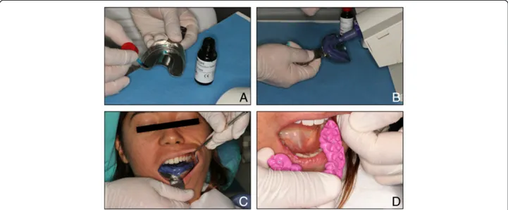
The effectiveness and clinical outcomes of the conven-tional impression technique was evaluated by measuring the total treatment time, including the individual steps (Figure 1): A) tray selection, B) adhesive application, C) upper/lower impression, D) bite registration. The treat-ment time was measured in seconds and recorded for each step by a second operator (R.T. & H.B.). Immedi-ately after the impressions were made, the attitudes and perceptions of the subjects towards the conventional im-pression technique were evaluated using a standardized questionnaire. The subjects’ perceived source of stress was also evaluated using the State-Trait Anxiety Scale immediately after the impression technique.
Digital impressions
A digital impression appointment was scheduled for the same patients 2–3 weeks following the conventional
impressions. The digital impressions were performed with the chairside dental CAD-CAM system (Cerec OMNICAM, Sirona Dental GmBH, Wals Bei Salzburg, Austria). The digital impression electronic data constitu-ents of the virtual models for both arches and bite regis-tration were recorded. All digital scanning procedures were carried out according to the manufacturer’s guide-lines and performed by the same operator (EY).
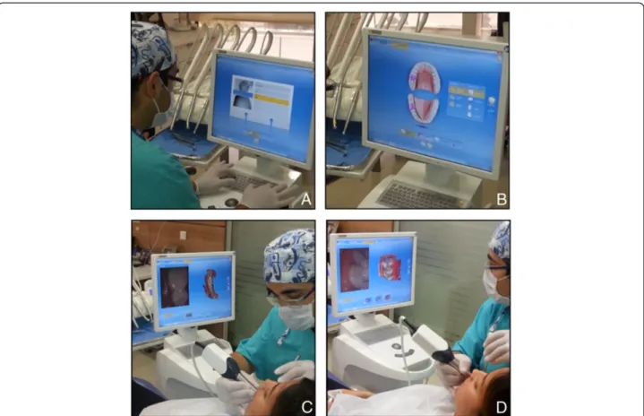
The effectiveness and clinical outcomes of the digital im-pression technique were evaluated by measuring the total treatment time, including the individual steps (Figure 2): A) entering patient information (including name, last name, date of birth, B) laboratory prescription (including shade of restoration, material choice of restoration, form of restor-ation), C) upper/lower scan, and D) bite scan. Treatment time was measured in seconds and recorded for each step by a second operator (R.T. & H.B.). Immediately after the impressions were made, the attitudes and perceptions of the subjects towards the digital impression technique were evaluated using a standardized questionnaire. The subjects’ perceived source of stress was also evaluated using the State-Trait Anxiety Scale immediately after the impression technique.
The subjects were also asked to answer a 9-item com-parative questionnaire including the following research questions: Which was the preferred impression nique? Which was the recommended impression tech-nique? Which impression technique was more efficient? Which impression technique would be most comfortable regarding impression techniques?
Reliability and validity of questionnaires
The questionnaires used in this study were pre-tested, revised, and retested before use. A pilot questionnaire
was tested on a representative sample of 10 patients. Test-retest reliability was performed to test the reliability and internal consistency of the questionnaires. The Cronbach Alpha reliability coefficient of the scales were found as 0.921, and 0.982, respectively. The adaptation, reliability and validity of the Turkish version of the State-Trait Anxiety Scale were evaluated by Öner and Le Compte in 1983 [41].
Statistical analysis
Statistical analysis by the Wilcoxon Signed-Rank Test, with p = 0.05 as the level for statistical significance, was performed to evaluate the differences in effectiveness and clinical outcomes between conventional and digital impression techniques, using the SPSS 15.0 for Windows statistical software (SPSS Inc., Chicago, IL, USA).
The attitudes and perceptions of the subjects on both impression techniques were assessed with a self-administrated questionnaire using a Visual Analog Scale (VAS) ranging from 0 to 100. The data were analyzed statistical by the Wilcoxon Signed-Rank Test, with p = 0.05 as the level for statistical significance, using the SPSS 15.0 statistical software (SPSS Inc., Chicago, IL, USA).
The subjects’ preferences for the impression techniques were assessed with a 9-item comparative questionnaire, and the distribution of the answers were evaluated by de-scriptive analysis using the SPSS 15.0 statistical software (SPSS Inc., Chicago, IL, USA).
Results
The evaluation of the effectiveness and clinical outcomes for both impression techniques are presented in Table 1. The mean overall treatment times were statistically
Figure 1 Conventional impression technique. Conventional impression technique. A) Adhesive application, B) Impression tray loading, C) Upper and lower arches impression, D) Bite registration.
significantly different (p < 0.001), and comparison of the mean impression times indicated a statistically significant difference (p < 0.001). The mean tray selection time for the conventional impression technique and the mean time for entering patient information for the digital impression technique were not statistically significant (p > 0.05). The mean adhesive application time for the conventional im-pression technique was statistically significantly different (p < 0.001) from the mean time for entering the laboratory prescription time for the digital impression technique. The difference between the mean bite registration time for the conventional technique and the mean bite scan time for the digital technique was statistically significant (p < 0.001).
Outcomes of conventional impressions
The mean overall treatment time of the conventional impression technique was 605.38 ± 23.66 s. The mean treatment times of the individual steps of the conven-tional impression technique was as follows: Mean tray selection time, 18.87 ± 2.42 s; mean adhesive application time, 27.75 ± 3.12 s. The mean conventional impression time of the upper and lower jaws was 240.70 ± 16.38 s and the mean bite registration time was 91.96 ± 10.74 s.
Outcomes of digital impressions
The mean overall treatment time of the digital impression technique was 248.48 ± 23.48 s. The mean treatment times
Figure 2 Digital impression technique. A) Entering patient information, B) Laboratory prescription, C) Upper and lower arches scanning, D) Bite scanning.
Table 1 Scores of clinical efficiency outcomes of impression techniques
Efficiency Conventional Digital P-value
Tray selection/Patient information 18,87 ± 2,42 19,08 ± 3,57 >0.05
Adhesive application/Laboratory prescription 27,75 ± 3,12 13,63 ± 1,98 <0.001*
Upper impression/Upper scan 240,70 ± 16,38 102,14 ± 17,77 <0.001*
Lower impression/Lower scan 226,10 ± 10,89 98,94 ± 10,56 <0.001*
Bite registration/Bite scan 91,96 ± 10,74 14,68 ± 3,82 <0.001*
Total treatment time 605,38 ± 23,66 248,48 ± 23,22 <0.001*
of the individual steps of the digital impression technique were as follows: the mean time for entering patient infor-mation, 19.08 ± 3.57 s, and the mean time for entering the laboratory prescription time, 13.63 ± 1.98 s. The mean digital impression time for the upper and lower jaws was 98.94 ± 10.56 s and the mean bite scan time was 14.68 ± 3.82 s.
Patients’ preferences and self concerns
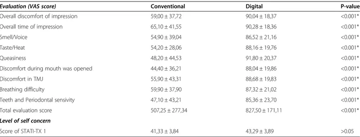
The evaluation scores and the level of concerns of the subjects regarding the impression techniques are pre-sented in Table 2. The mean scores of the subjects’ evaluation criteria regarding the two impression tech-niques were significantly different (p < 0.001). The sub-jects’ level of self concern were evaluated by scores of STATI-TX 1. The mean scores were not statistically significant (p > 0.05). All the subjects preferred the digital impression technique (p < 0.001), and patients’ preferences regarding the impression techniques, ac-cording to the 9-item comparative questionnaire, are listed in Table 3.
Discussion
In this clinical trial, according to the clinical scenario, the digital impression technique was more efficient than the conventional impression technique. Thus, the first null hypothesis was rejected. The subjects also preferred the digital impression technique rather than the conven-tional impression technique because of its comfort. Thus, the second null hypothesis was also rejected.
The study population was standardized and homoge-nized by including subjects who had no experience with conventional or digital impressions in their dental his-tory. To investigate the clinical outcomes of the two im-pression techniques, homogenizing the study population is an acceptable clinical research method to optimize ob-jectivity and minimize bias. This approach is important to avoid reporting the bias of patients who had previous experience with the dental impression procedure.
In this present study, we focused primarily on the effi-ciency of the two impression techniques and the prefer-ence of the patients under controlled clinical conditions. Future investigations should include the assessment of
Table 2 Participants’ evaluation scores and level of self concerns about impression techniques
Evaluation (VAS score) Conventional Digital P-value
Overall discomfort of impression 59,00 ± 37,72 90,04 ± 18,37 <0.001*
Overall time of impression 65,10 ± 41,55 90,28 ± 18,36 <0.001*
Smell/Voice 54,90 ± 39,04 86,52 ± 21,16 <0.001*
Taste/Heat 54,20 ± 28,06 88,16 ± 19,76 <0.001*
Queasiness 48,20 ± 44,53 91,80 ± 20,37 <0.001*
Discomfort during mouth was opened 44,40 ± 36,21 88,04 ± 19,86 <0.001*
Discomfort in TMJ 55,90 ± 43,31 88,68 ± 19,83 <0.001*
Breathing difficulty 59,90 ± 37,90 87,32 ± 21,02 <0.001*
Teeth and Periodontal sensivity 47,10 ± 43,21 85,36 ± 23,70 <0.001*
Total evaluation score 507,25 ± 277,34 827,50 ± 171,11 <0.001*
Level of self concern
Score of STATI-TX 1 41,33 ± 3,84 43,29 ± 3,89 >0.05
All data are presented as mean ± SD. Visual Analog Scale (VAS). *Statistical significance level p-0.05.
Table 3 Participants’ preferences about impression techniques according to the 9-item questionnaire
Preferences Conventional Digital
Which impression technique do you prefer in case of one more time for impression procedure? %0 %100 Which impression technique is more comfortable from point of comparison of two impression procedure? %0 %100 Which impression technique do you suggest in case of a friends’ need for impression making? %0 %100 Which impression technique do you prefer from point of time involved with impression procedure? %0 %100 Which impression technique do you prefer from point of feeling taste/smell or voice/heat during impression procedure? %0 %100 Which impression technique do you prefer from point of the size of the intraoral scanner/impression tray used in your
mouth during impression procedure?
%0 %100
Which impression technique do you prefer from point of having tooth/gingival sensitivity during impression procedure? %0 %100 Which impression technique do you prefer from point of having difficulty in breathing during impression procedure? %0 %100 Which impression technique do you prefer from point of having gagging reflex during impression procedure? %0 %100
the accuracy of the impressions produced by experi-enced versus non-experiexperi-enced operators, comparison of using scanning powders versus non-powder scanning, and comparison of full arch and partial impressions.
There are some limitations of this study. The study was designed as a comparative-controlled clinical trial, and the sequence of the evaluation of the two impres-sion techniques was chosen for psychological reasons. There is a 2–3-week interval between the two evaluation appointments. This time period was deemed sufficient to erase from memory an event or a process. The evalu-ation process focuses on the outcomes of the impression techniques by means of total treatment time in seconds, and the study does not analyze any differences in preci-sion of the two imprespreci-sion techniques.
Another limitation of the study was that only one op-erator performed the impression techniques to avoid the possible inter-operator error, such as the prolonged pro-cessing time taken by an inexperienced operator. The main purpose of the study was to focus on the patients’ perceptions and comfort in using different impression techniques. Evaluation by a second operator was not preferred because of main purpose of the study. Further investigations are planned to evaluate the perceptions of patients treated by different dental specialties and oper-ator experience to the digital impression technique.
The last limitation of this study is that it ignored the time factors involved in the conventional impression technique, such as pouring and mounting the cast, trim-ming the dies, painting the die spacer, etc. By eliminating these steps, time for the traditional workflow would be reduced significantly. Furthermore, the digital impres-sion technique and digital workflow are designed as the “digital working model” directly from the intraoral scan, without any additional factors. By virtually eliminating the intermediate processes, error accumulation in treat-ment and in the manufacturing cycle is no longer an issue.
The results of this study have revealed clinical evi-dence that the digital impression technique can be ap-plied successfully for the impressions of restorative procedures based on clinical outcomes and the patients’ preferences. However, this study was performed in a clinical scenario that excluded the effect of actual treat-ment conditions, perceived dental anxiety and stress as-sociated with treatment. This is an additional limitation of this study.
The major advantage of digital impressions is reducing the chair time. The mean total treatment time (p < 0.001) and the subjects’ evaluation scores (p < 0.001) regarding the impression techniques were significantly different (Tables 1 and 2). Improving the level of the patients’ comfort and treatment acceptance (p < 0.001) were other advantages of the digital impression techniques (Tables 1
and 2). Digital impressions tend to reduce repeat visits and retreatment, while increasing treatment effectiveness [42]. Patients will benefit from more comfort and a pleas-ant experience in the dentist’s chair.
The results of study indicate that the efficiency out-comes of the digital impression technique were higher than that of the conventional impression technique, with respect to treatment time taken up and the perceptions of the subjects. The effectiveness and clinical outcomes of both impression techniques (Table 1) were evaluated by recording the treatment time of each step in seconds, and were significantly different from each other (p < 0.001). The scores of the evaluation criteria regarding the two impression techniques (Table 2) that affect the subjects’ perception differed from one another in a statistically significant manner (p < 0.001).
The differences in the level of treatment comfort eval-uated by the subjects, including breathing difficulty, queasiness, discomfort in the TMJ, and discomfort while the mouth was kept open were statistically significant (p < 0.001). Thus, the digital impression technique is more patient-friendly than the conventional impression technique. The results of this study present the major reasons why the subjects preferred the digital impression technique instead of the conventional impression tech-nique (Table 3).
Conclusions
Within the limitations of this study, the following con-clusions can be drawn:
1. The digital impression technique was more efficient than the conventional impression technique. The overall treatment time for the conventional impression technique was longer than that for the digital impression technique. Thus, the first null hypothesis was rejected.
2. When compared with the conventional impression technique, the digital impression technique was accepted as the preferred and effective technique, according to the subjects’ perception. Thus, the second null hypothesis was rejected.
3. The treatment comfort of the digital impression technique was higher than that of the conventional impression technique when it was performed by an experienced operator.
Competing interests
The authors declare that they have no competing interests. Authors’ contributions
EY is the designed and carried out the clinical study, collected the data for analysis , performed the statistical analysis and drafted the manuscript. HK participated in the design of the study and interpretation of data. RT and HB were collected the data for analysis. All authors read and approved the final manuscript.
Received: 25 September 2013 Accepted: 29 January 2014 Published: 30 January 2014
References
1. De La Cruz JE, Funkenbusch PD, Ercoli C, Moss ME, Graser GN, Tallents RH: Verification jig for implant supported prosthesis: a comparison of standard impressions with verification jigs made of different materials. J Prosthet Dent 2002, 88:329–336.
2. Mormann WH, Brandestini M, Lutz F: The Cerec system: computer-assisted preparation of direct ceramic inlays in 1 setting. Quintessenz 1987, 38:457–470.
3. Luthardt R, Weber A, Rudolph H, Schone C, Quaas S, Walter M: Design and production of dental prosthetic restorations: basic research on dental CAD/CAM technology. Int J Comput Dent 2002, 5:165–176.
4. Otto T, Schneider D: Long-term clinical results of chairside CEREC CAD/ CAM inlays and onlays: a case series. Int J Prosthodont 2008, 21(1):53–59. 5. Wiedhahn K, Kerschbaum T, Fasbinder DF: Clinical long-term results
with 617 CEREC veneers: a nine-year report. Int J Comput Dent 2005, 8:233–246.
6. Sjögren G, Molin M, Van Dijken JW: A 10-year prospective evaluation of CAD/CAM-manufactured (CEREC) ceramic inlays cemented with a chem-ically cured or dual-cured resin composite. Int J Prosthodont 2004, 17 (2):241–246.
7. Posselt A, Kerschbaum T: Longevity of 2328 chairside CEREC inlays and onlays. Int J Comput Dent 2003, 6:231–248.
8. The glossary of prosthodontic terms. J Prosthet Dent 2005, 94(1):10–92. http://www.ncbi.nlm.nih.gov/pubmed/16080238.
9. Herbst D, Nel JC, Driessen CH, Becker PJ: Evaluation of impression accuracy for osseointegrated implant supported superstructures. J Prosthet Dent 2000, 83(5):555–561.
10. Walker MP, Ries D, Borello B: Implant cast accuracy as a function of impression techniques and impression material viscosity. Int J Oral Maxillofac Implants 2008, 23(4):669–674.
11. Lee H, Ercoli C, Funkenbusch PD, Feng C: Effect of subgingival depth of implant placement on the dimensional accuracy of the implant impression: an in vitro study. J Prosthet Dent 2008, 99(2):107–113. 12. Lee H, So JS, Hochstedler JL, Ercoli C: The accuracy of implant
impressions: a systematic review. J Prosthet Dent 2008, 100(4):285–291. 13. Wee AG: Comparison of impression materials for direct multi-implant
im-pressions. J Prosthet Dent 2000, 83(3):323–331.
14. Brosky ME, Pesun IJ, Lowder PD, Delong R, Hodges JS: Laser digitization of casts to determine the effect of tray selection and cast formation technique on accuracy. J Prosthet Dent 2002, 87(2):204–209. 15. Burns J, Palmer R, Howe L, Wilson R: Accuracy of open tray implant
impressions: an in vitro comparison of stock versus custom trays. J Prosthet Dent 2003, 89(3):250–255.
16. Ceyhan JA, Johnson GH, Lepe X: The effect of tray selection, viscosity of impression material, and sequence of pour on the accuracy of dies made from dual-arch impressions. J Prosthet Dent 2003, 90(2):143–149. 17. Chee W, Jivraj S: Impression techniques for implant dentistry. Br Dent J
2006, 201(7):429–432.
18. Vigolo P, Majzoub Z, Cordioli G: Evaluation of the accuracy of three techniques used for multiple implant abutment impressions. J Prosthet Dent 2003, 89(2):186–192.
19. Vigolo P, Fonzi F, Majzoub Z, Cordioli G: An evaluation of impression techniques for multiple internal connection implant prostheses. J Prosthet Dent 2004, 92(5):470–476.
20. Rudd RW, Rudd KD: A review of 243 errors possible during the fabrication of a removable partial denture: part II. J Prosthet Dent 2001, 86(3):262–276.
21. Rudd RW, Rudd KD: A review of 243 errors possible during the fabrication of a removable partial denture: part III. J Prosthet Dent 2001, 86(3):277–288.
22. Alhouri N, McCord JF, Smith PW: The quality of dental casts used in crown and bridgework. Br Dent J 2004, 197(5):261–264.
23. Powers J: Gypsum products and investments. In Craig’s Restorative Dental Materials. Edited by Powers J. St Louis: Mosby; 2006:313–336.
24. Duke P, Moore BK, Haug SP, Andres CJ: Study of the physical properties of type IV gypsum, resin-containing, epoxy die materials. J Prosthet Dent 2000, 83:466–473.
25. Powers J: Impression materials. In Craig’s Restorative Dental Materials. Edited by Powers J. St Louis: Mosby; 2006:269–312.
26. Wöstmann B, Rehmann P, Balkenhol M: Influence of impression technique and material on the accuracy of multiple implant impressions. Int J Prosthodont 2008, 21(4):299–301.
27. Birnbaum N, Aaronson HB, Stevens C, Cohen B: 3D digital scanners: a high-tech approach to more accurate dental impressions. Inside Dentistry 2009, 5(4). Available from: http://www.insidedentistry.net.
28. Kim SY, Kim MJ, Han JS, Yeo IS, Lim YJ, Kwon HB: Accuracy of dies captured by an intraoral digital impression system using parallel confocal imaging. Int J Prosthodont 2013, 26(2):161–163.
29. Christensen GJ: Impressions are changing: deciding on conventional, digital or digital plus in-office milling. JADA 2009, 140:1301–1304. 30. Syrek A, Reich G, Ranftl D, Klein C, Cerny B, Brodesser J: Clinical evaluation
of all-ceramic crowns fabricated from intraoral digital impressions based on the principle of active wavefront sampling. J Dent 2010, 38:553–559. 31. Henkel GL: A comparison of fixed prostheses generated from
conventional vs digitally scanned dental impressions. Compend Contin Educ Dent 2007, 28:422–424.
32. Brawek PK, Wolfart S, Endres L, Kirsten A, Reich S: The clinical accuracy of single crowns exclusively fabricated by digital workflow the comparison of two systems. Clin Oral Investig 2013, 17(9):2119–2125.
33. Seelbach P, Brueckel C, Wöstmann B: Accuracy of digital and conventional impression technique and workflow. Clin Oral Investig 2013, 17(7):1759–1764. 34. Luthardt RG, Loos R, Quaas S: Accuracy of intraoral data acquisition in
comparison to the conventional impression. Int J Comput Dent 2005, 8:283–294.
35. Güth JF, Keul C, Stimmelmayr M, Beuer F, Edelhoff D: Accuracy of digital models obtained by direct and indirect data capturing. Clin Oral Investig 2013, 17:1201–1208.
36. Karl M, Shubinski P, Taylor T: Effect of intraoral scanning on the passivity of fit of implant-supported fixed partial prostheses. Quintessence Int 2012, 43:555–563.
37. Mehl A, Ender A, Mörmann W, Attin T: Accuracy testing of a new intraoral 3D camera. Int J Comput Dent 2009, 12:11–28.
38. Ender A, Melh A: Full arch scans: conventional versus digital impressions. An in-vitro study. Int J Comput Dent 2011, 14:11–21.
39. van der Meer WJ, Andriessen FS, Wismeijer D, Ren Y: Application of intra-oral dental scanners in the digital workflow of implantology. PLoS One 2012, 7:e43312.
40. Lee SJ, Gallucci GO: Digital vs. conventional implant impressions: efficiency outcomes. Clin. Oral Impl. Res 2013, 24(1):111–115. 41. Öner N, Le Compte A: Handbook of state-trait anxiety inventory. Istanbul:
Bogazici University; 1985.
42. Polido WD: Digital impressions and handling of digital models: the future of dentistry. Dental Press J Orthod 2010, 15(5):18–22.
doi:10.1186/1472-6831-14-10
Cite this article as: Yuzbasioglu et al.: Comparison of digital and conventional impression techniques: evaluation of patients’ perception, treatment comfort, effectiveness and clinical outcomes. BMC Oral Health 2014 14:10.
Submit your next manuscript to BioMed Central and take full advantage of:
• Convenient online submission
• Thorough peer review
• No space constraints or color figure charges
• Immediate publication on acceptance
• Inclusion in PubMed, CAS, Scopus and Google Scholar
• Research which is freely available for redistribution
Submit your manuscript at www.biomedcentral.com/submit