NOVEL SAR REDUCTION
METHODS FOR MAGNETIC
RESONANCE IMAGING
A THESIS
SUBMITTED TO THE DEPARTMENT OF ELECTRICAL AND ELECTRONICS ENGINEERING
AND THE INSTITUTE OF ENGINEERING AND SCIENCES OF BILKENT UNIVERSITY
IN PARTIAL FULLFILMENT OF THE REQUIREMENTS FOR THE DEGREE OF
DOCTOR OF PHILOSOPHY
By
Yiğitcan Eryaman
March 2011
I certify that I have read this thesis and that in my opinion it is fully adequate, in scope and in quality, as a thesis for the degree of doctor of philosophy
Prof. Dr. Ergin Atalar (Supervisor)
I certify that I have read this thesis and that in my opinion it is fully adequate, in scope and in quality, as a thesis for the degree of doctor of philosophy
Prof. Dr. Yusuf Ziya İder
I certify that I have read this thesis and that in my opinion it is fully adequate, in scope and in quality, as a thesis for the degree of doctor of philosophy
Prof. Dr. Ayhan Altıntaş
I certify that I have read this thesis and that in my opinion it is fully adequate, in scope and in quality, as a thesis for the degree of doctor of philosophy
I certify that I have read this thesis and that in my opinion it is fully adequate, in scope and in quality, as a thesis for the degree of doctor of philosophy
Prof. Dr. Mark Ladd
Approved for the Institute of Engineering and Sciences:
Prof. Dr. Levent Onural
ABSTRACT
NOVEL SAR REDUCTION
METHODS FOR MAGNETIC RESONANCE
IMAGING
Yiğitcan Eryaman
Ph.D. in Electronics Engineering Supervisor: Prof. Dr. Ergin Atalar
March 2011
In this thesis, novel methods are presented, which can be used to reduce the heating of the human body due to radiofrequency fields in magnetic resonance imaging (MRI). The proposed methods depend on the modification of the electric field distribution for reducing the specific absorption rate (SAR). These methods can be used to reduce the local SAR in the vicinity of metallic devices and the whole-volume average SAR, as shown by electromagnetic field simulations and phantom, animal and patient experiments. These results can improve the safety of MRI scans performed on patients with metallic implants and MRI-guided interventional procedures. Additionally, by reducing the whole body average SAR, safer and faster MRI scans can be performed.
ÖZET
MANYETİK REZONANS GÖRÜNTÜLEME’DE
ÖZGÜL SOĞRULMA HIZINI AZALTMAK
İÇİN YENİ YÖNTEMLER
Yiğitcan Eryaman
Elektrik ve Elektronik Mühendisliği Bölümü Doktora Tez Yöneticisi: Prof. Dr. Ergin Atalar
Mart 2011
Bu tezde, insan vücudunun Manyetik Rezonans Görüntüleme (MRG) esnasında radyofrekans (RF) alana bağlı ısınmasını azaltmak için kullanılabilecek yeni yöntemler sunulmaktadır. Önerilen yöntemler Özgül Soğrulma Hızı’nın (ÖSH) azaltılması için elektrik alanın değiştirilmesine dayanır. Elektromanyetik benzetimler, fantom, hayvan ve insan deneyleri ile gösterildiği üzere bu yöntemler metal cihazlarin yakinlarinda oluşan yerel ÖSH’nın ve tüm vücut ortalama ÖSH’nın azaltılması için kullanılabilir. Bu sonuçlar vücudunda implant taşıyan hastalarda yapılan MRG taramalarının ve MRG rehberliğindeki girişimsel ugyulamaların güvenliğini arttırabilir. Ayrıca tüm vücut ÖSH’nın azaltılması suretiyle daha güvenli ve hızlı MRG taramaları da yapılabilir.
Anahtar Kelimeler: Manyetik Rezonans Görüntüleme(MRG), Özgül Soğrulma
Acknowledgements
Thanks to many people who supported me during the past 4 years of my PhD study. Thanks to them, I was able to continue research persistently, and I was able to finish this thesis which I hope will be a useful source for the future graduate students working in the field of MRI. Among many people, I want to thank first to Prof. Ergin Atalar who guided me with his wisdom, experience, and humanity through this exciting journey of science. He is a great mentor and I owe him a debt of gratitude for everything that he taught me.
I also want to thank my jury members, Prof. Ayhan Altıntaş, Prof. Murat
Eyüboğlu, Prof. Ziya İder and especially Prof. Mark Ladd who came from
Germany to attend my jury. Their valuable comments improved the scientific quality of this thesis.
I want to thank my friends in National Magnetic Resonance Research Center (UMRAM) Esra Abacı, Emre Kopanoğlu, Burak Akın, Volkan Açıkel,
Taner Demir and Haldun Özgür Bayındır for their friendship and all the
scientific collaborations that we made.
I want to thank Erdem Ulusoy for his “muhabbet” along all the “rakı” that we consumed. I also want to thank him for his musical companionship during the Tunali Hilmi Recitals.
I want to thank Kıvanç Köse for reminding me that there are many important aspects of life other than research.
I also want to thank my love Gözde Karagöz who simply made me a happier person. Thanks for sharing the burden.
I thank my family who supported me whenever I needed them.
Finally I want to thank everyone who made Ankara actually a pleasant city to live. It was a true miracle.
Table of Contents
1. INTRODUCTION……….. 1
2. MINIMUM SAR FOR RF SHIMMING BY ALLOWING SPATIAL PHASE VARIATION 2.1 Preface……….... 5 2.2 Introduction………... 5 2.3 Theory……… 6 2.4 Simulations ….….……….….….….….….… 9 2.5 Results ……….….……….….….….….…....10 2.6 Discussions……….19 2.7 Conclusion……… 20
3. REDUCTION OF IMPLANT RF HEATING THROUGH MODIFICATION OF TRANSMIT COIL ELECTRIC FIELD 3.1 Preface………21
3.2 Introduction….….….….….….….….….….….….….….……….21
3.3 Theory..….……….…23
3.3.1 Implant-Friendly RF Coil ………..…. 23
3.3.2 Transmit Field Optimization ………..… 25
3.4 Experiments and Simulations..………29
3.4.1 Implant - Friendly RF Coil ..………..…. 29
3.4.2 Transmit Field Optimization………..…. 31
3.4.2.1 Quadrature Birdcage Coil……..………...32
3.4.2.2 Linear Birdcage Coil…….……..………...32
3.4.2.3 Implant - Friendly Coil….……..………...32
3.4.2.4 Implant – Friendly Homogenous Coil...………...33
3.5 Results………....………34
3.5.1 Implant - Friendly RF Coil ..………..…. 34
3.5.2 Transmit Field Optimization………..…. 36
3.5.2.1 Quadrature Birdcage Coil……..………...37
3.5.2.2 Linear Birdcage Coil…….……..………...37
3.5.2.3 Implant - Friendly Coil….……..………...38
3.5.2.4 Implant – Friendly Homogenous Coil...………...38
3.6 Discussions………....……….... 40
3.5 Conclusion………....………. 42
4. REDUCTION OF RF HEATING OF METALLIC DEVICES BY USING A TWO-CHANNEL TRANSMIT ARRAY SYSTEM 4.1 Introduction….….….….….….….….….….….….….….……….43
4.2 Theory..….……….…45
4.2.1 Monitoring RF Induced Current Artifacts……..…………..….48
4.2.2 Finding a Safe Excitation Pattern………..… 49
4.3 Experiments………..……… 50
4.3.1 Phantom Experiments………..…... 50
4.3.2 Animal Experiments………..….. 53
4.3.3 Patient Experiments.………..….. 54
4.4.1 Phantom Experiments………..…... 54
4.4.2 Animal Experiments………..….. 57
4.4.3 Patient Experiments.………..….. 58
4.5 Discussion..………..……….. 59
4.6 Conclusions………..………..61
5. REDUCTION OF RF HEATING OF METALLIC DEVICES THROUGH MULTI-CHANNEL EXCITATION 5.1 Preface……….….….….….….….….….….….….….….………. 62
5.2 Introduction….….….….….….….….….….….….….….……….62
5.3 Theory..….……….…63
5.4 Simulations………..……….. 65
5.5 Results……..………..………66
5.6 Discussions and Conclusions..………..69
6. EFFECT OF PHASE VARIATION OF THE ELECTRIC FIELD ON METALLIC LEAD HEATING 6.1 Preface……….….….….….….….….….….….….….….………. 71
5.2 Introduction….….….….….….….….….….….….….….……….71
5.3 Theory..….……….…72
5.4 Simulations and Experiments………..74
5.5 Results……..………..………76
5.6 Conclusion………..……….. 81
7. CONCLUSIONS..……….….….….….….….….….….….….….….………82
APPENDIX ………... 83
List of Figures:
Figure 2.1
Homogenous body phantom model with uniform electromagnetic properties is shown. Target transmit profile is obtained by constraining the field at 45 sample points which forms a circular region of radius 8 cm.Figure 2.2
Optimum transmit sensitivity for imaging a single point of interest is shown. Since target profile consists of a single point, sensitivity profile solutions are not homogenous.Figure 2.3
Optimum transmit sensitivity for imaging a target profile is shown. Since target profile consists of multiple points, sensitivity solutions are homogenous. Phase throughout the profile is assumed constant.Figure 2.4
Optimum transmit sensitivity for imaging a target profile is shown. Since target profile consists of multiple points, sensitivity solutions are homogenous. Phase throughout the profile is also optimized to minimize the average SAR.Figure 2.5
The transmit sensitivity due to optimum solution sampled in radial direction. Transmit sensitivity profile is homogenous in magnitude as seen in Panel a. The phase variation is tolerated and in order to minimize average SAR as seen in Panel b.Figure 2.6
The z component of the electric field in z=0 transverse plane (1.5T) Uniform phase solution is shown in Panel a, optimum solution is shown in Panel b.Figure 2.7
The z component of the electric field in z=0 transverse plane (3.0T). Uniform phase solution is shown in Panel a, optimum solution is shown in Panel b.Figure 2.8
The z component of the electric field in z=0 transverse plane (4.7T). Uniform phase solution is shown in Panel a, optimum solution is shown in Panel b.Figure 2.9
The z component of the electric field in 0 half plane (1.5 T). Uniform phase solution is shown in Panel a, optimum solution is shown in Panel b.Figure 2.10
The z component of the electric field in half plane (3.0 T). 0 Uniform phase solution is shown in Panel a, optimum solution is shown in Panel b.Figure 2.11
The z component of the electric field in half plane (4.7 T). 0 Uniform phase solution is shown in Panel a, optimum solution is shown in Panel b.Figure 3.1
Gel phantoms with straight and curved wires. Fiber-optic temperature measurements were performed near the tips of the lead wires. (a) and (b), quadrature excitation; (c) and (d), linear excitation under the minimum heating condition; (e) and (f), linear excitation under the maximum heating condition.Figure 3.2
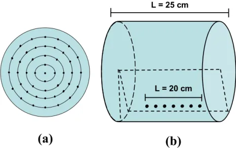
To ensure homogeneous excitation, the coil transmit sensitivity was constrained to unity at 45 sample points, forming a circular region with a diameter of 15 cm on the transverse plane (Panel a ). The electric field was constrained to zero at seven sample points on a straight line whose distances to the phantom surface were 1 cm (Panel b ).Figure 3.3
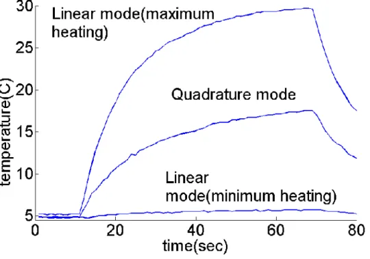
Temperature rise as a function of time measured for a straight wire with three modes: the minimum heating linear mode, the maximum heating linear mode and the quadrature mode. Final temperature increases of 0.8°C, 24.7°C, 12.1°C were observed with the minimum heating linear mode, the maximum heating linear mode and the quadrature mode, respectively.Figure 3.4
Temperature rise as a function of time measured for a curved wire with three modes: the minimum heating linear mode, the maximum heating linear mode and the quadrature mode. Final temperature increases of 0.3°C, 19.1°C and 9.2°C wereobserved with the minimum heating mode, the maximum heating linear mode and the quadrature mode, respectively.
Figure 3.5
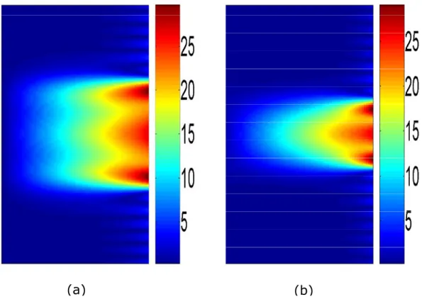
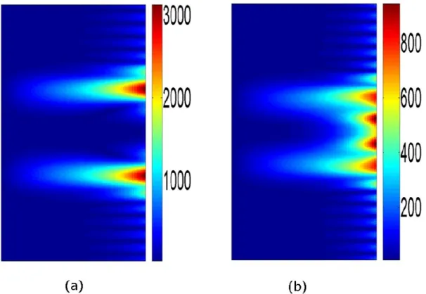
Transmit sensitivity (a, d), electric field in the trans-axial plane (b, e) and electric field in the 0 half-plane (c, f) generated by quadrature and linear coils. Note that all field solutions are in arbitrary units.Figure 3.6
Transmit sensitivity (a, d), electric field in the trans-axial plane (b, e) and electric field in the 0 half-plane (c, f) generated by implant-friendly coils and implant-friendly homogeneous coils. Locations of the implant lead are denoted by arrows in the figures. Note that all field solutions are in arbitrary units.Figure 4.1
A metallic lead with a shape that is confined inside the 0 plane experiences zero electric field. Therefore, no current flows on the lead conductor (Panel a). Similarly, for leads that extend slightly out of the zero-electric-field plane, the induced current on the lead can be made very small (Panel b).Figure 4.2
A metallic lead may have a shape that is confined in a large cylindrical volume. In that case linearly polarized excitation may be insufficient to completely reduce the tip current.Figure 4.3
Copper wire and DBS lead in in Panel a and Panel b have a shape that slightly extends out of the cylindrical angular plane. The copper wire in Panel c is confined in a larger cylindrical volume.Figure 4.4
A rectangular region approximately 1 cm deep was cut below the chest of the animal. Then, the copper wire and the temperature probe were placed under the cut section, Finally, the muscle and the skin layer were sewn back in place to cover all parts of the copper wire with living tissue.Figure 4.5
Theoretical and measured signal intensity curves of copper wire and DBS lead which was shown in Figure 4.3, Panel a and Panel b.Figure 4.6
Theoretical and measured signal intensity curves of copper wire which was shown in Figure 4.3, Panel cFigure 4.7
Theoretical and measured signal intensity curves of copper wire used in animal experiment.Figure 4.8
Figure 4.8 Brain images obtained with the quadrature (Panel a) and the safe excitation patterns (Panel b,c,d), using GRE sequence. ,5 10o was used inPanel b, 20, 16o was used in Panel c, 60, 85owas used in Panel d. The
sequence parameters are; Flip Angle=25 deg, TR=350 msec, TE=4 msec. By visual inspection it can be seen that all images have similar image homogeneity.
Figure 5.1
Uniform phantom model and transmit coil array used in simulations. The conductivity, relative permittivity, and relative permeability of the medium is chosen as 0.5 S/m, 70, 1 respectively.Figure 5.2
The transmit sensitivity solutions in the transverse plane due to quadrature, linear and optimized excitations are shown.Figure 5.3
Longitudinal component of the electric field due to quadrature, linear and optimized excitations, is shown in the transverse plane (x marks the location of the straight metallic wire in the transverse plane).Figure 5.4
Longitudinal component of the electric field due to quadrature, linear and optimized excitations, is shown in the in the angular plane / 6 (the location of the metallic wire is shown by black straight line)Figure 5.5
The variation of the tangential component of the electric field along the wire due to quadrature, linear and optimized solutions is plotted.Figure 6.1
A lead with a helical geometry is placed inside a uniform head phantom model. The length and radius of the phantom L and r are chosen as 22cm and 7.5 cm respectively. The radius of the helix formed by the lead d is chosen as 6 cmFigure 6.2
A straight lead inside the uniform phantom model is shown. Transmit array elements are fed by currents with same magnitudes but varying phases. With this method an incident electric field is obtained whose phase is changing linearly along the lead.Figure 6.3
A straight lead inside the uniform head phantom model is shown. The lead is exposed to quadrature birdcage coil excitation.Figure 6.4
The phase variation of the electric field along the helical and straight leads is plotted.Figure 6.5
Temperature variation recorded at the lead tips in 1.5 TFigure 6.6
Temperature variation recorded at the lead tips in 3.0 TFigure 6.7
The phase variation of the incident electric field in 3.0 T is shown for different Figure 6.8
The SAR reduction in tip A and SAR amplification in tip B with respect to case is shown for 1.5 T.0Figure 6.9
The SAR reduction in tip A and SAR enhancement in tip B with respect to case is shown for 3.0 T.0List of Tables
Table 1
Reduction in SAR with respect to uniform magnitude-phase solution is shown.Table 2
Maximum tip temperature is shown for each experiment. Notably, using the safest excitation pattern reduced the tip temperature substantially with respect to that of the quadrature excitation.Table 3
SAR reduction at the tips of the helical lead with respect to straight leadAs it can be seen from the table, the results obtained from experiments and simulations are in agreement for both 1.5 T and 3.0 T.
1. INTRODUCTION
Magnetic Resonance Imaging (MRI) is a safe imaging technology that provides many clinical benefits. Basically, MRI is performed by exciting magnetic spins with radiofrequency (RF) pulses and receiving the response generated by these spins as they relax into their original state. This response is spatially encoded by using the gradient fields and converted to an actual image.
Although it is not desirable, the body is exposed to an electric field during the RF excitation of the spins. This electric field may cause heat dissipation as it penetrates the conductive medium of body tissues. The specific absorption rate (SAR) is used as a measure of the electromagnetic (EM) power dissipated in a given volume. Current regulations set limits [1] on both the average whole body SAR and the peak local SAR in order to ensure the safety of patients. Furthermore, regulations limiting the maximum temperature increase in different parts of the body are also included.
The average-volume SAR is a factor that limits the maximum power that can be used for the RF excitation. The reduction of the average SAR is important for two reasons. First, an approximately quadratic relationship exists between the SAR and the operating frequency of the MRI. The frequency in MR scanners has increased over the years in order to benefit from the SNR advantages obtained at higher field strengths. Because SAR limits have remained the same, SAR reduction techniques must be developed in order to ensure patient safety in the higher field strengths. Second, the average SAR increases as the duration of the RF pulse decreases. Therefore, if fast scans with short RF pulses are to be implemented, the SAR should be reduced.
The SAR may depend on both the transmit coil design and the sequence parameters. Accordingly, there are approaches that focus on tailoring the RF excitation and redesigning the MR sequences in order to reduce the average SAR [2-4]; other methods are based on designing new transmit coil geometries and RF shimming [5]. Methods that adopt both of these approaches for SAR
reduction have also been investigated [6]. Transmit arrays are promising due to the freedom of exciting the spins with multiple elements simultaneously [7]. With that freedom, the SAR problem can be solved, along with the problem of RF field inhomogeneity. In most of the applications, a spatially homogenous RF excitation is usually desirable in order to obtain high-quality MR images. The RF field homogeneity is especially reduced in higher field strengths where the wavelength is comparable to the body size. RF shimming [8] and transmit sense [6] methods can be used with transmit arrays to solve the homogeneity problem while handling the SAR problem simultaneously.
In the presence of metallic devices, the local SAR becomes more important for safety. The current clinical MR scanners operate safely within the SAR limits. However, when metallic devices are present in the patient, the SAR amplification near the device can be dangerous [9-12]. When the local SAR is amplified, the temperature may increase excessively in the vicinity of the device, and tissue damage and burns may occur [13]. To prevent this, patients carrying metallic devices are not allowed into the MR scanner; considering that there are many people in the world who have metallic implants, the importance of this problem can be appreciated. There is also a risk related to local heating around metallic devices in interventional procedures. With the advent of interventional MRI, it is now possible to perform catheterization and biopsy procedures under MRI guidance; however, RF safety problems introduce additional risks with these procedures [14-17]. A solution that directly addresses this safety problem may increase the quality of life of millions of people.
In general, for both diagnostic and interventional procedures, the reduction of RF heating of metallic devices is crucial for patient safety. Many solutions to this problem have been proposed in the literature. In most of these works, the device or its long conductor extension is modified electrically in order to prevent heating [18-21]. Although this approach is promising, in some applications, such as MR-guided biopsy procedures [22], it may be impractical to make a modification to the device. Furthermore, in the case of implanted devices, such as pacemakers and deep brain stimulators (DBS), the replacement
of the device with a safer one may not always be convenient for patients. Thus, instead of modifying the device, the electromagnetic (EM) field surrounding the device can be modified to reduce the RF heating, such that replacing the metallic device with a safer version is no longer necessary.
The modification to the electric field should be made in order to reduce the RF heating only, and the resulting MR image quality should not be affected. In this thesis, methods to achieve this task are investigated. By modification of the EM field distribution, the RF heating of the human body was shown to be reduced. In Chapter 2, this approach is demonstrated to minimize the average SAR while keeping the magnitude of the transmit sensitivity unchanged. In addition, it is shown that, by releasing the phase constraint of the transmit sensitivity, the average SAR can be reduced even further. The optimum field solution that results in the minimum average SAR was also calculated [23].
Different RF excitation methods are used to reduce the RF heating due to implanted devices in the third chapter of the thesis. Evidence is provided that, a linearly polarized coil can be used to safely scan a patient with an implant [24]. The zero electric-field plane of the coil was coincided with the implant in order to prevent the tip heating of the metallic device. Furthermore, the transmit sensitivity characteristics of the coil were preserved with respect to a quadrature coil. This approach required either rotating the coil or the patient in order to reduce the electric field in the vicinity of the device. In Chapters 4 and 5, a similar task was achieved by using a two-channel and a multi-channel transmit array [25, 26], where the patient or the coil remained constant and the EM field was altered. In Chapter 4, a method to monitor the induced current on a metallic device is also presented. The method was based on measuring the induced current artifacts in the MR images: the safest two-channel excitation that cancelled the current on the device was found, and the RF heating was minimized. The application of a similar method to the multi-channel excitation is discussed in Chapter 5, with the average SAR and homogeneity issues also being addressed. Lastly, in Chapter 6, the effect of the phase variation of the electric field on the heating of the metallic device is investigated [27]. It is
shown that a linear-phased electric field variation would amplify the heating at one of the tips of a metallic wire and reduce the heating at the other one. The lead/wire geometry was modified in order to achieve such a condition. Additionally, a similar idea was tested with transmit arrays, which were used to generate an electric field whose phase changed linearly along the lead. A demonstration of the tip SAR amplification and reduction is provided.
2. MINIMUM SAR FOR RF SHIMMING BY
ALLOWING SPATIAL PHASE VARIATION
2.1 Preface
The content of this chapter was presented (in part) in a conference publication [23], reference: Eryaman Y, Tunç C A., Atalar E “Minimum SAR for RF Shimming By Allowing Spatial Phase Variation” Proc Intl Soc Mag Reson Med 18(2009):4777.
2.2 Introduction
The Specific Absorption Rate (SAR) is a patient safety parameter that should be seriously considered in MRI procedures. There are many studies in the literature in which the whole body average SAR has been minimized under conditions of satisfying a target transmit sensitivity [5,6]. In one of these studies, the SAR due to a given multi-channel transmit coil was minimized with respect to the phase and the magnitude of the excitation currents of the individual channels [5]. In another work, the ultimate value of the SAR for transmit sense was calculated by the optimization of the field inside a homogenous body model [6]. In these studies, the target transmit sensitivities were chosen in order to obtain a uniform magnitude field distribution. However, reducing the SAR by relaxing the phase constraints of the target profile was not investigated. In the study presented here, by keeping the magnitude distribution of the target transmit profile within a given boundary, the phase distribution was optimized to obtain the true ultimate SAR for the MRI coils. It was shown that it was possible to reduce the whole body SAR by orders of magnitude up to 30, while realizing a desired magnitude distribution for target sensitivity. For this purpose, the related optimization problem was solved by Particle Swarm Optimization (PSO) [3]. PSO is a search algorithm that can easily be implemented and used to solve optimization problems with various constraints. By using PSO, the optimum EM field distribution that satisfied the above conditions was calculated. Using well-known techniques [6], the current distributions that can generate the optimum field inside the
body model can also be calculated. These optimum current paths can be used to optimize RF coils and to obtain a minimum whole body SAR.
2.3 Theory
The average whole body SAR for a homogenous body model depends on the volume integral of the magnitude square of the electric field distribution, as shown below:
(1)
In this expression, is the conductivity, and M is the total body mass.
During MRI, it is usually desirable to obtain a uniform transmit sensitivity profile. In order to achieve that, the forward polarized field component, Hf , should be constrained on desired locations in the body. For any given point of interest ( , ) , the expression 0 0 for theHf in cylindrical coordinates is shown below:
(2)
where Hand H are the magnetic field components in the radial and angular directions,
respectively.
In order to make a general formulation for the problem, the cylindrical basis expansion can be formulated to express the field, as follows:
(3)
The expression for each separate mode, Emn, can be written as Emn Emnmne ejm j zz
, where and z are the angular and z coordinates in the cylindrical coordinate system, respectively, and m and n are integer variables representing the expansion modes. E is mn
a 3x2 matrix and is a function of , the radial coordinate, but not or z, and mn
is a 2 / body SAR M
E dv
mn m n E E
( ) j f H H jH e 2x1 vector whose elements are the constants that multiply the basis functions and
[ ]T
mn A Bmn mn
.
The E matrix and its components are shown below:mn
(4)
Using the basis expansion, the forward polarized field component, Hf , can also be expressed, as follows:
(4)
where each separate mode for Hf can be expressed as shown below:
(5)
Here, denotes the complex conductivity of the medium. ' and zn are the wave n
numbers along the longitudinal and radial directions, respectively, which can be calculated as 2
0[ 0 ]
j j
, where L is the length of the cylinder.
For the case where the number of points of interest is equal to k, the whole constraint on the values of Hf can be written in the following matrix form:
(6) ( ) 0 1 ( ) ' ( ) 1 ' ( ) ( ) ' m n m n m n m n m n J J J jJ J mn E ( ) ( ) mn f fmn mn H r
H r ( 1) 1 ' ( ) , ( ) 2 2 j m z f mn m j H r J e B cwhere c, the desired transmit sensitivity profile, is represented by a kx1 vector whose elements are equal to the desired Hf values at each point of interest. is a column vector that contains the weighting coefficients (A and mn B ) for each separate mode. mn
Lastly, B is the transmit sensitivity matrix whose elements are equal to the basis functions of Hf evaluated at the desired point of interests.
The average whole body SAR in the homogenous body model can be written as follows:
(7)
where R is a Hermitian matrix and can be computed by using the following expression mn
[6]:
(8)
The average whole body SAR can be expressed in a shorter form, as R , where R is
the electric field cross-correlation matrix whose block diagonals are equal to R .mn
After the definition of required variables, the SAR minimization problem can then be expressed as follows:
. (9)
The solution for this problem can easily be found by using the Lagrange optimizer method, as shown below:
(10)
In MRI, the magnitude distribution of the transmit field profile is usually desired to be homogenous. However, the phase distribution is a free parameter that can be optimized in
( / ) mnH mn mn mn SAR M
R 0 2 rbody H mn mn mn R L E E d
minaa R Bc 1 1 ( ) avg SAR c BR B cD= 0.2 m
L=1 m
order to obtain a minimum whole body SAR. If the S matrix is defined as S(BR B1 ) 1,
then the minimization problem above can be expressed as follows:
(11)
where k is the number of sample points in the target transmit field profile, and is the tolerance for the magnitude of Hf .
As will be shown later, the solution of this problem will significantly decrease the SAR while preserving the magnitude distribution of the transmit sensitivity.
2.4 Simulations
In all solutions, a homogenous body model diameter of 0.2 meters and a length of 1 meter were assumed (Figure 2.1).
Figure 2.1 The homogenous body phantom model with uniform electromagnetic properties is shown. The target transmit profile is obtained by constraining the field at 45 sample points, which forms a circular region of radius 8 cm.
minaac Sc
The relative permeability and permittivity for the phantom were chosen as 1 and 70, respectively. The conductivity was assumed to change linearly with the frequency, and it was taken as 0.4, 0.8 and 1.2 S/m for 1.5 T, 3.0 T and 4.7 T, respectively.
As an initial solution, the target profile was assumed to include only a single point of interest. This point was chosen as the center of the cylinder. For a single-point profile, varying the phase of Hf for that point of interest did not have an effect on the minimum whole body SAR. The optimum coil sensitivity and the minimum whole SAR were calculated for 1.5 T, 3.0 T and 4.7 T.
To obtain a homogenous transmit sensitivity, multiple numbers of points were used for the target profile. First, a zero phase profile was assumed, and the corresponding optimum sensitivity and minimum SAR were calculated. Then, in order to generate the optimum phase distribution, the PSO algorithm [3] was used to solve the problem. A MATLAB (version 7.0, MathWorks Inc., Natick, MA) program was written to implement the algorithm. For the target transmit field profile, Hf was constrained to have a uniform magnitude at 45 sample points, which formed a circular region of a radius of 8 cm. Figure 2.1 shows the location of sample points that Hf constrained. The phase distribution is optimized by using the particle swarm optimization method in order to minimize the whole body SAR. The simulations were made for 3 field strengths, 1.5 T, 3.0 T and 4.7 T, and for 3 different tolerance values, 0, 0.03 and 0.1. Note that the transmit sensitivity was permitted to fluctuate in the interval [1,1]. The SAR reduction with respect to a zero phase transmit profile was calculated. The optimum Hf
distribution was calculated for the 0 case at 3 field strengths. Electric field maps related to the zero phase and optimum phase solutions were also generated.
2.5 Results
For the PSO solutions, the number of particles, the constriction factor and the cognitive and social rates for velocity updates were chosen, as explained in a previous work [40]. The fitness function was defined as the ratio of the average SAR. Each simulation lasted
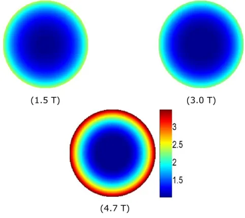
(1.5 T) (3.0 T)
(4.7 T)
for 50-100 seconds using a PC with an AMD Athlon™ 64 Processor and 2.41 GHz 4 GB RAM.
As an initial solution, the target profile was assumed to include only a single point of interest. This point was chosen as the center of the cylinder and sets a lower bound for all of the other solutions. As will be shown, larger profiles consisting of multiple locations will result in larger SARs when compared with the solution found by using a single point of interest. Figure 2.2 shows the sensitivity variation in the z=0 transverse plane at 3 field strengths. Because SAR will depend on sequence parameters, such as flip angle and TR, the SAR value for each solution is reported here in arbitrary units.
Figure 2.2 The optimum transmit sensitivity for imaging a single point of interest is shown. Because the target profile consists of a single point, the sensitivity profile solutions are not homogenous.
The SAR values obtained by each solution were 1, 6.51 and 29.66 arbitrary units (au). For these solutions, the relationship between the average SAR and field strength was
(1.5 T) (3.0 T)
(4.7 T)
Uniform circular target profile Uniform circular
target profile Uniform circular target profile
expected to be quadratic. However, it was also assumed that the conductivity increased with the field strength. Therefore, the increase in the SAR due to the field strength was larger than a quadratic increase.
As the second solution, a uniform magnitude target profile was assumed; the phase of the target profile was assumed to be constant as well. The sensitivity of the optimum solution is shown in Figure 2.3.
Figure 2.3 The optimum transmit sensitivity for imaging a target profile is shown. Because the target profile consists of multiple points, the sensitivity solutions are homogenous. The phase throughout the profile was assumed to be constant.
As expected, an almost perfectly homogenous sensitivity solution was obtained in the transverse plane for all of the field strengths. However, this homogeneity resulted in an increase in the average SAR. The SAR values obtained for these solutions were 4.30, 452 and 53,287 au for 1.5 T, 3.0 T and 4.7 T, respectively. This increase can easily be
(1.5 T) (3.0 T) (4.7 T) Uniform circular target profile Uniform circular target profile Uniform circular target profile
explained by the modal expansion of the EM field inside the phantom model. The modal expansion of a uniform profile in the space requires using modes with large indices. These modes exhibit a fast spatial variation in both the angular and longitudinal directions. Therefore, their SAR contribution to the solution is significantly higher than the modes with slower spatial variation. Therefore, an increase in SAR due to a homogenous target profile constraint is physically unavoidable. It should also be noted that among the infinitely many solutions satisfying the uniform phase-constant magnitude sensitivity profiles, these solutions have the minimum average SAR.
However, to alleviate this SAR problem, a different approach can be implemented. Although it is desirable to obtain a uniform magnitude distribution, the phase variation of the sensitivity is not a constraint. For this purpose, the optimization problem mentioned in Equation 11 is solved. The resulting transverse sensitivity solutions are shown in Figure 2.4.
Figure 2.4 The optimum transmit sensitivity for imaging a target profile is shown. Because the target profile consists of multiple points, the sensitivity solutions are homogenous. The phase throughout the profile is also optimized to minimize the average SAR.
-10
-5
0
5
10
0.8
0.9
1
1.1
1.5 T
3.0 T
4.7 T
TARGET PROFILE-10
-5
0
5
10
-200
-150
-100
-50
0
50
4.7 T
3.0 T
1.5 T
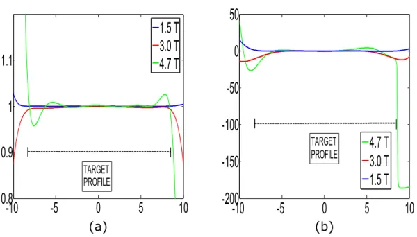
TARGET PROFILE (a) (b)Similar to the previous solution, the magnitude variation was kept constant through the target profile. The phase variation, however, was optimized to minimize the average SAR. Figure 2.5 shows the magnitude and the phase of the transmit sensitivity sampled in the radial direction.
Figure 2.5 The transmit sensitivity due to the optimum solution sampled in the radial direction. The transmit sensitivity profile is homogenous in magnitude, as seen in Panel a. The phase variation is tolerated in order to minimize the average SAR, as seen in Panel b.
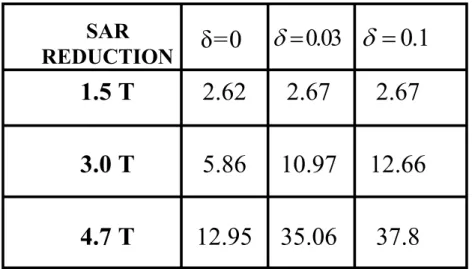
The SAR due to the optimum solutions was 1.64, 77.3 and 4027 au for 1.5 T, 3.0 T and 4.7 T, respectively. Clearly, by releasing the phase constraints, a reduction with respect to the uniform phase solution was achieved. Similarly, by releasing the magnitude constraint, a reduction in SAR can be realized. For this purpose, the magnitude of the field was permitted to fluctuate in the interval [1,1], as indicated in equation 11. Three different values, 0, 0.03 and 0.1, were used for the solutions. Table 1 shows the
37.8
35.06
12.95
4.7 T
12.66
10.97
5.86
3.0 T
2.67
2.67
2.62
1.5 T
SAR
REDUCTION
δ=0
0.03
0.1
amount of the reduction in the SAR with respect to the uniform magnitude-uniform phase solution.
Table 1 Reduction in the SAR with respect to the uniform magnitude-uniform phase solution is shown.
As expected, as the constraints are released, the number of freedoms increases, and the SAR can be reduced further. Note that a maximum reduction of 37.8 was obtained with
0.1
. With this solution magnitude, the sensitivity was permitted to fluctuate between 0.9 and 1.1.
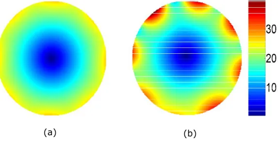
It should be noted that the field solutions are also critical to understand and design better RF coils with less average SAR. For this purpose, electric field variation should also be investigated. The z component of the electric field of both the uniform phase solutions and the optimum solutions were calculated for 1.5 T, 3.0 T and 4.7 T and are shown in Figure 2.6, Figure 2.7 and Figure 2.8, respectively. is assumed for all of the solutions.0
The longitudinal component of the field can be used to identify the coil geometry and the current paths on the coil.
(a) (b)
(a) (b)
Figure 2.6 The z component of the electric field in the z=0 transverse plane (1.5 T). The uniform phase solution is shown in Panel a, and the optimum solution is shown in Panel b.
Figure 2.7 The z component of the electric field in the z=0 transverse plane (3.0 T). The uniform phase solution is shown in Panel a, and the optimum solution is shown in Panel b.
(a) (b)
Figure 2.8 The z component of the electric field in the z=0 transverse plane (4.7 T). The uniform phase solution is shown in Panel a, and the optimum solution is shown in Panel b.
In order to understand the geometry of the RF coil, the longitudinal variation of the electric field should also be investigated. Figure 2.9, Figure 2.10 and Figure 2.11 show this variation for both the uniform phase and optimum solutions.
(a) (b)
Figure 2.9 The z component of the electric field in the half plane (1.5 T). The uniform 0 phase solution is shown in Panel a, and the optimum solution is shown in Panel b.
(a) (b)
Figure 2.10 The z component of the electric field in the half plane (3.0 T). The uniform 0 phase solution is shown in Panel a, and the optimum solution is shown in Panel b.
Figure 2.11 The z component of the electric field in the half plane (4.7 T). The uniform 0 phase solution is shown in Panel a, and the optimum solution is shown in Panel b. is 0 assumed.
2.6 Discussion
From Figure 2.5, Figure 2.6 and Figure 2.7, it can be seen that transmit arrays can be used to realize field variations. The electric field due to each channel element is visible in Figure 2.5. The number of channels can be identified by a visual inspection of the results. Because the field varies in the angular direction, the coil elements can be fed with the appropriate excitation currents to approximate these field solutions.
Furthermore, it can be seen from Figure 2.9, Figure 2.10 and Figure 2.11 that the electric field has a variation in the longitudinal direction. This may be achieved by modulating the amplitude of the conductor currents or the geometry of the coil in a longitudinal direction, such that the optimum field is approximated. Transmit array elements can be placed in a longitudinal direction with the same motivation as well. In all cases, the
solutions show that the effective length of the coil varies from one-third to one-half of the body length for different field strengths.
2.7 Conclusion
In this work, the minimum average whole body SAR for RF shimming was calculated. The EM field of a transmit coil was optimized in order to achieve a homogenous transmit sensitivity and minimum whole body SAR. The reduction in SAR due to relaxing the phase and magnitude constraints was calculated. The optimum coil sensitivities and electric field magnitude distributions are also presented.
3. REDUCTION OF IMPLANT RF HEATING
THROUGH MODIFICATION OF TRANSMIT
COIL ELECTRIC FIELD
3.1 Preface
The content of this chapter was published in a journal paper[24] Reference: Eryaman, Y., Akin, B. and Atalar, E. Reduction of implant RF heating through modification of transmit coil electric field. Magnetic Resonance in Medicine, n/a. doi: 10.1002/mrm.22724))
3.2 Introduction
Magnetic resonance imaging (MRI) is known as a very safe imaging technology. However, because of the possibility of inducing excessive currents on the metallic wires, MRI scanning is generally not performed on people with metallic implants such as pacemakers. A radio-frequency (RF) electric field, although undesirable, is often generated in the body during the excitation of spins with RF magnetic field pulses. Power absorbed by the body under this electric field is determined by the specific absorption rate (SAR) and needs to be kept at a level that is safe to the patients. If a patient with a metallic implant is examined using MRI, a very significant SAR amplification may occur around the implant, which may cause excessive body heating and burns. Due to this well-known problem, patients with metallic implants are currently not allowed inside the MRI scanners.
Previous studies [9,10] have assessed the implant heating problem via both in vitro and in vivo approaches. Mathematical models have also been presented [11,12] and the validity of these models has been further verified by comparison with the experimental data. A detailed analysis of the problem was conducted in [11] by solving the bio-heat equation with Green’s function and the linear
system theory. The maximum steady-state temperature increase in the tissue near a transmitter catheter antenna was calculated. In [12], a parameter called the “safety index” was introduced, which combines the effect of the SAR gain of the implant lead and the bio-heat transfer process to measure the in vivo temperature changes. Variations of the safety index with respect to the length and radius of the implant lead, the thickness of the insulation, tissue conductivity and permittivity were also investigated. These studies presented a good model of tissue heating caused by metallic wires in RF fields. In another study, experimental methods were developed to measure and monitor the RF-induced currents inside implants [29].
Modifications of the implant leads and wires for reducing the RF-induced heating were investigated in other studies. In two of these studies, a series of chokes was added to the coaxial cables [18,19] to reduce the currents generated on the cable shield. In another study [20], the effects of coiled wires on induced heating were investigated. By introducing air gaps and lowering the parasitic capacitance, the self-resonance frequency of a coiled wire was shifted to the operating frequency. This change increased the impedance of the wire and thus reduced the RF heating. However, all of these designs are based on modifying the lead wires or cables, which makes it difficult to produce mechanically robust leads. In addition, for patients who already have pacemakers, replacing the original leads with these modified safer leads may not always be feasible. For these reasons, modifications of the implant lead designs or the catheters may not always be the most appropriate solution for the MRI-induced RF heating of metallic wires.
The relationship between the electric field distribution and the temperature increase of the implant leads was recently investigated [30]. It was found that tissue heating depends on the orientation of the lead with respect to the direction of the electric field. To identify the worst-case scenario, an optimization-based approach was used in [31] to calculate the EM field that could produce the maximum heating at the wire tip. However, studies to optimize the EM transmitter field to minimize the implant heating were not carried out.
In this study, we showed that the transmitter coil field used in MRI could be optimized to steer the electric field away from the implant lead and thus prevent heating. As demonstrated experimentally, a linearly polarized birdcage coil could be used for this purpose. Although this approach preserved the homogeneous transmit sensitivity characteristics of the coil, it caused a doubling of the whole-body SAR. To alleviate this problem and achieve uniform sensitivity, we further modified the field of the transmitter coil to minimize both whole-body SAR and the implant heating. Details of this approach are described in the following sections.
3.3 Theory
3.3.1) Implant-Friendly RF Coil
In the standard quadrature birdcage coils, the electric field is uniform in the angular direction but varies roughly linearly in the radial direction [32]. Therefore, an implant lead placed at the edge of the body experiences a high electric field, which induces currents both on the lead and in the body and eventually causes local SAR amplification. The electric field distribution of a standard forward polarized quadrature birdcage coil in an infinitely long homogeneous model can be approximated by ignoring the end-ring currents as follows (see the appendix for a detailed derivation):
(12)
where E ,z E,E are the longitudinal, angular and radial components of the electric field, respectively; Hf is the transmit sensitivity of the coil; is the Larmor frequency; and0 are the permeability and permittivity, respectively; is the conductivity of the homogeneous model; and are the radial and angular coordinates in the cylindrical coordinate system, respectively; and j is the imaginary number defined by . Similarly, the electrical field of a 1 linearly polarized coil can be expressed as follows:
0 , 0, 0 2 f j z H E e E E
(13)
Note that j indicates a 90-degree phase shift with respect to a real field
expression. As can be seen from the above equations, linear and quadrature birdcage coils have similar transmit sensitivity yet different electric field distributions. The transmit sensitivities of each coil are approximately uniform in the transverse plane. (Please see the appendix for the detailed derivation.) It can be noted from the above equation that the electric field is zero over the entire 0 plane. This plane can be steered into any angular direction by either changing the feeding location or simply rotating the linear birdcage coil. The same task can also be performed by controlling the amplitudes of the currents fed into the two ports of a quadrature birdcage coil. For example, if port-1 and port-2 are set in such a way as to make the corresponding electric fields zero at the 0 and / 2 planes, respectively, then the excitation currents with relative amplitudes of cos and 0 sin at ports 1 and 2 would generate a zero 0 electric field plane at .Note that 0 plane covers both0 and 0
0
half planes. If an implant lead lies on the zero electric field plane, there will be no induced currents on the lead. Because setting the electric field to zero makes the perpendicular component of the magnetic field vanish at the same plane, this method also intrinsically prevents the H-field coupling.
Using a linear birdcage coil solves the heating problem for an arbitrarily shaped implant lead when the lead is located in the zero electric field plane. Despite this modification of the electric field distribution, the transmit sensitivity is not significantly disturbed. However, as previously shown [33], linear birdcage coils are not efficient for RF transmission when the volume average SAR is considered. For linear excitation, a reverse polarized field component co-exists with the forward polarized component, and the whole-volume average SAR per unit flip angle is doubled.
0 sin , 0, 0
z f
3.3.2) Transmit Field Optimization
To alleviate the doubling problem of the whole-volume average SAR, a general formulation was developed. Because transmit sensitivity is determined by the forward polarized component of the magnetic field and SAR is determined by the electric field distribution, the above-mentioned problem can be solved by optimizing the electromagnetic field of the coil [6]. In this study, we successfully demonstrated the feasibility of such a strategy by obtaining the desired electromagnetic field distribution in the body. Although the work is not trivial, once an optimum electromagnetic field is identified, it is possible to design a coil that produces the desired field. The design of such an optimum implant-friendly coil is left for a future study.
First, we assumed that the optimization would be conducted in a uniform cylindrical object. This assumption simplified the formulation but could also be used with the other geometries. Because a cylindrical object is assumed, the cylindrical basis functions were used to expand the optimum field solution that minimized the whole volume average SAR [34,35]:
(14)
where and z are the angular and z coordinates in the cylindrical coordinate
system, respectively, and m and n respectively denote the index of the circumferential and longitudinal modes used in the basis expansion. E is a 3x2 mn matrix that contains the electric field basis functions for the , and z components as shown below:
(15) z j z jm mn m n E e e
mn E ( ) 0 1 ( ) ' ( ) 1 ' ( ) ( ) ' m n m n m n m n m n J J J jJ J mn Emn
E is a function of , the radial coordinate, but not of or z.mn
is a 2x1 vector whose elements are the constants that multiply the basis functions and
[ ]T
mn A Bmn mn
.
The transmit coil sensitivity can be expressed by evaluating the forward polarized field, which can be written in the summation form as
(16)
Each separate mode for Hf can be expressed as follows:
(17) where zn 2 n L , 2 2 2 n zn
and ' j [35]. Here, denotes the '
complex conductivity of the medium, and and zn are the wave numbers n
along the radial and longitudinal directions, respectively, which can be calculated as 2
0[ 0 ]
j j
. L is the length of the cylinder.
For k points of interest, the whole summation in the Hf expression can be written in the following matrix form:
(18)
where c, the desired transmit sensitivity profile, is represented by a kx1 vector whose elements are equal to the desired Hf values at each point of interest. is a column vector that contains the weighting coefficients (Amn B ) for each mn
separate mode. H is the transmit sensitivity matrix whose elements are equal to the basis functions of Hf evaluated at the desired points of interest. H is a
(2 )
k M N matrix, where M and N denote the total number of
( 1) ( ) ( ) j m j znz fmn mn f mn H r
H r e e 1 1 ' ( ) , ( ) 2 zn fmn m n n j H r J H ccircumferential (m) and longitudinal (n) modes that are used in the basis expansion. Implementing different field variations would require using different combinations of the cylindrical modes. To express a field exhibiting a rapid spatial change in the circumferential or the longitudinal direction, one must use higher-order modes in that particular direction. Therefore, to characterize an arbitrary EM field with this expansion, an infinite number of modes is required. For practical purposes, the number of modes is truncated in our study
The desired target transmit sensitivity is one of the linear constraints for minimizing the average SAR. A separate constraint also exists on the electric field to reduce the implant heating.
To achieve the zero-implant-heating condition, the components of the electric field that are parallel to the lead should be set to zero. Therefore, the induced current on the lead wire will be zero. This condition can also be expressed as a linear constraint, similar toHf , as shown below:
(19)
where 0is a px1 vector with all of its elements equal to zero, and p denotes the number of sample points where the electric field is set to zero. E is a
(2 )
k M N matrix, where M and N denote the total number of circumferential (m) and longitudinal (n) modes that are used in the basis expansion. E matrix contains the basis functions forE ,z Eand Eevaluated at
the desired zero electric field locations.
The constraints on Hf and the components of the electric field can be combined into a single matrix equation: F e, where F and e are formed by concatenating the matrices B, E and the vectors c and0, respectively.
While it is desirable to set the magnetic and electric field to certain values at points of interest, the specific absorption rate (SAR) needs to be under control. The expression for the average SAR is:
0 E
2 /
body
SAR M
E dv
(20)where the conductivity, M is the total body mass and dvis the differential volume element . With the cylindrical mode expansion for a homogeneous body model, the resulting relation can be written as:
(20)
where R is a Hermitian matrix and can be computed using the following mn
expression [35]:
(21)
where rdenotes the radius of the homogeneous model. The average SAR can be expressed in a more compact form as R , where R is the electric field cross correlation matrix whose block diagonals are equal toR .mn
Among the infinite number of solutions satisfying F e, the solution with the minimum volume average SAR can be found by minimizing R . The solution for can then be defined as:
(22)
The minimum whole body SAR value can be computed as:
(23)
These equations give the minimum possible SAR under the conditions of the desired transmit sensitivity and zero electric field near the implant. They also
( / ) mnH mn mn mn SAR M
R 0 2 r H mn mn mn R L E E d
1 ( 1 ) 1 opt R F FR F e 1 1 min ( ) SAR e FR F egive the corresponding weights for the cylindrical expansion modes. Although this solution does not directly specify the type of coil to be used, uniquely opt determines the EM field of the optimum coil. The significance of this result can be appreciated by experiments and simulations as explained in the next section.
3.4 Experiments and Simulations
3.4.1) Implant-Friendly RF Coil
To demonstrate the proposed theory, heating of metallic wires with both linear and quadrature excitation was tested. A phantom head model of 16 cm in diameter and 25 cm in length was prepared with commercially available gel (Dr Oetker Jello, Izmir, Turkey). To measure conductivity and relative permittivity, a cylindrical transmission line setup was used. By measuring the impedance at the end of the line and using the lossy transmission line impedance equations, the conductivity and relative permittivity were calculated [36]. A conductivity of 0.51 S/m and relative permittivity of 70 were obtained with 2.4 g/l of salt in the gel solution.
Heating experiments were performed with a straight wire and a curved wire, as shown in Figure 3.1. Both of the wires were tested with quadrature and linear excitation.
Figure 3.1 Gel phantoms with straight and curved wires. Fiber-optic temperature measurements were performed near the tips of the lead wires. (a) and (b), quadrature excitation; (c) and (d), linear excitation under the minimum heating condition; (e) and (f), linear excitation under the maximum heating condition.
The body coil of the Siemens 3.0 T Trio system was used in all experiments. A gradient echo sequence with a 4-msec TR and a 45-degree flip angle was used to scan the phantoms. A peak SAR value of 4.4 W/kg was obtained by finding the initial slope of the temperature rise and then multiplying it by the specific heat capacity of the gel, which was measured as 4100 J/kg/deg by using the KD2 Pro Thermal Properties Analyzer (Decagon Devices Inc, WA, USA). The temperature measurement was conducted at a depth of 1 cm from the phantom surface.
When the phantom was scanned with the quadrature excitation, the temperature variations near the wire tips were recorded using a Neoptix ReFlex signal conditioner equipped with T1 fiber optic temperature sensors (Neoptix Inc, Quebec City, Canada). The fiber optic probes were placed in a specific way so as to ensure contact with the wire tips. The temperature data for each lead were obtained from different scans. To ensure a fair comparison, the gel phantom was kept in the refrigerator and allowed to reach the same initial temperature (5.5 °C). This low initial temperature, rather than the room temperature of 19 °C, was chosen to prevent the gel from melting because it would be exposed to high heat during the experiment. The rate of temperature increase caused by heat conduction from the surface was approximately 2 °C/hour, which was significantly lower than that caused by the applied electric field.
To obtain a linearly polarized excitation, one of the ports was disconnected. The orientation of the phantom was adjusted to make the location of the lead coincide with the zero electric field plane. Once the temperature data under this condition (minimum heating condition) was collected, the phantom was rotated 90 degrees to position the lead in the maximum electric field plane. Similar steps were taken for the measurement of curved wires.
In all of the experiments, a single temperature probe was used to eliminate probe calibration errors and measurement errors caused by improper probe placement.
3.4.2) Transmit Field Optimization
The linearly polarized birdcage coils may solve the RF heating problem of the implant leads. As previously mentioned, a linear birdcage coil can generate a whole-volume averaged SAR that is twice that generated by a quadrature birdcage coil, which may be unacceptable for certain applications. Therefore, alternative implant-friendly strategies that can guarantee similar or better MR image homogeneity need to be identified.
As previously mentioned, instead of designing novel coils, we tried to optimize the electric field distributions of currently available coils via simulation.
The optimization was conducted on a cylindrical head model with a conductivity of 0.5 S/m, relative permittivity of 70, diameter of 16 cm and length of 25 cm. Four separate optimum field solutions were computed under four different sets of conditions, as given below.
3.4.2.1) Quadrature birdcage coil
The field distribution of an ideal quadrature coil was obtained using the above-mentioned optimization algorithm but with no constraint on the electric field. In this calculation, only a single point at the center of the object was chosen as the point of interest. Due to angular symmetry, the solution contained a single circumferential mode that corresponded to the field of a forward polarized birdcage coil. The whole-head averaged SAR calculated using this method can be considered as the minimum SAR one can obtain with a birdcage coil.
3.4.2.2) Linear birdcage coil
The field of the linearly polarized birdcage coil was directly constructed from the previous solution by introducing a reverse circular polarization mode. The conjugates of the field expansion coefficients calculated for the quadrature coil were used for the reverse polarized mode. According to our theory, this solution should contain a zero electric-field plane. If this field coincides with the plane of the implant lead, no implant heating will be observed. Although this linearly polarized coil may be regarded as an implant-friendly coil, the whole-head average SAR obtained using this solution is twice as large as that of the quadrate birdcage coil. Therefore, a better solution is needed.
3.4.2.3) Implant-friendly coil
To minimize the electric field around the implant, the exact location of the implant lead needs to be known. For demonstration purposes, a 20-cm straight implant lead is assumed to be placed 1 cm away from the surface in the longitudinal direction (Figure 3.2).
