Journal of the Hellenic Veterinary Medical Society
Vol. 70, 2019
Assessment of plasma nitric oxide concentration and erythrocyte arginase activity in dairy cows with
traumatic reticuloperitonitis
KIRBAS A. Department of Internal
Medicine, Faculty of
Veterinary Medicine, Ataturk University, Erzurum/Turkey
BAYDAR E. Department of Internal
Medicine, Faculty of Veterinary Medicine, Balıkesir University, Balıkesir/Turkey
KANDEMIR F,M, Department of Biochemistry,
Faculty of Veterinary
Medicine, Ataturk University, Erzurum/Turkey
https://doi.org/10.12681/jhvms.22230
Copyright © 2020 A. KIRBAS, E. BAYDAR, F,M,
KANDEMIR
To cite this article:
Research article
Ερευνητικό άρθρο
J HELLENIC VET MED SOC 2019, 70(4): 1833-1840ΠΕΚΕ 2019, 70(4): 1833-1840
ABSTRACT. The aim of this study was to evaluate plasma nitric oxide (NO) concentrations, erythrocyte arginase
(ARG) activity, plasma fibrinogen (Fb) and serum iron (Fe) levels and some biochemical parameters in dairy cows with traumatic reticuloperitonitis (TRP). The animal material of the study consisted of 14 Swiss Brown cows diagnosed
with TRP (TRP group) between 4-8 years old brought to Firat University Animal Hospital Clinics and 14 healthy Swiss Brown cows (control group) aged 4-8 years obtained from dairy farms in different regions. Blood samples were taken from the vena jugularis of the animals. Concentrations of plasma NO, Fb, erythrocyte ARG activity, and some biochemical markers were determined after the serum and plasma of the receiving blood were separated. While the NO (318.9±5.8 vs. 270.3±9.6 μmol/L) concentrations of the TRP group were found to be significantly higher than the control group (P<0.001), the erythrocyte ARG activity (29.5±0.5 vs. 35.2±1.0 U/hb) was found to be higher in the control group (P<0.001). It was also observed that total protein (TP) (6.6±0.5 vs. 7.8±0.1 g/dL) (P<0.05) and Fb (914.3±68.6 vs. 265.4±19.8 mg/dL) (P<0.001) concentrations were higher in the TRP group, compared to the control group, while albumin (ALB) (1.9±0.2 vs. 3.1±0.1 g/dL) and Fe (47.00±5.29 vs.106.79±9.44 μg/dL) concentrations were significantly lower than the control group (P<0.001). In addition, a positive correlation was found between NO and Fb concentrations and between erythrocyte ARG activity and Fe concentrations. As a result, it was determined that NO concentrations were increased and erythrocyte ARG activity was not significant in dairy cows with TRP. In addition, increased plasma Fb concentration and decreased serum Fe concentration were determined in dairy cows with TRP. This study demonstrated that plasma NO, Fb and serum Fe concentrations in dairy cows with TRP may be useful markers for prognosis.
Keywords: Arginase, dairy cows, iron, nitric oxide, traumatic reticuloperitonitis
Assessment of plasma nitric oxide concentration and erythrocyte arginase
activity in dairy cows with traumatic reticuloperitonitis
A. Kirbas1*, E. Baydar2, F.M. Kandemir3
1Department of Internal Medicine, Faculty of Veterinary Medicine, Ataturk University, Erzurum/Turkey 2Department of Internal Medicine, Faculty of Veterinary Medicine, Balıkesir University, Balıkesir/Turkey
3Department of Biochemistry, Faculty of Veterinary Medicine, Ataturk University, Erzurum/Turkey
Corresponding Author:
Akin Kirbas, Ataturk University, Faculty of Veterinary Medicine, Department of Internal Medicine, 25240, Erzurum, Turkey Email address: [email protected]; [email protected]
Date of initial submission: 28.03.2019 Date of revised submission: 14.07.2019 Date of acceptance: 03.08.2019
J HELLENIC VET MED SOC 2019, 70(4) ΠΕΚΕ 2019, 70(4)
1834 A. KIRBAS, E. BAYDAR, F.M. KANDEMIR
INTRODUCTION
T
raumatic reticuloperitonitis (TRP) is a common disease in adult cattle caused by the ingestion and migration of foreign bodies in the reticulum. Perfo-ration of the wall of the reticulum allows leakage of ingesta and bacteria, which contaminate the peritone-al cavity, resulting in locperitone-al or diffuse peritonitis (Con-stable 2010; Con(Con-stable et al., 2017). Especially pica occurring as a result of malnutrition is a disease risk factor for TRP (Ocal et al., 2008). These foreign bod-ies, such as nails or pieces of wire, perforate the wall of the reticulum and cause various complications, including reticulitis, peritonitis, pericarditis, pleuri-tis, hepatipleuri-tis, and septicaemia (Ward and Ducharme 1994; Constable 2010; Braun et al., 2018). The clin-ical signs of cattle with TRP are variable, depending on the severity, duration, and involvement of other organs. Fever, increased heart and respiratory rates, anorexia, dehydration, decreased milk production, weight loss, ruminal atony, tympani, abdominal ten-sion, abdominal pain and grunting are the most com-mon clinical signs observed in cattle with TRP (Ward and Ducharme 1994; Constable 2010; Constable et al., 2017).Nitric oxide (NO) is released from a variety of cells. It is generated from the terminal guanidine ni-trogen atom of L-arginine by NO synthase (Marletta 1989; Gokce and Woldehiwet 2002). NO is an impor-tant molecule involved in physiological and patholog-ical processes in animals. It can be protective or haz-ardous for organs or tissues in which it is present in biological fluids (Zelnickova et al., 2008). It has been reported that NO has pro-inflammatory and injurious effects on several systems (Van Der Vielt et al., 2000; Sharma et al., 2007). NO is known to play a major role in the primary defence against several species of bacteria (Degroote and Fang 1999; Nisbet et al., 2007; Hanedan et al., 2017), viruses (Issi et al., 2010; Kandemir et al., 2011) and parasites (Kontas and Sal-manoglu 2006; Hanedan et al., 2015). NO also regu-lates the motility of the rumen and reticulum in cattle (Onaga et al., 2001). The activity of NO in cellular defence mechanisms includes participation in tissue injury and the mediation of inflammatory processes and apoptosis (Boucher et al., 1999; Wallace 2005).
Arginase (ARG) is the final enzyme of the urea cycle, and catalyze the hydrolysis of L-arginine to or-nithine and urea. ARG has two isoforms. While ARG I is localized in the cytoplasm, ARG II is found in the mitochondria (Kepka-Lenhart et al., 2008). Although
the urea cycle is present only in hepatocytes, the ARG enzyme is seen in many other cells. The liver has the highest content and it is active in the urea cycle to transform ammonia to non-toxic components (Spec-tor et al., 1982; Fuentes et al., 1994). It has been re-ported to be present serves special functions, such as polyamine synthesis and the production of the pro-line required for protein biosynthesis, in addition to its functions in the urea cycle (Ozcelik and Ozdemir 2003).
Although there are studies on biochemical (Balikci and Gunay 2004; Bozukluhan and Gokce 2007a; Kir-bas et al., 2015; Braun et al., 2018) and hematological parameters (Bozukluhan and Gokce 2007a; Kirbas et al., 2015; Braun et al., 2018), coagulation profile (Gokce et al., 2007), and some acute phase protein concentrations (Bozukluhan and Gokce 2007b; Kir-bas et al., 2015) in cattle with TRP, there are not enough studies on erythrocyte ARG activity and plas-ma NO concentration. Therefore, in this study, we aimed to determine plasma NO concentration, eryth-rocyte ARG activity and fibrinogen (Fb) levels as well as serum iron (Fe) concentration and some biochemi-cal parameters in TRP disease frequently encountered in dairy cattle.
MATERIALS AND METHODS
Animals. 14 Swiss brown breed dairy cows with
TRP referred to the Veterinary Teaching Hospital School of Veterinary Medicine, Firat University, were included in the study as an experimental group (TRP). 14 clinically healthy Swiss brown breed dairy cows were obtained from the dairy farm of the different re-gion as a control group (CG). The animals in the TRP and CG consisted of cows in the lactation period and have given birth 4 to 6 times on average. All cows were adult ageing 4 to 8 years old. This study was conducted in accordance with ethical rules.
Clinical examination and diagnosis. The
diag-nosis of TRP was determined according to clinical, ferroscopy (Hauptner Ferroscope, Art-Nr 39500; H. Hauptner & Richard Herberholz GmbH & Co. KG, Solingen, Germany) and ultrasonographic findings and responses to pain tests. The CG consisted of cows with a negative response to these findings and tests. In the clinical examination of cows with TRP and healthy cows; rectal temperature (RT), heart (HR) and respiration (RR) rates and rumen contractions (RC) numbers were determined. It was detected that sick animals were brought to Veterinary Teaching Hospital
School of Veterinary Medicine, Firat University 1-2 days after clinical findings appeared. For the cows in the TRP group, slaughtering was recommended for those with Fb levels above 1000 mg/dl and conserv-ative and platform treatments were recommended for those with Fb levels below 1000 mg/dl.
Sampling. Blood samples were taken only
be-fore treatment. Blood samples were taken from the jugular veins into vacuum tubes with anticoagulant (EDTA, 3.6 mg K2E, Vacutainer, BD-Plymouth, UK) for plasma analyses and without anticoagulant tubes (Vacutainer, BD-Plymouth, UK) for serum analyses. Plasma and serum samples were separated by centrif-ugation at 3000 g for 10 minutes at room temperature and stored at -80°C until analyses. In addition, blood samples were also collected into vacuum tubes with heparin (Lithium heparin, Vacutainer, BD-Plymouth, UK) for the determination of ARG analysis.
Biochemical assays
Plasma total NO. A commercial NO detection kit
(Enzo Life Science, Switzerland) was used for meas-uring plasma total NO level. The kit involves the en-zymatic conversion of nitrate to nitrite, by the enzyme nitrate reductase, followed by the colourimetric de-tection of nitrite as a coloured azo dye product of the Griess reaction that absorbs visible light at 540 nm.
Erythrocyte ARG activity. The erythrocyte ARG
activity was determined using the thiosemicarbazide diacetyl-monoxime urea (TDMU) method (Geyer and Dabich 1971). The haemoglobin amount necessary for the determination of the erythrocyte ARG activity was ascertained with the Drabkin method depending on the cyanmethemoglobin formation (Drabkin and Austin 1932). In the present study, 1 unit of the en-zyme was defined as the amount of enen-zyme generat-ing 1 μmol urea from L-arginine in 1 hour at 37°C and stated as specific activity urea/hour/g haemoglobin.
Plasma fibrinogen (Fb). Plasma Fb
concentra-tions were measured using the heat-precipitation method and were measured using a refractometer (Beijing, China) (Coles 1986).
Serum biochemistry. Serum enzyme activities
al-kaline phosphatase (ALP), aspartate aminotransferase (AST), total bilirubin (TBIL), albumin (ALB), total protein (TP), and iron (Fe) concentrations were de-termined with commercial test kits by a biochemistry autoanalyzer (Beckman Coulter, AU5800, USA). The concentration of total globulin (GLOB) was
calcu-lated by subtracting the ALB concentration from the TP concentration (Roussel et al., 1997; Gokce et al., 2007).
Statistical analysis
Statistical analysis was performed using SPSS® (SPSS 16.0, Chicago, IL, USA) program package. Distribution of the data within groups was evaluat-ed using a Shapiro-Wilk test. Parametrically distrib-uted groups were compared using T-test (Independ-ent-Samples T-Test). Levene’s test was used to test whether variances were homogenous. Correlation be-tween parameters was performed by Pearson Correla-tion test. Data were expressed as the mean ± standard error of the mean (SEM). The significance degree be-tween two groups was determined to be P<0.05.
RESULTS
Clinical signs. Mean values of clinical signs of
dairy cows with TRP and CG were shown in Table 1. Body temperature (P<0.05), respiratory rate and heart rate of cows with TRP significantly increased compared to the control group (P<0.001) and the numbers of rumen contraction significantly decreased (P<0.001).
Additional findings of the cows with TRP were anorexia, grunting, constipation, tympani, ruminal stasis, impaction, abdominal pain and tension. Ferros-copy detected metallic foreign bodies with different response to the device (as 10-30 µA) around the retic-ulum and the cranio-ventral region of the rumen in the left side of the cows with TRP.
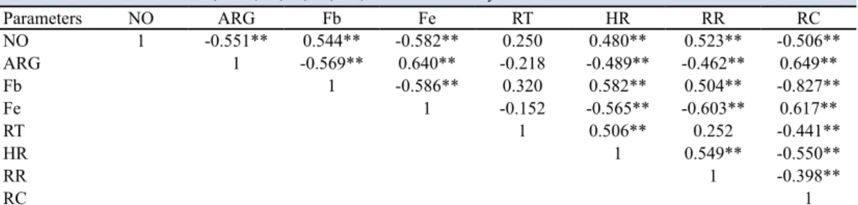
While there was a positive correlation between NO concentrations and heart and respiratory rates of TRP group, there was a negative correlation between NO concentrations and rumen contractions. In addi-tion, there was a positive correlation between erythro-cyte ARG activity and rumen contractions of the TRP group and a negative correlation between erythrocyte ARG activity and heart and respiratory rates (Table 4).
Biochemical findings
ARG activity, NO, Fb and Fe concentrations.
The mean values of erythrocyte ARG activity, NO, Fb and Fe concentrations of dairy cows in TRP and CG were given in Table 2. In the TRP group, plasma NO (318.9±5.8/μmol/L-270.3±9.6/μmol/L) and Fb (914.3±68.6/mg/dL and 265.4±19.8/mg/dL) concen-trations were higher than CG (P<0.001). However, in
J HELLENIC VET MED SOC 2019, 70(4) ΠΕΚΕ 2019, 70(4)
1836 A. KIRBAS, E. BAYDAR, F.M. KANDEMIR
the TRP group, the serum Fe concentrations (47.0±5.3/ μg/dL and 106.8±9.4/μg/dL) and erythrocyte ARG activity (29.52± 0.5/Ug hemoglobin and 35.2±1.0/ Ug hemoglobin) were lower than CG (P<0.001). In addition, in the TRP group, a positive correlation was found between plasma NO and Fb concentrations and between erythrocyte ARG activity and serum Fe concentrations. However, a negative correlation was
determined between plasma NO concentrations and erythrocyte ARG activity (Table 4).
Serum biochemistry. The serum biochemical
pa-rameters of dairy cows with TRP and CG were shown in Table 3. Concentrations of TBIL, TP (P<0.05) and AST activity of TRP group were found higher than in CG (P<0.001), but concentrations of ALB of TRP group were determined lower than in CG (P<0.001).
Table 1. Mean values and standard error of the mean of clinical signs in dairy cows with TRP and control group.
Parameters Control group (n=14) TRP group (n=14) P value
RT (°C) 38.5±0.1 39.0±0.1 <0.05
HR (beat/min) 69.3±1.7 86.6±3.5 <0.001
RR (breaths/min) 23.1±0.6 27.7±0.9 <0.001
RC (cyle/5 min) 8.7±0.2 4.6±0.4 <0.001
RT: rectal temperature; HR: heart rate; RR: respiration rate; RC: rumen contraction
Table 2. Mean values and standard error of the mean of erythrocyte ARG activity, NO, Fb and Fe concentrations in dairy cows with TRP and control group.
Parameters Control group (n=14) TRP group (n=14) P value
ARG (U/g haemoglobin) 35.2±1.0 29.52±0.5 <0.001
NO (μmol/l) 270.3±9.6 318.9±5.8 <0.001
Fb (mg/dl) 265.4±19.8 914.3±68.6 <0.001
Fe (μg/dl) 106.8±9.4 47.0±5.3 <0.001
ARG: arginase; NO: nitric oxide; Fb: fibrinogen; Fe: iron.
Table 3. Mean values and standard error of the mean of serum biochemical parameters in dairy cows with TRP and control group.
Parameters Control group (n=14) TRP group (n=14) P value
AST (U/l) 60.5±6.4 90.1±4.2 <0.001 ALP (U/l) 58.0±9.6 96.8±27.7 -TBIL (mg/dl) 0.2±0.0 0.4±0.1 <0.05 ALB (g/dl) 3.1±0.1 1.9±0.2 <0.001 GLOB (g/dl) 4.9±0.2 4.9±0.5 -TP (g/dl) 7.8±0.1 6.6±0.5 <0.05
AST:aspartate transaminase; ALP:alkhaline phosphatase; TBIL:total bilirubin; ALB: albumin; GLOB: globulin; TP: total protein; -: P>0.05.
Table 4. Correlation between NO, ARG, Fb, Fe, RT, HR, RR and RC in dairy cows with TRP.
Parameters NO ARG Fb Fe RT HR RR RC NO 1 -0.551** 0.544** -0.582** 0.250 0.480** 0.523** -0.506** ARG 1 -0.569** 0.640** -0.218 -0.489** -0.462** 0.649** Fb 1 -0.586** 0.320 0.582** 0.504** -0.827** Fe 1 -0.152 -0.565** -0.603** 0.617** RT 1 0.506** 0.252 -0.441** HR 1 0.549** -0.550** RR 1 -0.398** RC 1
NO: nitric oxide; ARG: arginase; Fb: fibrinogen; Fe: iron; RT: rectal temperature; HR: heart rate; RR: respiration rate; RC: rumen contraction; **: P<0.01
DISCUSSION
Traumatic reticuloperitonitis progresses in cattle as reticulitis, acute local and diffuse peritonitis, or chronic local and diffuse peritonitis. Besides, depend-ing on contamination stages to surrounddepend-ing organs, inflammation in reticulum and complications may also occur (Constable 2010; Constable et al., 2017). Progress of inflammation in the reticulum from acute process to chronic process makes treatment and heal-ing process difficult. Therefore, the aim of this study was to determine plasma NO concentration, erythro-cyte ARG activity and Fb levels as well as serum Fe concentration and some biochemical parameters in the dairy cows with TRP.
In the present study, a significantly different rec-tal temperature, respiratory and heart rates and rumen contractions were detected in the TRP group com-pared to the control group. It can be said that this
sit-uation may be due to the local or widespread inflam-mation in the reticulum region.
Nitric oxide is a signaling molecule that plays a key role in the pathogenesis of inflammation (Sharma et al., 2007). It involves in immune responses by cy-tokine-activated macrophages, which release NO in high concentrations (Wallace 2005). In consequence, large amounts of NO are synthesized, exceeding the physiological NO production by up to 1000-fold (Wallace 2005; Sharma et al., 2007). It was reported that NO concentrations in animals with bacterial (Nis-bet et al., 2007; Li et al., 2010; Hanedan et al., 2017), viral (Kandemir et al., 2011; Bozukluhan et al., 2013) and parasitic diseases (Kontas and Salmanoglu 2006) increased compared to healthy controls. It was deter-mined that NO concentrations increased in cattle with TRP (Atakisi et al., 2010) and traumatic pericarditis (Ozkan et al., 2012). The presence of pathogens, such as bacteria, and mucosal trauma resulting from TRP irritate the reticulum wall and stimulate mucosal NO production, thereby increasing NO synthesis (Yag-murca et al., 2009). The higher NO concentrations detected in the cows with TRP in the present study might be due to stimulation of the reticular mucosa by trauma caused by foreign bodies and possibly by entry of bacteria into the peritoneal cavity.
Arginase is a key enzyme of the urea cycle, an es-sential metabolic pathway for the removal of highly toxic ammonium ions resulting from protein degra-dation (Sharma et al., 2007). ARG activity is reduced by NO in the inflammatory process. It has been de-termined that ARG activity increases according to
healthy animals in some viral (Issi et al., 2010; Kan-demir et al., 2011), bacterial (KanKan-demir et al., 2013) and parasitic (Hanedan et al., 2015) diseases of cattle. In the present study was determined that ARG activi-ty of TRP group was significantly lower than control group (Table 2). These findings supported the hypoth-esis that increased NO concentration in the inflamma-tory process decreased ARG activity.
Fb is one of the acute phase proteins (APPs) used to evaluate the inflammatory process in cattle (Cole et al., 1997; Jones and Allison 2007). Fb has been used for many years in inflammatory and traumatic diseases. It is characterized by a significant increase in response to trauma and infection. Plasma Fb con-centrations in cattle increase within two days after trauma, inflammation and infection (Cole et al., 1997; Hirvonen and Pyörala 1998; Jones and Allison 2007). Hirvonen and Pyörälä (1998) have stated that Fb is useful for distinguishing TRP from other gastrointes-tinal diseases and pre-determination of the healing process of abdominal disorders. Gokce et al. (2007) stated that TRP is indicative of hyperfibrinogenemia. Therefore, Fb concentration is known to be useful for the diagnosis of TRP (Bozukluhan and Gokce 2007b; Kirbas et al., 2015). Kirbas et al. (2015) stated the increase in the Fb concentration was associated with the severity of inflammation process. Similarly, in this study, the Fb concentration of cows in the TRP group was significantly higher than in the control group (Ta-ble 2).
It was reported that Fe deficiencies were triggered by cytokines in the time of the inflammatory response. It is stated that Fe concentrations decrease during the acute phase response (APR) in the organism due to inflammation in horses (Borges et al., 2007), dogs (Torrente et al., 2015), adult cattle (Baydar and Dabak 2014) and calves (Aydogdu et al., 2018). Baydar and Dabak (2014) stated that serum Fe concentration in cattle with mastitis and TRP is significantly decreased compared to the control group and serum Fe concen-tration may be a useful parameter for the determina-tion of inflammadetermina-tion. Borges et al. (2007) reported that the decrease in serum Fe concentration in horses is a sensitive marker of acute, subacute and chronic systemic inflammation, and the change in Fe concen-tration may be a useful parameter for monitoring re-sponse to treatment. Torrente et al. (2015) stated that serum Fe concentrations might also be a useful mark-er for the detection of acute inflammation in dogs with systemic inflammatory response syndrome (SIRS).
J HELLENIC VET MED SOC 2019, 70(4) ΠΕΚΕ 2019, 70(4)
1838 A. KIRBAS, E. BAYDAR, F.M. KANDEMIR
In a recent study was indicated that serum Fe con-centrations were significantly reduced in calves with SIRS compared to the control group, and serum Fe concentrations could be a useful parameter for the de-termination of inflammatory response in calves with SIRS (Aydogdu et al., 2018). Similarly, in the present study, was determined that the Fe concentrations of the TRP group were significantly lower than that of control group (Table 2). Thus, it was determined that Fe could be a useful marker in the monitoring of the inflammatory process in cows with TRP.
Changes in TP, GLOB and ALB concentrations were expected in response to inflammation during the clinical form of TRP. In previous studies, TP concen-trations have been determined as normal (Ozdemir 1989; Balikci and Gunay 2004; Kirbas et al., 2015; Braun et al., 2018), low (Batmaz 1990; Braun et al., 2018) or high (Ok and Aslan 1994; Gokce et al., 2007; Bozukluhan and Gokce 2007a; Braun et al., 2018) under these circumstances. In this study, TP concen-trations of TRP group were low. Reticular abscess associated with TRP have been found to result in hy-perglobulinemia (Balikci and Gunay 2004). Ok and Aslan (1994) stated that total globulin concentrations decreased during the disease as a result of protein migration into the inflammatory area. In this present study, mean GLOB concentrations in the TRP group were not found to be statistically different than the control group. The decrease in ALB may be linked
to the synthesis of APPs (Kirbas et al., 2015), star-vation, malnutrition and/or digestive failure (Balikci and Gunay 2004; Bozukluhan and Gokce 2007a). In this study, mean ALB concentrations in TRP group were different from control group. This result may reflect that hepatic ALB synthesis was affected by APR synthesis. Statistically significant differences in serum AST activity and TBIL concentration of TRP group compared to control group were detected (Ta-ble 3). These findings could be indicate that hepato-cyte integrity of the liver was impaired in the cows with TRP.
In conclussion, it was determined that NO con-centrations were increased and erythrocyte ARG ac-tivity was not significant in dairy cows with TRP. In addition, increased plasma Fb concentration and de-creased serum Fe concentration were determined in dairy cows with TRP. This study demonstrated that plasma NO, Fb and serum Fe concentrations in dairy cows with TRP may be useful markers for prognosis.
ACKNOWLEDGEMENTS
Study abstract was present as poster presentation in the 1st International Herd of Health and Manage-ment Congress, October 14-17, 2018, Antalya, Tur-key.
CONFLICT OF INTEREST
REFERENCES
Atakisi E, Bozukluhan K, Atakisi O, Gokce HI (2010) Total oxidant and antioxidant capacities and nitric oxide levels in cattle with traumatic reticuloperitonitis. Vet Rec 167: 908-909.
Aydogdu U, Coskun A, Yildiz R, Guzelbektes H, Sen I (2018) Changes of hematological parameters and serum iron levels in calves with systemic inflammatory response syndrome. Eurasian J Vet Sci 34: 56-59.
Balikci E, Gunay C (2004) The comparison of some clinical, hemato-logical, biochemical and electrocardiographic findings at before and after ruminatomy in cows with traumatic reticuloperitonitis. Fırat Univ J Health Sci 18: 13-19.
Batmaz H (1990) Klinik olarak normal sığırlar ile retiküloperitoni-tis travmatikalı sığırların teşhis ve prognozunda serum protein elektroforezi ve SGOT, SGPT ve LDH enzim düzeyleri üzerine karşılaştırmalı araştırmalar. Turk J Vet Anim Sci 14: 476-479. Baydar E, Dabak M (2014) Serum iron as an indicator of acute
inflam-mation in cattle. J Dairy Sci 97: 222-228.
Braun U, Warislohner S, Torgerson P, Nuss K, Gerspach C (2018) Clinical and laboratory findings in 503 cattle with traumatic retic-uloperitonitis. BMC Vet Res 14: 66.
Borges AS, Divers TJ, Stokol T, Mohammed OH (2007) Serum iron and plasma fibrinogen concentrations as indicators of systemic inflammatory diseases in horses. J Vet Intern Med 21: 489-494. Boucher JL, Moali C, Tenu JP (1999) Nitric oxide biosynthesis, nitric
oxide synthase inhibitors and arginase competition for L-arginine utilization. Cell Mol Life Sci 55: 1015-1028.
Bozukluhan K, Gokce HI (2007a) Investigation of clinical, haemato-logical, and biochemical parameters in cattle with traumatic retic-uloperitonitis or with traumatic pericarditis. J Fac Vet Med Erciyes Univ 4: 97-106.
Bozukluhan K, Gokce HI (2007b) Investigation of some acute phase proteins in cattle with traumatic reticuloperitonitis or with trau-matic pericarditis. J Fac Vet Med Erciyes Univ 4: 107-113. Bozukluhan K, Atakisi E, Atakisi O (2013) Nitric oxide levels, total
antioxidant and oxidant capacity in cattle with foot-and-mouth-disease. Kafkas Univ Vet Fak Derg 19: 179-36.
Cole DJ, Roussel AJ, Whitney MS (1997) Interpreting a bovine CBC: Evaluating the leukon and acute-phase proteins. Vet Med 92: 470-478.
Coles EH: Veterinary Clinical Pathology. 4th ed. W.B.Saunders
Copm-pany, Philadelphia, PA and London, UK, 1986, 90–91.
Constable PD: Traumatic reticuloperitonitis. In: Khan C (ed.). The Merck Veterinary Manual, 10th ed. Merck & Co., Inc., Whitehouse,
USA, 2010, 209-211.
Constable PD, Hinckliff KW, Done SH, Grunberg W: Veterinary Med-icine. 11th ed. W.B.Saunders Copmpany, London, 2017, 482-490.
Degroote MA, Fang FC: Antimicrobial properties of nitric oxide. In: Fang FC (ed.). Nitric Oxide and Infection. Kluwer Academic/Ple-num,1999, 231- 261.
Drabkin DL, Austin JM (1932) Spectrophotometric studies, spec-trophtometric constants for common haemoglobin derivatives in human, dog and rabbit blood. J Biol Chem 98: 719-733. Fuentes JM, Campo ML, Soler G (1994) Kinetics and inhibition by
some aminoacids of lactating rat mammary gland arginase. Arch Int Physiol Biochem Biophys 102: 255-258.
Geyer JW, Dabich D (1971) Rapid method for determination of argi-nase activity in tissue homogenates. Anal Biochem 39: 412-417. Gokce HI, Woldehiwet Z (2002) Production of tumor necrosis
fac-tor-alpha (TNF-α) and reactive nitrogen intermediates by ovine
peripheral blood leucocytes stimulated by Ehrlichia (Cytoecetes)
phagocytophila. J Comp Pathol 126: 202-211.
Gokce HI, Gokce G, Cihan M (2007) Alterations in coagulation pro-files and biochemical and haematological parameters in cattle with traumatic reticuloperitonitis. Vet Res Commun 31: 529-537. Hanedan B, Kirbas A, Kandemir FM, Ozkaraca M, Kilic K, Benzer F
(2015) Arginase activity and total oxidant/antioxidant capacity in cows with lung cystic echinococcosis. Med Weter 71: 167-170. Hanedan B, Kirbas A, Kandemir FM, Aktas MS, Yildiz A (2017)
Eval-uation of arginase activity, nitric oxide, and oxidative stress status in sheep with Contagious agalactiae. Acta Vet Hung 65: 394-401. Hirvonen J, Pyörälä S (1998) Acute-phase response in dairy cows with
surgically treated abdominal disorders. Vet J 155: 53-61. Issi M, Kandemir FM, Basbug O, Gul Y, Ozdemir N (2010) Saliva and
erythrocyte arginase activity in beef cattle with foot-and mouth disease. YYU Vet Fak Derg 21: 91-93.
Jones ML, Allison RW (2007) Evaluation of the ruminant complete blood cell count. Vet Clin North Am Food Anim Pract 23: 377-402.
Kandemir FM, Issi M, Benzer F, Gul Y, Basbug O, Ozdemir N (2011) Plasma nitric oxide concentrations and erythrocyte arginase ac-tivities in lambs with contagious ecthyma. Revue Méd Vét 162: 275-278.
Kandemir FM, Yuksel M, Ozdemir N, Deveci H (2013) A different ap-proach to diagnosis of subclinical mastitis: milk arginase activity. Vet Arhiv 83: 603-610.
Kepka-Lenhart D, Ash DE, Morris SM (2008) Determination of mam-malian arginase activity. Method Enzymol 440: 221‐230. Kirbas A, Ozkanlar Y, Aktas MS, Ozkanlar S, Ulas N, Erol HS (2015)
Acute phase biomarkers for inflammatory response in dairy cows with traumatic reticuloperitonitis. Isr J Vet Med 70: 23-29. Kontas T, Salmanoglu B (2006) Tumour necrosis factor-α, adenosine
deaminase and nitric oxide levels in cattle babesiosis before and after treatment. Bull Vet Inst Pulawy 50: 485-487.
Li D, Liu Y, Li Y, Lv Y, Pei X, Guo D (2010) Significance of nitric oxide concentration in plasma and uterine secretes with puerperal endometritis in dairy cows. Vet Res Commun 34: 315–321. Marletta MA (1989) Nitric oxide: biosynthesis and biological
signifi-cance. Trends Biochem Sci 14: 488-492.
Nisbet C, Yarim GF, Ciftci A, Cenesiz S, Ciftci G (2007) Investigation of serum nitric oxide and malondialdehyde levels in cattle infected with Brucella abortus. Ankara Univ Vet Fak Derg 54: 159-163. Ocal N, Gokce G, Gucu AI, Uzlu E, Yagci BB, Ural K (2008) Pica as a
predisposing factor for traumatic reticuloperitonitis in dairy cattle: serum mineral concentrations and hematological findings. J Anim Vet Adv 7: 651-656.
Ok M, Aslan V (1994) The importance of blood proteins and glutar-aldehyde coagulation test in the diagnosis and prognosis of cattle with traumatic reticuloperitonitis. Eurasian J Vet Sci 10: 90-95. Onaga T, Okada H, Hagiwara S, Nagasiima C, Inoue H, Orczynski
W, Kato S (2001) Effects of nitric oxide donor and nitric oxide synthase inhibitor on ruminal contractions in conscious sheep. Res Vet Sci 71: 189-195.
Ozcelik M, Ozdemir N (2003) Some biochemical properties of argin-ase in sheep mammary tissue. Turk J Vet Anim Sci 27: 719-725. Ozdemir H (1989) Retikuloperitonitis travmatika olgularında klinik
ve hematolojik çalışmalar ile serum protein fraksiyonları üzerine araştırmalar. Turk J Vet Anim Sci 13: 213-231.
Ozkan C, Altug N, Kaya A, Basbugan Y (2012) Serum nitric oxide concentrations in cattle with traumatic pericarditis. YYU Vet Fak Derg 23: 131-135.
J HELLENIC VET MED SOC 2019, 70(4) ΠΕΚΕ 2019, 70(4)
1840 A. KIRBAS, E. BAYDAR, F.M. KANDEMIR
chemistry profile: Part: 1. Vet Med 92: 551-558.
Sharma JN, Al-Omran A, Parvathy SS (2007) Role of nitric oxide in inflammatory diseases. Inflammopharmacol 15: 252–259. Spector EB, Rice SCH, Moedjeno S, Bernard B, Cederbaub SD (1982)
Biochemical properties of arginase in human adult and fetal tis-sues. Biochem Med 28: 165-175.
Torrente C, Manzanilla EG, Bosch L, Fresno L, Rivera Del Alamo M, Andaluz A, Saco Y, Ruiz De Gopegui R (2015) Plasma iron, C-re-active protein, albumin, and plasma fibrinogen concentrations in dogs with systemic inflammatory response syndrome. J Vet Emerg Crit Care 25: 611-619.
Van Der Vielt A, Eiserich JP, Cross CE (2000) Nitric oxide: a
pro-in-flammatory mediator in lung disease? Respir Res 1: 67-72. Wallace JL (2005) Nitric oxide as a regulator of inflammatory
process-es. Mem Inst Oswaldo Cruz, 100 (Suppl. I): 5-9.
Ward JL, Ducharme NG (1994) Traumatic reticuloperitonitis in dairy cattle. J Am Vet Med Assoc 204: 874-877.
Yagmurca M, Ucar M, Fadillioglu E, Erdogan H, Ozturk F (2009) The effects of nitric oxide on rat stomach injury induced by acetylsali-cylic acid. Turk J Med Sci 39: 13-19.
Zelnickova P, Matiasovic J, Pavlova B, Kudlackova H, Kovaru F, Fal-dyna M (2008) Quantitative nitric oxide production by rat, bovine and porcine macrophages. Nitric Oxide 19: 36-41.