273
http://journals.tubitak.gov.tr/biology/ © TÜBİTAK
doi:10.3906/biy-2002-79
Mesenchymal stem cell derived extracellular vesicles: promising immunomodulators
against autoimmune, autoinflammatory disorders and SARS-CoV-2 infection
Özlem BULUT, İhsan GÜRSEL*
Therapeutic Oligodeoxynucleotide Research Laboratory (THORLAB), Department of Molecular Biology and Genetics, Faculty of Science, İhsan Doğramacı Bilkent University, Ankara, Turkey
* Correspondence: [email protected]
1. Introduction
1.1 Mesenchymal stem/stromal cells (MSCs)
Mesenchymal stem cells, also known as mesenchymal stromal cells (MSCs), are multipotent cells with self-renewal capacity and the ability to differentiate into mesenchymal lineages such as osteogenic, chondrogenic and adipogenic (Pittenger et al., 1999). MSCs can also give rise to other cell types including neurons (Arthur et al., 2008) and hepatocytes (Snykers et al., 2009). According to International Society for Cellular Therapy’s minimal criteria, apart from the self-renewal and tri-lineage differentiation capacities, MSCs are characterized byexpressing surface markers CD73, CD90 and CD105 while they lack CD14, CD19, CD34, CD45 and class II major histocompatibility complex (MHC) molecules (Dominici et al., 2006).
Although MSCs were first identified inthe bone marrow (Friedenstein et al., 1970), since then they have been isolated from various sources including peripheral blood, umbilical cord tissue Wharton’s jelly, umbilical cord blood, dental pulp, adipose tissue, amniotic fluid, endometrium, placenta and menstrual blood (Ding et al., 2011; Hass et al., 2011). MSCs of different sources vary in differentiation capacities, gene expressions and secretomes (El Omar et al., 2014). For instance, adipose tissue-derived MSCs (AD-MSCs) and umbilical cord-derived MSCs (UC-(AD-MSCs) from Wharton’s jelly are stronger immunosuppressors compared to bone marrow-derived MSCs (BM-MSCs) (Melief et al., 2013; Li et al., 2014). MSCs have long been an interest for regenerative medicineowing to their exceptional differentiation capacity. In the past two decades, their ability to interact with the immune system
Abstract: Discovery of novel and broad-acting immunomodulators is of critical importance for the prevention and treatment of
disorders occurring due to overexuberant immune responseincluding SARS-CoV-2 triggered cytokine storm leading to lung pathology and mortality during the ongoing viral pandemic. Mesenchymal stem/stromal cells (MSCs), highly regarded for their regenerative capacities, also possessesremarkable immunoregulatory functions affecting all types of innate and adaptive immune cells. Owing to that, MSCs have been heavily investigated in clinic for the treatment of autoimmune and inflammatory diseases along with transplant rejection. Extensive research in the last decaderevealed that MSCs carry out most of their functions through paracrine factors which are soluble mediators and extracellular vesicles (EVs). EVs, including exosomes and microvesicles, are an efficient way of intercellular communication due to their unique ability to carry biological messages such as transcription factors, growth factors, cytokines, mRNAs and miRNAs over long distances. EVs originate through direct budding of the cell membrane or the endosomal secretion pathway and they consist of the cytosolic and membrane components of their parent cell. Therefore, they are able to mimic the characteristics of the parent cell, affecting the target cells upon binding or internalization. EVs secreted by MSCs are emerging as a cell-free alternative to MSC-based therapies. MSC EVs are being tested in preclinical and clinical settings where they exhibit exceptional immunosuppressivecapacity. They regulate the migration, proliferation, activation and polarization of various immune cells, promoting a tolerogenic immune response while inhibiting inflammatory response. Being as effective immunomodulators as their parent cells, MSC EVs are also preferable over MSC-based therapies due to their lower risk of immunogenicity, tumorigenicity and overall superior safety. In this review, we present the outcomes of preclinical and clinical studies utilizing MSC EVs as therapeutic agents for the treatment of a wide variety of immunological disorders.
Keywords: Mesenchymal stem cells, extracellular vesicles, exosomes, inflammation, autoimmunity, COVID-19
Received: 27.02.2020 Accepted/Published Online: 04.05.2020 Final Version: 21.06.2020
and to modulate immune responses has also attracted a great amount of attention.
1.2. Extracellular vesicles (EVs)
Extracellular vesicles (EV), main two classes of which are microvesicles and exosomes, are small vesicles forming by direct budding of the plasma membrane or originating from endosomes, respectively (Stahl and Raposo, 2019). Microvesicles, the bigger class of EVs, can range between 100 and 1000 nm in diameter while exosomes are much smaller and usually in the range of 30 and 100 nm in diameter (Minciacchi et al., 2015). EVs partially enclose the cell’s cytosol with a lipid bilayer and may contain cytosolic or transmembrane proteins, amino acids, lipids, mitochondrial or genomic DNAs, mRNAs, miRNAs and long non-coding RNAs of the parent cell (Maas et al., 2017). EVs are a conserved and highly efficient form of intercellular communication utilized by prokaryotic and eukaryotic cells. In mammalians, they can be found in every bodily fluid including blood, urine, saliva, cerebrospinal fluid, synovial fluid, bronchoalveolar fluid, nasal fluid, amniotic fluid, uterine fluid, breast milk, seminal plasma and bile where they carry out various physiological roles (Yanez-Mo et al,, 2015).
EVs are critical for homeostasis and theyalsogreatly impactdisease pathogenesis and immune defense (Yuana et al., 2013). Due to their unique ability to transmit critical biological information over long distances, EVs have lately been attractive targets for therapeutic and diagnostic purposes.Although most existing preclinical and clinical studies investigate EVs as biomarkers for diagnostic and prognostic purposes, the number of studies utilizing EVs as therapeutic agents has been rapidly growing (Wiklander et al., 2019). In this review, the encompassing term “EV” will be used in cases when the distinct EV types exosomes and microvesicles have not been separated and the vesicle population mentioned in the studies include both types of EVs.
2. MSCs as immunomodulators
MSCs are promising therapeutic agents for autoimmune and inflammatory diseases as well as transplant rejection, even though they may promote inflammation in certain cases depending on the factors present in the environment. During tissue injury or inflammation, danger-associated molecular patterns (DAMPs) or pathogen-associated molecular patterns (PAMPs) trigger the recruitment and response of MSCs through Toll-like receptors (TLRs) (Bernardo and Fibbe, 2013). Mouse and human MSC types differentially expressvarious TLRs, most prominently TLR3 and TLR4. An MSC classification was proposedto term MSCs with proinflammatory and antiinflammatory behaviors as MSC1 and MSC2, respectively (Waterman et al., 2010). Upon TLR4 stimulation, MSCs shift to MSC1
state and produce higher amounts of cytokine IL-6 and chemokines CCL5 (RANTES), CXCL8 and CXCL10, thereby trigger immune cell recruitment and activation. On the other hand, upon TLR3 stimulation, MSCs are skewed to the MSC2 state and produce immunosuppressive molecules prostaglandin E2 (PGE2) and indoleamine 2,3-dioxygenase (IDO). PGE2 inhibits IL-2 production from T cells and also suppress granulocyte, natural killer (NK) cell, macrophage and dendritic cell (DC) functions (Kalinski, 2012). IDO is important for tryptophan catabolism and it leads to generation of suppressive tryptophan catabolites that have cytotoxic effects on T-cells (Terness et al., 2002).Through such soluble mediators, MSCs modulate both innate and adaptive arms of immunity.
2.1. MSCs and innate immunity
MSCs are able to regulate variousfacets of innate immunity. Upon microbial challenge, MSCs secrete CXCL8 and macrophage migration inhibitory factor (MIF) that cause neutrophilaccumulationand consequently microbial clearance (Brandau et al., 2010). On the other hand, they producesuperoxide dismutase 3 (SOD3) that reduces superoxide anion levels, inhibits neutrophil extracellular trap (NET) formation and the release of tissue-damaging proteases in order to restrain excess tissue damage mediated by neutrophils (Jiang et al., 2016). MSCs also secrete chemokines such as CCL2 (MCP-1), CCL3 and CCL12 that promote monocyte and macrophage migration to wound sites which leads to enhanced healing (Chen et al., 2008).
Moreover, MSCs restrict allergic inflammation by inhibiting IgE secretion, cytokine production and degranulation of mast cells in a PGE2, transforming growth factor β (TGFβ) and prostaglandin-endoperoxide synthase 2 (COX2)-dependent manner (Brown et al., 2011; Kim et al., 2015). MSCs downregulate activating NK receptors such as NKG2D, inhibit IL-2 induced NK cell proliferation, their cytotoxic activity and cytokine production through IDO and PGE2 (Spaggiari et al., 2008). Upon exposure to MSCs, NK cells also gain CD73 expression which leads to production of adenosine that has antiinflammatory effects (Cronstein, 1994; Chatterjee et al., 2014).
In DCs, MSCs downregulate CD40, CD80, CD86 and HLA-DR expressions, inhibit their differentiation from monocytes andmaturation, limitcytokine producing and T-cell activating capacities (Zhang et al., 2004). Also, MSCs skew mature DCs into an IL-10-producing regulatory phenotype in an IL-10, suppressor of cytokine signaling 3 (SOCS3) and Jagged-2-dependent manner (Zhang et al., 2009; Liu et al., 2012). IL-6 secreted by MSCs was also suggested to upregulate SOCS1 and drive a similar tolerogenic DC phenotype (Deng et al., 2014).
SOCS1 upregulation caused impaired TLR4 signaling and inhibited DC maturation. Another study identified PGE2 as the key mediator produced by MSCs that inhibits DC maturation (Spaggiari et al., 2009).
Alternatively activated M2 macrophages are another regulatory cell type promoted by MSCs. Macrophages upregulate CD206 and arginase 1 expression, increase IL-4 and IL-10 secretion, reducemonocyte chemoattractant protein 1 (MCP-1), tumor necrosis factor α (TNFα), IL-1β and inducible nitric oxide synthase (iNOS) production when cocultured with MSCs (Zhang et al., 2010; Cho et al., 2014). Mechanisms that have been shown to contribute to MSC-based induction of antiinflammatory M2 phenotype include TSG-6-mediated NF-κB suppression (Choi et al., 2011), PGE2 engagement with EP2 and EP4 receptors on macrophages (Nemeth et al., 2009) and lactate-mediated metabolic reprogramming of macrophages (Selleri et al., 2016). IL-1Ra, another antiinflammatory cytokine secreted by MSCs, was also identified to play a role in M1 to M2 polarization (Luz-Crawford et al., 2016).
2.2. MSCs and adaptive immunity
Suppressive actions of MSCs also involve the adaptive arm of the immune system. MSCs cause cell cycle arrest and thus block CD4+ and CD8+ T-cell proliferation (Di Nicola et al., 2002; Glennie et al., 2015), induce T-cell apoptosis via FasL-Fas engagement (Akiyama et al., 2012) and IDO-mediated tryptophan catabolism (Plumas et al., 2005), promote regulatory T cell (Treg) generation and IL-10 production while preventing Th1 and Th17 differentiation of CD4+ T-cells (Luz-Crawford et al., 2013). However, T-cell suppressing abilities of MSCs require presence of inflammatory cytokines such as IFNγ, TNFα, 1α or IL-1β in the vicinity. These cytokines trigger the production of T-cell attracting chemokines and iNOSfrom MSCs so that T-cells migrate towards the MSCs and get inactivated by nitric oxide (NO) produced by iNOS (Ren et al., 2008). IDO production from MSCs also increase upon exposure to IFNγ, IL-6 and TNFα (Crop et al., 2010). Of note, IDO is the key mediator of MSC-mediated immunosuppression in humans and other primates, while iNOS takes on the same rolefor rodent MSCs (Su et al., 2014).
Similar to their effects on T-cells, MSCs also block B-cell proliferation via cell cycle arrest, inhibit their antibody production, and hinder their chemotactic abilities by downregulating chemokine receptors that they expresssuch as CCR7, CXCR4 and CXCR5 (Corcione et al., 2006). MSCs also compromise B-cell viability by regulating ERK1/2 and p38 pathways (Tabera et al., 2008). IL-1Ra, which contributes to MSC-induced M2 macrophage polarization, is also important for preventing B-cell differentiation into plasma cells (Luz-Crawford et al., 2016). Another mechanism through which MSCs suppress B-cell function is the engagement of programmed
death 1 1) receptor on B cells with PD-1 ligand (PD-L1)on MSCsupon direct contact between the cells and theresulting suppression is more potent in the presence of IFNγ (Schena et al., 2010). Lastly, MSCs are also able to promote the generation of IL-10 producing regulatory B-cells (Bregs) (Franquesa et al., 2015).
2.3. Clinical use of MSCs
MSCs have been heavily investigated in clinical trials for many conditions. Currently there are over 900 completed, ongoing and planned trials related to MSCs (Clinicaltrials.gov, 2019)1. The wide range of these trials involve cardiovascular, rheumatological, neurological, hematological, autoimmune and inflammatory disorders along with transplant rejection. Due to their far-reaching immunomodulatory capacities, MSCs have shown great promise for the treatment of immunological diseases.
BM-MSC treatment could regress graft-versus-host disease (GvHD), a severe transplant rejection that occurs after hematopoietic stem cell (HSC) transplants, irrespective of MHC compatibility of the receiver and the MSC donor (Le Blanc et al., 2008). MSC-administered GvHD patients had higher Treg numbers without an increased risk of tumor relapse or viral infections (Zhao et al., 2015). When patients undergoing kidney transplants were inoculated with autologous BM-MSCs, incidence of acute rejection and opportunistic infections were lower while renal function recovery was faster (Tan et al., 2012). Calcineurin inhibitors are used as immunomodulating agents for transplantation but they have nephrotoxic effects. Addition of BM-MSC treatment allowed the use of lower doses of these nephrotoxic substances upon renal transplantation with no side effects (Pan et al., 2016). When cotransplanted with pancreatic islet cells to type 1 diabetes patients, MSCs enhanced graft vascularization while reducing autoimmune reaction through T cell and DC inhibition (Figliuzzi et al., 2014).
MSCs also showed remarkable immunosuppressive potential in inflammatory and autoimmune disorders. Intrafistular autologous BM-MSC injection to fistulizing Crohn’s disease patients promoted mucosal healing and reduced disease activity which were attributable to a local and systemic increase in Treg numbers due to MSC treatment (Ciccocioppo et al., 2011). Intravenous (IV) injection of allogeneic BM-MSCs to treatment-refractory patients with luminal Crohn’s disease also led to significant clinical improvement without causing any adverse effects (Forbes et al., 2014). A single subcutaneous injection of UC-MSCs could decrease circulating eosinophil numbers and IgE levels and lead to clinical improvement in atopic
1U.S. National Library of Medicine (2019). ClinicalTrials.gov
[online]. Website https://clinicaltrials.gov/ct2/results?cond=& term=mesenchymal+stem+cells&cntry=&state=&city=&dist= [accessed 31 August 2019].
dermatitis patients (Kim et al., 2017). Moreover, allogeneic BM-MSC infusion for drug-resistant severe systemic lupus erythematosus (SLE) decreased the rates of organ dysfunction and disease relapse (Wang et al., 2013). In amyotrophic lateral sclerosis (ALS) and multiple sclerosis (MS) patients, intrathecal or IV BM-MSC injection increased Treg numbers, reduced lymphocyte proliferation and downregulated the expression of activation and maturation markers such as CD40, CD83 and HLA-DR on DCs (Karussis et al., 2010).
Contribution of MSCs in infectious diseases is also being investigated. On one hand, literature findings revealed that these cells might play a critical role in spreading the disease during onset of bacterial invasion. On the other hand, data indicates that pathogen driven immune exacerbation could be alleviated by these cells and could act as an effective tool for treatment of infectious diseases (Marrazzo et al., 2019). MSCs were shown to be effective against endotoxin induced sepsis, or acute lung injury or even microbe triggered pneumonia (Marrazzo et al., 2019). In the case of the current coronavirus disease 2019 (COVID-19) pandemic triggered by SARS-CoV-2 infection, however, the infection leads to lung pathology (Liue et al., 2020) and utilization of MSCs to control the clinical outcomes were shown to be effective on patients with COVID-19 pneumonia (Leng et al., 2020). In a pilot study conducted in China, TNFα levels were significantly decreased while regulatory DCs and IL-10 levels were elevated in MSC-treated patients. SARS-CoV-2 enters to
target cells via surface receptor ACE2 and also in some cells requiresTMPRSS2 (Hoffmann et al., 2020). Since MSCs are negative for both ACE2 and TMPRSS2, they would be free from COVID-19 infection, thus could be safe and beneficial for the patients with COVID-19 (Zhao, 2020). As of April 9, 2020, out of 583 registered clinical trials, 24 are being conducted with MSCs against COVID-19 (Chinese Clinical Trial Registry, 2020)2.
3. MSC EVs as the cell-free alternative to MSC-based therapeutics
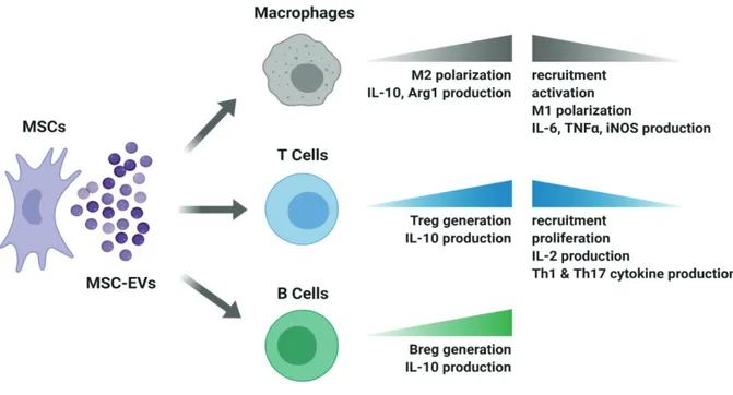
The remarkable capacity of MSCs for tissue repair and regeneration has long been attributed to their differentiation ability to other cell types upon environmental cues. However, later it became clear that MSCs exert most of their reparative, angiogenic and immunosuppressive functions through paracrine mediators involving soluble factors and EVs (Caplan and Dennis, 2006). Therefore, MSC-derived EVs, particularly exosomes, are now of great interest as a cell-free alternative to MSC-based therapeutic approaches. Since EVs are able to carry biological messengers such as growth factors, cytokines and miRNAs produced by the parent cell which could shape the behavior of immune cells in the environment (Figure 1), MSC EVs are expected to exert clinically valuable immunomodulatory and regenerative actions similar to their parent cells.
2Chinese Clinical Trial Register (2020). Chinese Clinical Trial
Registry (ChiCTR) [online]. Website www.chictr.org.cn [ac-cessed 9 April 2020].
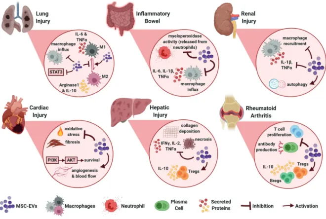
Therapeutic efficacy of MSC EVs was first reported in a murine myocardial ischemia/reperfusion (I/R) injury model where exosomes from human embryonic stem cell (ESC)-derived MSCs were cardioprotective and able to reduce the infarct size (Lai et al., 2010). Since then, various animal disease models proved that MSC EVs have regenerative and immunosuppressive effects (Figure 2) and use of MSC EVs progressed to clinical trials for the treatments of acute ischemic stroke, refractory macular holes, type 1 diabetes mellitus and bronchopulmonary dysplasia (Clinicaltrials.gov, 2019)3.
3.1. MSC EVs in cardiac injury
Following the abovementioned first I/R injury study, in another murine model, exosomes secreted by human ESC-derived MSCs reduced oxidative stress, increased myocardial viability through the activation of PI3K/Akt pathway, and consequently enhanced cardiac function (Arslan et al., 2013). BM-MSC EVs or exosomes also promoted angiogenesis and provided enhanced blood flow in myocardial infarction models while inhibiting T-cell proliferation (Bian et al., 2014; Teng et al., 2015). In
3U.S. National Library of Medicine (2019). ClinicalTrials.gov
[online]. Website https://clinicaltrials.gov/ct2/results?cond=&t erm=mesenchymal+stem+cell+exosome&cntry=&state=&city= &dist= [accessed 30 August 2019].
an interesting study, rats with myocardial infarction were treated with cardiac stem cells (CSCs) that were previously exposed to BM-MSC exosomes in culture. CSCs exposed to MSC exosomes led to greater improvement of cardiac function and reduction of fibrosis compared to regular CSC treatment (Zhang et al., 2016).
3.2. MSC EVs in renal injury
MSC-EVs also proved to be beneficial for the treatment of renal injuries. BM-MSC exosomes were protective in a murine renal I/R injury model and the protective effect was dependent on the chemokine receptor CCR2 carried on exosome surface which could sequester the chemokine CCL2 and therefore impair macrophage recruitment and activation (Shen et al., 2016). In a study of cisplatin-induced nephrotoxicity, UC-MSC exosomes reduced the levels of proinflammatory cytokines IL-1β and TNFα while promoting autophagy (Wang et al., 2017).
3.3. MSC EVs in hepatic injury
MSC-EVs were reported to alleviate hepatic inflammation and fibrosis. Exosomes from human UC-MSCs were shown to reduce hepatic inflammation while decreasing TGFβ levels and collagen deposition in a murine liver fibrosis model (Li et al., 2013). In mice with concanavalin A (ConA)-induced liver injury, repeated BM-MSC exosome
injection reduced IFNγ, IL-1, IL-2, TNFα expression, increased Treg numbers and alleviated necrosis in the liver (Tamura et al., 2016).
3.4. MSC EVs in lung injury
Immunomodulatory properties of MSC-EVs are also therapeutic for lung injury. In mice withhypoxic pulmonary hypertension, BM-MSC exosomes decreased macrophage influx, suppressed inflammatory mediators such as MCP-1 and inhibited STAT3 signaling (Lee et al., 20MCP-12). UC-MSC exosomes improved lung structure and function in mice with bronchopulmonary dysplasia by promoting an M1-M2 shift in macrophages evidenced by reduced IL-6, TNFα, CCL5 and increased arginase 1 (Willis et al., 2018). Inhibition of the inflammatory M1 macrophage phenotype and promotion of M2 phenotype was also proposed as the mechanism by which AD-MSC exosomes alleviated inflammation of white adipose tissue in obese mice. Exosome treatment to these mice led to higher arginase 1 and IL-10 expression while limiting IL-6, IL-12 and TNFα (Zhao et al., 2018). Furthermore, although there are no studies using MSC EVs in a preclinical or clinical model of COVID-19, given the promise of intravenous MSC injection for alleviating SARS-CoV-2 infection, it would be worthwhile to investigate the potential of MSC EVs to repair lung injury in COVID-19 patients.
3.5. MSC EVs in autoinflammatory disorders
MSC-EVs have been showing promise for the treatment of various autoimmune disorders. AD-MSC exosomes were utilized in a murine type-1 autoimmune diabetes mellitus model where they were able to reduce IFNγ and IL-17 levels while upregulating IL-4, IL-10 and TGFβ along with higher number of Treg cells in spleen (Nojehdehi et al., 2018). In another study that combined type-1 diabetes and experimental autoimmune uveoretinitis (EAU) models, MSC EVs prevented disease onset and inhibited the activation of antigen-presenting cells as well as secretion of Th1 and Th17 cytokines (Shigemoto-Kuroda et al., 2017). Another supporting EAU study revealed that UC-MSC exosomes reduce Th1 and Th17 in the eye, prevented macrophage and T cell infiltration, thereby mitigating disease intensity (Bai et al., 2017).
3.6. MSC EVs in inflammatory bowel disease
MSC EVs are also attractive therapeutic agents for inflammatory bowel disease. BM-MSC exosomes reduced tissue damage, limited myeloperoxidase activity in colon, decreased IL-1β, TNFα and iNOS levels in the tissue in a rat model of 2,4,6-trinitrobenzene sulphonic acid (TNBS)-induced colitis (Yang et al., 2015). Likewise, in a mouse model of dextran sulfate sodium (DSS)-induced colitis, upon UC-MSC exosome treatment, macrophage infiltration to the tissue was reduced along with downregulation of IL-1β, IL-6, IL-7, TNFα and iNOS in colon tissue and spleen (Mao et al., 2017).
3.7. MSC EVs in other inflammatory conditions
UC-MSC EVs were able to reduce mortality in mice that received allogeneic HSC transplantation. Treated mice had fewer T cells and lower serum levels of IFNγ, IL-2, TNFα and higher levels of IL-10 (Wang et al., 2016). In mice with spinal cord injury, UC-MSC exosomes led to lower levels of IFNγ, IL-6, TNFα, macrophage inflammatory protein-1 (MIP-1α) and improved the recovery of animals (Sun et al., 2018). More recently, potential of MSC exosomes for alleviating rheumatic diseases was also revealed. In a collagen-induced arthritis (CIA) study, BM-MSC exosomes inhibited antibody production and T-cell proliferation in vitro while enhancing Breg and Treg populations in vivo where they were able to alleviate disease progression (Cosenza et al., 2018).
3.8. MSC EV therapeutics in humans
Despite numerous publications of preclinical studies depicting MSC EVs as safe and remarkably effective therapeutic agents for a wide-range of disorders, there are only two reports of clinical success utilizing MSC EVs thus far. The first one was a one-patient trial where BM-MSC exosomes were administered to a therapy-refractory GvHD patient (Kordelas et al., 2014). IFNγ, IL-1β and TNFα produced by the patient’s peripheral blood mononuclear cells (PBMCs) were reduced more than 50% after the third exosome injection. Clinical symptoms of the patient improved exceptionally and were stable for 4 months although the patient later died of pneumonia. In the other study where 20 chronic kidney disease patients received UC-MSC EVs, improved clinical conditions were accompanied with reduced TNFα and elevated IL-10 and TGFβ levels in circulation (Nassar et al., 2016).
4. Conclusions
Outcomes of numerous preclinical and clinical studies presented in this review leave no doubt of the efficacy of MSC EVs as immunosuppressive agents similar to their parent cells. Although EV-based therapies are certainly attractive in terms of safety, there are a few concerns that should be addressed for proper clinical use. In the MSC EV studies, there is wide variation and lack of standardization regarding MSC expansion and exosome or EV purification methods. Moreover, as Kordelas et al. (2014) pointed out, EVs of MSCs from different donors might differ in terms of cargo and therefore immunomodulatory capacity. Thus, potency tests and protocol standardization are critical for mass-production of clinical-grade exosome or EV preparations.
MSC EVs appear to be beneficial for the treatment of GvHD, cardiovascular, renal, hepatic and pulmonary diseases, autoimmune diseases such as type 1 diabetes and uveitis, inflammatory bowel diseases and rheumatoid arthritis (Figure 2) through mechanisms including inhibition of T cell proliferation, Th1 and Th17
differentiation, macrophage recruitment, M1 polarization, as well as promotion of Treg cells, Breg cells and M2 polarization (Figure 1). However, further investigation is essential in order to fully reveal the mechanisms of immunomodulation and the EV cargo responsible for these cellular alterations specific for each clinical condition. Profiling of EV cargo with approaches such as proteomics, lipidomics and RNA-sequencing is crucial to fill the knowledge gap. Future in vitro and preclinical research and newly commencing clinical trials will surely establish MSC EVs as a novel, safe and effective therapeutic approach for
the treatment of immunological disorders. Lastly, in the course of the devastating COVID-19 pandemic, use of MSC derived EVs in the treatment of COVID-19 driven pneumonia and lung pathology should be investigated as a viable and safe strategy since MSCs are not subject to SARS-CoV-2 infection and corresponding EVs will be devoid of any virus or virus by-products.
Acknowledgments
Özlem Bulut was funded by TÜBİTAK project number 115S837. Figures were created with Biorender.com.
References
Akiyama K, Chen C, Wang D, Xu X, Qu C et al. (2012). Mesenchymal-stem-cell-induced immunoregulation involves FAS-ligand-/ FAS-mediated T cell apoptosis. Cell Stem Cell 10 (5): 544-555. doi: 10.1016/j.stem.2012.03.007
Arslan F, Lai RC, Smeets MB, Akeroyd L, Choo A et al. (2013). Mesenchymal stem cell-derived exosomes increase ATP levels, decrease oxidative stress and activate PI3K/Akt pathway to enhance myocardial viability and prevent adverse remodeling after myocardial ischemia/reperfusion injury. Stem Cell Research 10 (3): 301-312. doi: 10.1016/j.scr.2013.01.002 Arthur A, Rychkov G, Shi S, Koblar SA, Gronthos S (2008). Adult
human dental pulp stem cells differentiate toward functionally active neurons under appropriate environmental cues. Stem Cells 26 (7): 1787-1795. doi: 10.1634/stemcells.2007-0979 Bai L, Shao H, Wang H, Zhang Z, Su C et al. (2017). Effects of
mesenchymal stem cell-derived exosomes on experimental autoimmune uveitis. Scientific Reports 7 (1): 4323. doi: 10.1038/s41598-017-04559-y
Bernardo ME, Fibbe WE (2013). Mesenchymal stromal cells: sensors and switchers of inflammation. Cell Stem Cell 13 (4): 392-402. doi: 10.1016/j.stem.2013.09.006
Bian S, Zhang L, Duan L, Wang X, Min Y et al. (2014). Extracellular vesicles derived from human bone marrow mesenchymal stem cells promote angiogenesis in a rat myocardial infarction model. Journal of Molecular Medicine 92 (4): 387-397. doi: 10.1007/s00109-013-1110-5
Brandau S, Jakob M, Hemeda H, Bruderek K, Janeschik S et al. (2010) Tissue-resident mesenchymal stem cells attract peripheral blood neutrophils and enhance their inflammatory activity in response to microbial challenge. Journal of Leukocyte Biology 88 (5): 1005-10015. doi: 10.1189/jlb.0410207
Brown JM, Nemeth K, Kushnir-Sukhov NM, Metcalfe DD, Mezey E (2011). Bone marrow stromal cells inhibit mast cell function via a COX2-dependent mechanism. Clinical and Experimental Allergy 41 (4): 526-534. doi: 10.1111/j.1365-2222.2010.03685.x Caplan AI, Dennis JE (2006). Mesenchymal stem cells as trophic
mediators. Journal of Cellular Biochemistry 98 (5): 1076-84. doi: 10.1002/jcb.20886
Chatterjee D, Tufa DM, Baehre H, Hass R, Schmidt RE et al. (2014). Natural killer cells acquire CD73 expression upon exposure to mesenchymal stem cells. Blood 123 (4): 594-595. doi: 10.1182/ blood-2013-09-524827
Chen L, Tredget EE, Wu PY, Wu Y (2008). Paracrine factors of mesenchymal stem cells recruit macrophages and endothelial lineage cells and enhance wound healing. PloS One 3 (4): e1886. doi: 10.1371/journal.pone.0001886
Cho DI, Kim MR, Jeong HY, Jeong HC, Jeong MH et al. (2014) Mesenchymal stem cells reciprocally regulate the M1/ M2 balance in mouse bone marrow-derived macrophages. Experimental & Molecular Medicine 46 (1): e70. doi: 10.1038/ emm.2013.135
Choi H, Lee RH, Bazhanov N, Oh JY, Prockop DJ (2011). Anti-inflammatory protein TSG-6 secreted by activated MSCs attenuates zymosan-induced mouse peritonitis by decreasing TLR2/NF-kappaB signaling in resident macrophages. Blood 118 (2): 330-338. doi: 10.1182/blood-2010-12-327353 Ciccocioppo R, Bernardo ME, Sgarella A, Maccario R, Avanzini MA
et al. (2011). Autologous bone marrow-derived mesenchymal stromal cells in the treatment of fistulising Crohn’s disease. Gut 60 (6): 788-798. doi: 10.1136/gut.2010.214841
Corcione A, Benvenuto F, Ferretti E, Giunti D, Cappiello V et al. (2006). Human mesenchymal stem cells modulate B-cell functions. Blood 107 (1): 367-372. doi: 10.1182/ blood-2005-07-2657
Cosenza S, Toupet K, Maumus M, Luz-Crawford P, Blanc-Brude O et al. (2018). Mesenchymal stem cells-derived exosomes are more immunosuppressive than microparticles in inflammatory arthritis. Theranostics 8 (5): 1399-1410. doi: 10.7150/ thno.21072.
Cronstein BN (1994). Adenosine, an endogenous anti-inflammatory agent. Journal of Applied Physiology 76 (1): 5-13. doi: 10.1152/ jappl.1994.76.1.5
Crop MJ, Baan CC, Korevaar SS, Ijzermans JN, Pescatori M et al. (2010). Inflammatory conditions affect gene expression and function of human adipose tissue-derived mesenchymal stem cells. Clinical and Experimental Immunology 162 (3): 474-486. doi: 10.1111/j.1365-2249.2010.04256.x
Deng Y, Yi S, Wang G, Cheng J, Zhang Y et al. (2014). Umbilical cord-derived mesenchymal stem cells instruct dendritic cells to acquire tolerogenic phenotypes through the IL-6-mediated upregulation of SOCS1. Stem Cells and Development 23 (17): 2080-2092. doi: 10.1089/scd.2013.0559
Di Nicola M, Carlo-Stella C, Magni M, Milanesi M, Longoni PD et al. (2002). Human bone marrow stromal cells suppress T-lymphocyte proliferation induced by cellular or nonspecific mitogenic stimuli. Blood 99 (10): 3838-3843. doi: 10.1182/ blood.v99.10.3838
Ding DC, Shyu WC, Lin SZ (2011). Mesenchymal stem cells. Cell Transplantation 20 (1): 5-14. doi: 10.3727/096368910X Dominici M, Le Blanc K, Mueller I, Slaper-Cortenbach I, Marini
F et al. (2006). Minimal criteria for defining multipotent mesenchymal stromal cells. The International Society for Cellular Therapy position statement. Cytotherapy 8 (4): 315-317. doi: 10.1080/14653240600855905
El Omar R, Beroud J, Stoltz JF, Menu P, Velot E et al. (2014). Umbilical cord mesenchymal stem cells: the new gold standard for mesenchymal stem cell-based therapies? Tissue Engineering Part B, Reviews 20 (5): 523-544. doi: 10.1089/ten. TEB.2013.0664
Figliuzzi M, Bonandrini B, Silvani S, Remuzzi A (2014). Mesenchymal stem cells help pancreatic islet transplantation to control type 1 diabetes. World Journal of Stem Cells 6 (2): 163-172. doi: 10.4252/wjsc.v6.i2.163
Forbes GM, Sturm MJ, Leong RW, Sparrow MP, Segarajasingam D et al. (2014). A phase 2 study of allogeneic mesenchymal stromal cells for luminal Crohn’s disease refractory to biologic therapy. Clinical Gastroenterology and Hepatology 12 (1): 64-71. doi: 10.1016/j.cgh.2013.06.021
Franquesa M, Mensah FK, Huizinga R, Strini T, Boon L et al. (2015) Human adipose tissue-derived mesenchymal stem cells abrogate plasmablast formation and induce regulatory B cells independently of T helper cells. Stem Cells 33 (3): 880-891. doi: 10.1002/stem.1881
Friedenstein AJ, Chailakhjan RK, Lalykina KS (1970). The development of fibroblast colonies in monolayer cultures of guinea-pig bone marrow and spleen cells. Cell and Tissue Kinetics 3 (4): 393-403.
Glennie S, Soeiro I, Dyson PJ, Lam EW, Dazzi F (2005). Bone marrow mesenchymal stem cells induce division arrest anergy of activated T cells. Blood 105 (7): 2821-2827. doi: 10.1182/ blood-2004-09-3696
Hass R, Kasper C, Bohm S, Jacobs R (2011). Different populations and sources of human mesenchymal stem cells (MSC): A comparison of adult and neonatal tissue-derived MSC. Cell Communication and Signaling 9 (1): 12. doi: 10.1186/1478-811X-9-12
Hoffmann M, Kleine-Weber H, Krüger N, Mueller MA, Drosten C et al. (2020). The novel coronavirus 2019 (2019-nCoV) uses the SARS-coronavirus receptor ACE2 and the cellular protease TMPRSS2 for entry into target cells. BioRxiv. doi: 10.1101/2020.01.31.929042
Jiang D, Muschhammer J, Qi Y, Kugler A, de Vries JC et al. (2016) Suppression of neutrophil-mediated tissue damage-a novel skill of mesenchymal stem cells. Stem Cells 34 (9): 2393-2406. doi: 10.1002/stem.2417
Kalinski P (2012). Regulation of immune responses by prostaglandin E2. Journal of Immunology 188 (1): 21-28. doi: 10.4049/ jimmunol.1101029
Karussis D, Karageorgiou C, Vaknin-Dembinsky A, Gowda-Kurkalli B, Gomori JM et al. (2010). Safety and immunological effects of mesenchymal stem cell transplantation in patients with multiple sclerosis and amyotrophic lateral sclerosis. Archives of Neurology 67 (10): 1187-1194. doi: 10.1001/ archneurol.2010.248
Kim HS, Lee JH, Roh KH, Jun HJ, Kang KS et al. (2017). Clinical trial of human umbilical cord blood-derived stem cells for the treatment of moderate-to-severe atopic dermatitis: phase I/IIa studies. Stem Cells 35 (1): 248-255. doi: 10.1002/stem.2401 Kim HS, Yun JW, Shin TH, Lee SH, Lee BC et al. (2015) Human
umbilical cord blood mesenchymal stem cell-derived PGE2 and TGF-beta1 alleviate atopic dermatitis by reducing mast cell degranulation. Stem Cells 33 (4): 1254-1266. doi: 10.1002/ stem.1913
Kordelas L, Rebmann V, Ludwig AK, Radtke S, Ruesing J et al. (2014). MSC-derived exosomes: a novel tool to treat therapy-refractory graft-versus-host disease. Leukemia 28 (4): 970-973. doi: 10.1038/leu.2014.41
Lai RC, Arslan F, Lee MM, Sze NS, Choo A et al. (2010). Exosome secreted by MSC reduces myocardial ischemia/reperfusion injury. Stem Cell Research 4 (3): 214-222. doi: 10.1016/j. scr.2009.12.003
Le Blanc K, Frassoni F, Ball L, Locatelli F, Roelofs H et al. (2008). Mesenchymal stem cells for treatment of steroid-resistant, severe, acute graft-versus-host disease: a phase II study. Lancet 371 (9624): 1579-1586. doi: 10.1016/S0140-6736(08)60690-X Lee C, Mitsialis SA, Aslam M, Vitali SH, Vergadi E et al.
(2012). Exosomes mediate the cytoprotective action of mesenchymal stromal cells on hypoxia-induced pulmonary hypertension.Circulation 126 (22): 2601-2611. doi: 10.1161/ CIRCULATIONAHA.112.114173
Leng Z, Zhu R, Hou W, Feng Y, Yang Y et al. (2020). Transplantation of ACE2-mesenchymal stem cells improves the outcome of patients with COVID-19 pneumonia. Aging and Disease 11 (2): 216-228. doi: 10.14336/AD.2020.0228
Li T, Yan Y, Wang B, Qian H, Zhang X et al. (2013). Exosomes derived from human umbilical cord mesenchymal stem cells alleviate liver fibrosis. Stem Cells and Development 22 (6): 845-854. doi: 10.1089/scd.2012.0395
Li X, Bai J, Ji X, Li R, Xuan Y et al. (2014). Comprehensive characterization of four different populations of human mesenchymal stem cells as regards their immune properties, proliferation and differentiation. International Journal of Molecular Medicine 34 (3): 695-704. doi: 10.3892/ ijmm.2014.1821
Liu W, Zhang Q, Chen J, Xiang R, Song H et al. (2020). Detection of covid-19 in children in early January 2020 in Wuhan, China. New England Journal of Medicine 382 (14): 1370-1371. doi: 10.1056/NEJMc2003717
Liu X, Qu X, Chen Y, Liao L, Cheng K et al. (2012) Mesenchymal stem/stromal cells induce the generation of novel IL-10-dependent regulatory dendritic cells by SOCS3 activation. Journal of Immunology 189 (3): 1182-1192. doi: 10.4049/ jimmunol.1102996
Luz-Crawford P, Djouad F, Toupet K, Bony C, Franquesa M et al. (2016). Mesenchymal stem cell-derived interleukin 1 receptor antagonist promotes macrophage polarization and inhibits B cell differentiation. Stem Cells 34 (2): 483-492. doi: 10.1002/ stem.2254
Luz-Crawford P, Kurte M, Bravo-Alegria J, Contreras R, Nova-Lamperti E et al. (2013). Mesenchymal stem cells generate a CD4+CD25+Foxp3+ regulatory T cell population during the differentiation process of Th1 and Th17 cells. Stem Cell Research & Therapy 4 (3): 65. doi: 10.1186/scrt216
Maas SLN, Breakefield XO, Weaver AM (2017). Extracellular vesicles: unique intercellular delivery vehicles. Trends in Cell Biology 27 (3): 172-188. doi: 10.1016/j.tcb.2016.11.003
Mao F, Wu Y, Tang X, Kang J, Zhang B et al. (2017). Exosomes derived from human umbilical cord mesenchymal stem cells relieve inflammatory bowel disease in mice. BioMed Research International 2017: 5356760. doi: 10.1155/2017/5356760 Marrazzo P, Crupi AN, Alviano F, Teodori L, Bonsi L. (2019).
Exploring the roles of MSCs in infections: focus on bacterial diseases. Journal of Molecular Medicine 97 (4): 437-450. doi: 10.1007/s00109-019-01752-6
Melief SM, Zwaginga JJ, Fibbe WE, Roelofs H (2013). Adipose tissue-derived multipotent stromal cells have a higher immunomodulatory capacity than their bone marrow-derived counterparts. Stem Cells Translational Medicine 2(6): 455-463. doi: 10.5966/sctm.2012-0184
Minciacchi VR, Freeman MR, Di Vizio D (2015). Extracellular vesicles in cancer: exosomes, microvesicles and the emerging role of large oncosomes. Seminars in Cell & Developmental Biology 40: 41-51. doi: 10.1016/j.semcdb.2015.02.010
Nassar W, El-Ansary M, Sabry D, Mostafa MA, Fayad T et al. (2016). Umbilical cord mesenchymal stem cells derived extracellular vesicles can safely ameliorate the progression of chronic kidney diseases. Biomaterials Research 20: 21. doi: 10.1186/s40824-016-0068-0
Nemeth K, Leelahavanichkul A, Yuen PS, Mayer B, Parmelee A et al. (2009). Bone marrow stromal cells attenuate sepsis via prostaglandin E(2)-dependent reprogramming of host macrophages to increase their interleukin-10 production. Nature Medicine 15 (1): 42-49. doi: 10.1038/nm.1905
Nojehdehi S, Soudi S, Hesampour A, Rasouli S, Soleimani M et al. (2018). Immunomodulatory effects of mesenchymal stem cell-derived exosomes on experimental type-1 autoimmune diabetes. Journal of Cellular Biochemistry 119 (11): 9433-9443.
Pan GH, Chen Z, Xu L, Zhu JH, Xiang P et al. (2016). Low-dose tacrolimus combined with donor-derived mesenchymal stem cells after renal transplantation: a prospective, non-randomized study. Oncotarget 7 (11): 12089-12101. doi: 10.18632/oncotarget.7725
Pittenger MF, Mackay AM, Beck SC, Jaiswal RK, Douglas R et al. (1999). Multilineage potential of adult human mesenchymal stem cells. Science 284 (5411):143-147. doi: 10.1126/ science.284.5411.143
Plumas J, Chaperot L, Richard MJ, Molens JP, Bensa JC et al. (2005). Mesenchymal stem cells induce apoptosis of activated T cells. Leukemia 19 (9): 1597-1604. doi: 10.1038/sj.leu.2403871 Ren G, Zhang L, Zhao X, Xu G, Zhang Y et al. (2008). Mesenchymal
stem cell-mediated immunosuppression occurs via concerted action of chemokines and nitric oxide. Cell Stem Cell 2 (2): 141-150. doi: 10.1016/j.stem.2007.11.014
Schena F, Gambini C, Gregorio A, Mosconi M, Reverberi D et al. (2010). Interferon-gamma-dependent inhibition of B cell activation by bone marrow-derived mesenchymal stem cells in a murine model of systemic lupus erythematosus. Arthritis and Rheumatism 62 (9): 2776-2786. doi: 10.1002/art.27560 Selleri S, Bifsha P, Civini S, Pacelli C, Dieng MM et al. (2016).
Human mesenchymal stromal cell-secreted lactate induces M2-macrophage differentiation by metabolic reprogramming. Oncotarget 7 (21): 30193-30210. doi: 10.18632/oncotarget.8623 Shen B, Liu J, Zhang F, Wang Y, Qin Y et al. (2016). CCR2 positive
exosome released by mesenchymal stem cells suppresses macrophage functions and alleviates ischemia/reperfusion-induced renal injury. Stem Cells International 2016: 1240301. doi: 10.1155/2016/1240301
Shigemoto-Kuroda T, Oh JY, Kim DK, Jeong HJ, Park SY et al. (2017). MSC-derived extracellular vesicles attenuate immune responses in two autoimmune murine models: type 1 diabetes and uveoretinitis. Stem Cell Reports 8 (5): 1214-1225. doi: 10.1016/j.stemcr.2017.04.008
Snykers S, De Kock J, Rogiers V, Vanhaecke T (2009). In vitro differentiation of embryonic and adult stem cells into hepatocytes: state of the art. Stem Cells 27 (3): 577-605. doi: 10.1634/stemcells.2008-0963
Spaggiari GM, Capobianco A, Abdelrazik H, Becchetti F, Mingari MC et al. (2008). Mesenchymal stem cells inhibit natural killer-cell proliferation, cytotoxicity, and cytokine production: role of indoleamine 2,3-dioxygenase and prostaglandin E2. Blood 111 (3): 1327-1333. doi: 10.1182/blood-2007-02-074997
Spaggiari GM, Abdelrazik H, Becchetti F, Moretta L (2009). MSCs inhibit monocyte-derived DC maturation and function by selectively interfering with the generation of immature DCs: central role of MSC-derived prostaglandin E2. Blood 113 (26): 6576-6583. doi: 10.1182/blood-2009-02-203943
Stahl PD, Raposo G (2019). Extracellular vesicles: exosomes and microvesicles, integrators of homeostasis. Physiology 34 (3): 169-1677. doi: 10.1152/physiol.00045.2018
Su J, Chen X, Huang Y, Li W, Li J et al. (2014). Phylogenetic distinction of iNOS and IDO function in mesenchymal stem cell-mediated immunosuppression in mammalian species. Cell Death and Differentiation 21 (3): 388-396. doi: 10.1038/ cdd.2013.149
Sun G, Li G, Li D, Huang W, Zhang R et al. (2018). hucMSC derived exosomes promote functional recovery in spinal cord injury mice via attenuating inflammation. Materials Science & Engineering C 89: 194-204. doi: 10.1016/j.msec.2018.04.006 Tabera S, Perez-Simon JA, Diez-Campelo M, Sanchez-Abarca LI,
Blanco B et al. (2008). The effect of mesenchymal stem cells on the viability, proliferation and differentiation of B-lymphocytes. Haematologica 93 (9): 1301-1309. doi: 10.3324/haematol.12857 Tamura R, Uemoto S, Tabata Y (2016). Immunosuppressive effect of
mesenchymal stem cell-derived exosomes on a concanavalin A-induced liver injury model. Inflammation and Regeneration 36: 26. doi: 10.1186/s41232-016-0030-5
Tan J, Wu W, Xu X, Liao L, Zheng F et al. (2012) Induction therapy with autologous mesenchymal stem cells in living-related kidney transplants: a randomized controlled trial. Jama 307 (11): 1169-1177. doi: 10.1001/jama.2012.316
Teng X, Chen L, Chen W, Yang J, Yang Z et al. (2015). Mesenchymal stem cell-derived exosomes improve the microenvironment of infarcted myocardium contributing to angiogenesis and anti-inflammation. Cellular Physiology and Biochemistry 37 (6): 2415-2424. doi: 10.1159/000438594
Terness P, Bauer TM, Rose L, Dufter C, Watzlik A et al. (2002). Inhibition of allogeneic T cell proliferation by indoleamine 2,3-dioxygenase-expressing dendritic cells: mediation of suppression by tryptophan metabolites. The Journal of Experimental Medicine 196 (4): 447-457. doi: 10.1084/ jem.20020052
Wang B, Jia H, Zhang B, Wang J, Ji C et al. (2017). Pre-incubation with hucMSC-exosomes prevents cisplatin-induced nephrotoxicity by activating autophagy. Stem Cell Research & Therapy 8 (1): 75. doi: 10.1186/s13287-016-0463-4
Wang D, Zhang H, Liang J, Li X, Feng X et al. (2013). Allogeneic mesenchymal stem cell transplantation in severe and refractory systemic lupus erythematosus: 4 years of experience. Cell Transplantation 22 (12): 2267-2277. doi: 10.3727/096368911X582769c
Wang L, Gu Z, Zhao X, Yang N, Wang F et al. (2016). Extracellular vesicles released from human umbilical cord-derived mesenchymal stromal cells prevent life-threatening acute graft-versus-host disease in a mouse model of allogeneic hematopoietic stem cell transplantation. Stem Cells and Development 25 (24): 1874-1883. doi: 10.1089/scd.2016.0107 Waterman RS, Tomchuck SL, Henkle SL, Betancourt AM (2010).
A new mesenchymal stem cell (MSC) paradigm: polarization into a pro-inflammatory MSC1 or an Immunosuppressive MSC2 phenotype. PloS One 5 (4): e10088. doi: 10.1371/ journal.pone.0010088
Wiklander OPB, Brennan MA, Lotvall J, Breakefield XO, El Andaloussi S (2019). Advances in therapeutic applications of extracellular vesicles. Science Translational Medicine 11 (492). doi: 10.1126/scitranslmed.aav8521
Willis GR, Fernandez-Gonzalez A, Anastas J, Vitali SH, Liu X et al. (2018). Mesenchymal stromal cell exosomes ameliorate experimental bronchopulmonary dysplasia and restore lung function through macrophage immunomodulation. American Journal of Respiratory and Critical Care Medicine 197 (1): 104-116. doi: 10.1164/rccm.201705-0925OC
Yanez-Mo M, Siljander PR, Andreu Z, Zavec AB, Borras FE et al. (2015). Biological properties of extracellular vesicles and their physiological functions. Journal of Extracellular Vesicles 4 (1): 27066. doi: 10.3402/jev.v4.27066
Yang J, Liu XX, Fan H, Tang Q, Shou ZX et al. (2015). Extracellular vesicles derived from bone marrow mesenchymal stem cells protect against experimental colitis via attenuating colon inflammation, oxidative stress and apoptosis. PloS One 10 (10): e0140551. doi: 10.1371/journal.pone.0140551
Yuana Y, Sturk A, Nieuwland R (2013). Extracellular vesicles in physiological and pathological conditions. Blood Reviews 27 (1): 31-39. doi: 10.1016/j.blre.2012.12.002
Zhang B, Liu R, Shi D, Liu X, Chen Y et al. (2009). Mesenchymal stem cells induce mature dendritic cells into a novel Jagged-2-dependent regulatory dendritic cell population. Blood 113 (1): 46-57. doi: 10.1182/blood-2008-04-154138
Zhang QZ, Su WR, Shi SH, Wilder-Smith P, Xiang AP et al. (2010) Human gingiva-derived mesenchymal stem cells elicit polarization of m2 macrophages and enhance cutaneous wound healing. Stem Cells 28 (10): 1856-1868. doi: 10.1002/ stem.503
Zhang W, Ge W, Li C, You S, Liao L et al. (2004). Effects of mesenchymal stem cells on differentiation, maturation, and function of human monocyte-derived dendritic cells. Stem Cells and Development 13 (3): 263-271. doi: 10.1089/154732804323099190
Zhang Z, Yang J, Yan W, Li Y, Shen Z et al. (2016). Pretreatment of cardiac stem cells with exosomes derived from mesenchymal stem cells enhances myocardial repair. Journal of the American Heart Association 5 (1): e002856. doi: 10.1161/ JAHA.115.002856
Zhao H, Shang Q, Pan Z, Bai Y, Li Z et al. (2018). Exosomes from adipose-derived stem cells attenuate adipose inflammation and obesity through polarizing M2 macrophages and beiging in white adipose tissue. Diabetes 67 (2): 235-247. doi: 10.2337/ db17-0356
Zhao K, Lou R, Huang F, Peng Y, Jiang Z et al. (2015). Immunomodulation effects of mesenchymal stromal cells on acute graft-versus-host disease after hematopoietic stem cell transplantation. Biology of Blood and Marrow Transplantation 21 (1): 97-104. doi: 10.1016/j.bbmt.2014.09.030
Zhao RC. (2020). Stem cell-based therapy for coronavirus disease 2019. Stem Cells and Development. doi: 10.1089/scd.2020.0071