Original Article
The effect of Korean red ginseng on mesenchymal
stem cells from healthy and osteoporotic
human bone marrow
Ahmet Hamdi Kepekci1,2, Zehragul Ergul3,4, Alper Gultekin5, Erdal Karaoz3,4,6
1Meltem Hospital, Department of Otolaryngology, Istanbul, Turkey; 2Istanbul Yeni Yuzyil University, Vocational Health High School, Department of Audiometry, Istanbul, Turkey; 3Liv Hospital, Center for Regenerative Medicine and Stem Cell Manufacturing (LivMedCell), Istanbul, Turkey; 4Istinye University, Center for Stem Cell and Tissue Engineering Research and Practice, Istanbul, Turkey; 5Kocaeli Derince Training and Research Hospital, Department of Orthopaedics and Traumatology, Kocaeli, Turkey; 6Istinye University, Faculty of Medicine, Department of Histology and Embryology, Istanbul, Turkey
Received April 8, 2019; Accepted June 13, 2019; Epub June 15, 2019; Published June 30, 2019
Abstract: Background: Osteoporosis is a disease characterized by an increase in bone fragility as a result of de-creased bone mass and weakening of the bone structure. There are studies on the relationship between osteopo-rosis and hearing and balance system. The goal of this study was to compare the proliferation and osteogenesis induction properties of mesenchymal stem cells derived from healthy and osteoporotic individuals to better un-derstand the healing properties of Korean red ginseng (KRG) and Panax ginseng-Avena Sativa-Tribulus Terrestris mixture (PAT). Materials and methods: Osteoporotic and healthy MSCs were isolated successfully in culture condi-tions. The proliferation levels of cells treated with different doses of KRG and PAT were compared by Water-Soluble Tetrazolium-1 (WST-1) assay. Alkaline phosphatase (ALP) assay was performed by selecting the most effective KRG dose in proliferation. Results: Morphology of isolated cells and the expression of cell surface antigens have been detected as similar. The WST-1 assay showed that KRG was effective on the proliferation of osteoporotic cells. The levels of ALP in osteoporotic cells treated with KRG is increased depending on the differentiation day compared to healthy cells. Conclusion: KRG triggered an increase in intracellular ALP levels of osteoporotic MSCs. It suggests that KRG on osteoporotic cells is influential in stimulating osteogenesis and may be useful in osteoporotic patients. Keywords: Korean red ginseng, mesenchymal stem cells, osteoporotic human bone marrow
Introductıon
Panax ginseng is a plant that grows in Asia and called Korean red ginseng (KRG) after heat treatment to increase its biological activities [1]. It means “full healing” in Greek. KRG has been widely used throughout the world for heal-ing since old times and associated with antioxi-dant, anticancer and antidiabetic activities [2, 3]. In vivo studies to restore bone loss in osteo-porotic mouse models and in vitro studies to determine the protective effects against apop-tosis have been performed with Panax ginseng [1-4]. Avena sativa or so-called oats, which is rich in protein has been traded as grain for a long time. It has many bioactive components and has been used for medicinal purposes to
help balance the menstrual cycle and to treat dysmenorrhoea, osteoporosis, and urinary tract infections [5]. Tribulus Terrestris is an extract from a plant commonly used in the Indian and Chinese traditional systems of medicine for the improvement of general health and helps to synthesize proteins and provides a positive nitrogen balance. The leaves and roots of this plant are used for different medical purposes. Tribulus Terrestris is also popular as an aphrodi-siac [6]. In this study, we used PAT containing 22,4% Panax ginseng, 16,8% Avena sativa, 44,8% Tribulus Terrestris, 16% emulsifier and capsule which we used as a drug with KRG. Osteoporosis is the most common form of the metabolic bone disease which is characterized
by a decrease in bone mineral and matrix according to the normal value, therefore, bone fragility is increased [7-9]. Especially in women and after menopause, the incidence is increas-ing [10].
On the other hand, the studies investigating the relationship between otosclerosis and audio-logical profile [11, 12] benign paroxysmal posi-tional [13, 14] otosclerosis [15] are found in the literature. It has been a health problem that needs to be emphasized especially for societ-ies with an extending life span.
Bone marrow contains cells which have differ-entiating ability to osteoblasts, chondrocytes, adipocytes and referred to as mesenchymal stem cells (MSCs) [7, 16]. Techniques for the isolation, culture, and identification of these cells with various markers have been devel-oped. The isolation and culture of bone marrow (BM)-MSCs relies mainly on their ability to adhere to the bottom of the cell culture flasks. Isolated BM-MSCs have been shown to express specific cell surface markers that CD105 (trans-forming growth factor b receptor III), CD90 (thy-1), CD44 (hyaluronan receptor), CD73 (ecto 5 nucleotidase), CD146 (melanoma-cell adhe-sion molecule) and their expresadhe-sion of the hemopoietic markers including CD11b, CD45 and CD34 are missing [10, 17, 18].
In this study, we used osteoporotic human bone marrow-derived stem cells while using healthy human bone marrow-derived stem cells as a control group though bone marrow-derived stem cell studies are usually performed with healthy control groups. We aimed to determine the isolation, immunophenotypic properties of these cells and then the viability effects of KRG and PAT on these cells and Alkaline phospha-tase levels with differentiation day. The influen-tial effects in proliferation and stimulating osteogenesis were determined of KRG on osteoporotic human bone marrow mesenchy-mal stem cells.
Methods
Isolation, the culture of human bone marrow mesenchymal stem cells
Human bone marrow was used to isolate mes-enchymal stem cells in project scope. 1-2 mL human BM (hBM) samples were obtained from healthy and osteoporotic individuals. 1-2 ml
extra bone marrow was taken from the individu-als who came to the clinic for diagnosis and after the diagnosis, healthy bone marrow sam-ples were used as a control group.
The ethical declaration was obtained by KOU KAEK 20141106 research protocol code. Bone marrow samples delivered to the labora-tory were diluted 1:1 with PBS and centrifuged after spreading to the ficoll (Capricorn Scientific, Germany). After centrifugation, the collected buffy coat was washed with PBS and centri-fuged again. Basal DMEM-F12 medium was added to the pellet and the cells were filtered with a 70-micron cell strainer. Cells were washed by centrifugation and were cultivated in T25 culture flasks in a medium composed of DMEM-F12 medium (Gibco, USA), 10% FBS (Gibco, USA), 100 U/mL penicillin, 0.1 mg/mL streptomycin (Gibco, USA) in a 5% CO2 atmo-sphere at 37°C.
Flow cytometry analysis
Flow cytometry analysis of isolated cells was performed to assess the immune profile of MSCs. Cells were collected from culture dishes and resuspended in PBS after centrifugation. For each sample 3×10⁵ cells were incubated
with fluorescein isothiocyanate (FITC)-con- jugated antibodies against CD90, CD105, CD44, CD73, and CD45/34/11b/HLA DR/19 as a negative marker cocktail (ABclonal, USA) for 30 min at room temperature and the flow-cytometry analysis was performed using FACS-Calibur (BD Biosciences, Franklin Lakes, NJ, USA). The results were evaluated using the BD CellQuestTM software program.
WST-1 cell proliferation assay
KRG and PAT were weighed and dissolved in DMEM-F12 medium to prepare main stocks at a concentration of 2000 μg/ml. hBM-MSCs on passage 3 were seeded in a 96-well plate to be 1×10⁴ cells in each well after trypsinization.
KRG and PAT were diluted to concentrations of 15, 250, 1000 μg/ml and added to the medium as 2 replicates for each concentration. For day 1, 2 and 3, three plates were cultivated in the same condition, and 10 μl of Cell Proliferation Reagent WST-1 (Sigma-Aldrich, USA) was added to each plate after 1, 2 and 3 days. The plates exposed to the drug for 1, 2 and 3 days were incubated for 4 hours in incubator with WST-1
reagent and measured the absorbance of the samples at 480 nm. Charts were created according to OD values and the results were evaluated.
Alkaline phosphatase assay
Healthy and osteoporotic hBM-MSCs were seeded for osteogenic differentiation in 6-well plate to be 3×10⁵ cells in each well for control,
day 1, day 4, day 7, day 14 and day 21. 1000 μL/ml KRG was added to the differentiation medium and the differentiation medium was changed every 3 days. According to days, the cells were collected with a cell scraper from Petri dishes and precipitated in Eppendorf by centrifugation. The pellets were vortexed by adding the assay buffer from Alkaline Pho- sphatase Activity Colorimetric Assay Kit (BioVision, USA). After centrifugation, the super-natant was taken and protein samples were stored at -80°C. After the last 21 day cells were taken, the BCA Assay (Sigma-Aldrich, USA) was performed to quantitate all isolated proteins. Protein samples and standards were added with 2 replicates in 96-well plate. Mix solution formed with QA, QB, and Copper (II) sulfate were added to all the proteins and the plate was incubated for 1 hour at 60°C in dark. After incubation, the absorbance was measured at 562 nm and values were recorded.
measured in a microplate reader. To calculate ALP activation, the individual ALP values of all samples were divided by the protein values and graphs were created with the calculated values.
Results
Morphology of hBM-MSCs
MSCs isolation was successfully performed from bone marrow aspirates of healthy donors and osteoporosis patients and morphology was examined every day by phase-contrast micros-copy. From the earliest days of culture, the cells were observed as spindle-shaped. In the follow-ing days, it was observed that the cells were multiplying by forming colonies (Figure 1).
Flow cytometry analysis of hBM-MSCs
Flow cytometry analysis were performed with the immune profile of MSCs, and the isolated cells have positive expressions (Figure 2). Characterization studies have shown that the isolated cells have characteristic features of hBM-MSCs.
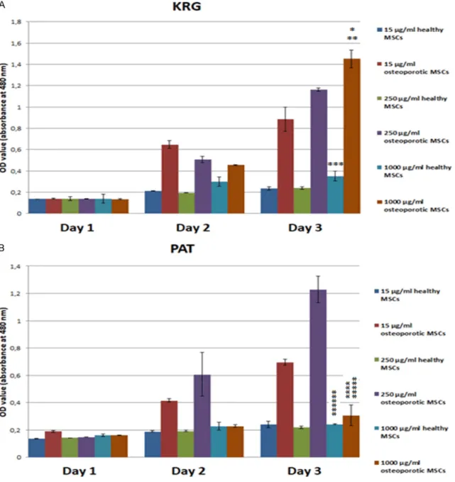
Effects of KRG and PAT on hBM-MSCs viability
In this study, we performed a WST-1 assay to compare proliferation levels by treating both
Figure 1. MSCs isolated from human bone marrow tissue were visualized by phase-contrast microscopy on day 6 (A, B) passage 0 (P0) and day 10 (C, D) P0 in adherent culture (Bars: 200 μm).
For the ALP assay 0, 4, 8, 12, 16, 20 μl of 1 mM pNPP solu-tions were used to create standards and 20 μl of any protein sample was used to create background control and 20 μl of each of our sam-ples were added as a dupli-cate in 96-well plate. Assay buffer was added to all sam-ples and ALP activity was stopped by adding stop solu-tion to the background con-trols. ALP enzyme was added to the standards and rapidly 5 mM pNPP solution was added to the samples and back-ground controls. The plate was incubated in dark at 25°C for 60 minutes and after incubation stop solution was added to all other wells except the background controls. The absorbance at 405 nm was
cells with KRG and PAT until day 3. There was
no significant effect of both drugs on the prolif- while KRG showed a more evident increase in proliferation of cells with the dose increase.
Figure 2. Flow cytometry analysis of hBM derived MSCs with the immune profile of MSCs (CD45/34/11b/HLA DR/19, CD90, CD105, CD44, CD73). Flow cytometry analysis of collected cells from culture dishes were per-formed. Positive expressions of CD90, CD105, CD44, CD73 and negative expressions of CD45/34/11b/HLA DR/19 marker cocktail indicate that iso-lated and analyzed cells have MSCs characteristic properties.
eration of both cells detected on day 1.
Firstly, when comparing the effect of KRG on the prolifera-tion of osteoporotic cells with dose increase, there is a regu-lar and significant increase on the 3rd day when there is an irregular increase on the 2nd day. In particular, 1000 μg/ml KRG significantly increased the proliferation of osteopo-rotic cells. Along with that sim-ilar effect is seen for healthy cells, but this increase is not as high as for osteoporotic cells.
However, PAT did not cause a regular increase in the prolif-eration of cells. The prolifera-tion of cells was increased on days 2 and 3 up to 250 μg/ml of dose and then decreased. In healthy cells, there was no clear effect after the 2nd day with a dose increase of PAT. As a result, according to PAT, KRG showed a more promi-nent increase on the prolifera-tion of cells with dose escala-tion, which was found to be significantly higher and more pronounced in osteoporotic cells than in healthy cells (Figure 3).
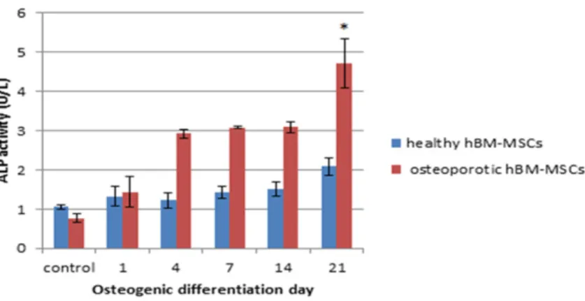
Alkaline phosphatase levels of hBM-MSCs
It is known that in osteoporo-sis the osteoblastic differenti-ation of MSCs is impaired and function of osteoblasts is decreased. Accordingly, ALP levels are also decreased. In this study, we attempted to improve this disorder by the addition of KRG.
According to WST-1 cell prolif-eration assay result, PAT showed no such increase,
Therefore the ALP Assay was performed with 1000 μg/ml KRG dose as the most appropriate dose for cell proliferation.
Osteogenic differentiation of MSCs was quanti-fied by monitoring ALP activity (Figure 4). Intracellular ALP was found to peak and there was greater calcium deposition when osteopo-rotic hBM-MSCs were incubated with KRG
rath-er than healthy IBM-MSCs. While the amount of ALP does not increase highly in healthy hBM-MSCs with differentiation day, there was a reg-ular increase in osteoporotic hBM-MSCs from day 1 with differentiation day, and markedly increased on day 21. As a result, it has been suggesting that KRG on osteoporotic cells is influential in proliferation and stimulating osteogenesis.
Figure 3. Evalution with column chart of WST-1 assay results with healthy and osteoporotic hBM derived MSCs depending on time (day 1, 2, 3) and concentration of KRG and PAT (15, 250, 1000 µg/ml). (*P<0.001 versus healthy MSCs with 1000 µg/ml KRG on day 3. **P<0.01 versus osteoporotic MSCs with 1000 µg/ml KRG on day 1. ***P<0.05 versus healthy MSCs with 1000 µg/ml KRG on day 1. ****P>0.10 versus healthy MSCs with 1000 µg/ ml PAT on day 3. *****P<0.10 versus osteoporotic MSCs with 1000 µg/ml PAT on day 1. ******P<0.01 versus healthy MSCs with 1000 µg/ml PAT on day 1) (KRG: Korean red ginseng, PAT: 22,4% Panax ginseng, 16,8% Avena sativa, 44,8% Tribulus Terrestris, 16% emulsifier and capsule.
Dıscussıon
MSCs derived from osteoporotic and healthy donors might be isolated from bone marrow and expanded in culture [16, 17]. These isolat-ed stem cells have similar morphology and express similar cell surface antigens as evi-denced by their reactivity with cell-specific monoclonal antibodies [17]. However, it has been shown that MSCs from osteoporotic patients have a lower rate of proliferation than healthy cells under in vitro conditions [19]. It has been shown in previous studies that KRG inhibits apoptosis of cells, increases expres-sion of osteogenic genes (ALP, OCN, OPN, Runx2, and BMPs) in vitro and increases bone formation in osteoporotic mice in vivo [1, 20]. Ginseng has also been shown to inhibit bone loss by inhibiting osteoclast differentiation, a process controlled by estrogen [21]. Ad- ditionally, there are many studies have shown that ginseng can reduce osteoporosis by stimu-lating ALP, Col-I, Runx2, inhibiting NF-kB, and increasing blood circulation [3].
In this study, we aimed to demonstrate the pos-sible effects of KRG and PAT on proliferation and in-vitro osteogenic differentiation process-es on MSCs from healthy and osteoporotic indi-viduals. For this purpose, we performed WST-1 cell proliferation assay and ALP assay by apply-ing KRG and PAT to healthy and osteoporotic MSCs.
the conclusion that the effect of KRG is more evident than PAT. Additionally, 1000 μg/ml KRG dose has been selected for ALP assay as the most appropriate dose for cell proliferation. ALP levels of healthy and osteoporotic cells were compared with differentiation days. However there was only a slight increase in healthy cells with differentiation days, increas-es in osteoporotic cells were more pronounced and intracellular ALP levels were significantly increased on day 21 of differentiation. These results showed that KRG can induce osteogen-esis with calcium deposition in MSCs derived from osteoporotic patients. Therefore, it is con-cluded that the use of KRG in patients stricken from osteoporosis due to menopause or other reasons may be useful however clinical trials are needed.
Conclusıon
MSCs derived from osteoporotic patients have significant differences in response to KRG treatment compared to control donors. This result suggests that KRG may be a new approach to the treatment of osteoporosis. Further studies are needed to elucidate the molecular mechanisms that explained the results of the increase in proliferation and cal-cium deposition of cells in this study. Illumination of this mechanism may lead to the development of new therapeutic strategies for the treatment of osteoporosis.
Figure 4. Effect of 1000 µg/ml KRG on ALP activity of hBM-MSCs. Cells were treated with 1000 µg/ml KRG and exposed to osteogenic differentiation me-dium for 1, 4, 7, 14, 21 days and exposed to normal DMEM-F12 meme-dium as a control group. ALP activity was analyzed and calculated by comparison with total protein levels. *P<0.01 versus osteoporotic control (KRG: Korean red ginseng).
As a result of the WST-1 assay, the proliferation of osteopo-rotic cells increased regularly by dose and day increase, especially on day 3. However, KRG is ineffective in healthy cells, and there is no signifi-cant increase in proliferation with the dose increase. Fu- rthermore in both PAT applied cells from healthy and osteo-porotic individuals, there was no regular increase in prolifer-ation by dose and day in- crease. PAT has no significant effect on the proliferation of both cells.
Alkaline phosphatase assay was performed with KRG from
Disclosure of conflict of interest None.
Address correspondence to: Erdal Karaoz, Liv Hospital, Center for Regenerative Medicine and Stem Cell Manufacturing (LivMedCell), Istanbul, Turkey. E-mail: [email protected]
References
[1] Kim J, Lee H, Kang KS, Chun KH and Hwang GS. Protective effect of Korean red ginseng against glucocorticoid-induced osteoporosis in vitro and in vivo. J Ginseng Res 2015; 39: 46-53.
[2] Kim HR, Cui Y, Hong SJ, Shin SJ, Kim DS, Kim NM, So SH, Lee SK, Kim EC and Chae SW. Effect of ginseng mixture on osteoporosis in ovariectomized rats. Immunopharmacol Im- munotoxicol 2008; 30: 333-45.
[3] Siddiqi MH, Siddiqi MZ, Ahn S, Kang S, Kim YJ, Sathishkumar N, Yang DU and Yang DC. Ginseng saponins and the treatment of osteo-porosis: mini literature review. J Ginseng Res 2013; 37: 261-8.
[4] Li XD, Liu ZY, Chang B, Liu DX, Chen B, Guo C, Wang YG, Xu JK, Huang DY, Du SX. Panax no-toginseng saponins promote osteogenic differ-entiation of bone marrow stromal cells through the ERK and P38 MAPK signaling pathways. Cell Physiol Biochem 2011; 28: 367-76. [5] Miraj S and Kiani S. Study of pharmacological
effect of avena sativa: a review. Der Pharmacia Lettre 2016; 8: 137-140.
[6] Gauthaman K and Adaikan P. Effect of tribulus terrestris on nicotinamide adenine dinucleo-tide phosphate-diaphorase activity and andro-gen receptors in rat brain. J Ethnopharmacol 2005; 96: 127-32.
[7] Pino AM, Rosen CJ and Rodríguez JP. In osteo-porosis, differentiation of mesenchymal stem cells (MSCs) improves bone marrow adipogen-esis. Biol Res 2012; 45: 279-87.
[8] Rosen CJ and Bouxsein ML. Mechanisms of disease: is osteoporosis the obesity of bone? Nat Clin Pract Rheumatol 2006; 2: 35-43. [9] Kiernan J, Hu S, Grynpas MD, Davies JE and
Stanford WL. Systemic mesenchymal stromal cell transplantation prevents functional bone loss in a mouse model of age-related osteopo-rosis. Stem Cells Transl Med 2016; 5: 683-93. [10] Román F, Urra C, Porras O, Pino AM, Rosen CJ
and Rodríguez JP. Real-time H2O2 measure-ments in bone marrow mesenchymal stem cells (MSCs) show increased antioxidant ca-pacity in cells from osteoporotic women. J Cell Biochem 2017; 118: 585-593.
[11] Kahveci O, Demirdal U, Yücedag F and Cerci U. Patients with osteoporosis have higher inci-dence of sensorineural hearing loss. Clin Otolaryngol 2014; 39: 145-9.
[12] Shafi OM, Lone ZA, Ali A and Ahmad R. Audiological profile in osteoporosis in kashmiri population. Indian Journal of Applied Research 2018; 7.
[13] Lee SY, Hong HS, Yang SC, Kim KS and Kim HJ. Is there a relationship between bone quality and hearing level? Otol Neurotol 2018; 39: e752-e756.
[14] Sacks D and Parham K. Preliminary report on the investigation of the association between BPPV and osteoporosis using biomarkers. Otol Neurotol 2015; 36: 1532-6.
[15] Clayton AE, Mikulec AA, Mikulec KH, Merchant SN and McKenna MJ. Association between os-teoporosis and otosclerosis in women. J Laryngol Otol 2004; 118: 617-21.
[16] Rodríguez JP, Astudillo P, Rios S and Pino AM. Involvement of adipogenic potential of human bone marrow mesenchymal stem cells (MSCs) in osteoporosis. Curr Stem Cell Res Ther 2008; 3: 208-18.
[17] Kern S, Eichler H, Stoeve J, Klüter H and Bieback K. Comparative analysis of mesenchy-mal stem cells from bone marrow, umbilical cord blood, or adipose tissue. Stem Cells 2006; 24: 1294-301.
[18] Pontikoglou C, Deschaseaux F, Sensebé L and Papadaki HA. Bone marrow mesenchymal stem cells: biological properties and their role in hematopoiesis and hematopoietic stem cell transplantation. Stem Cell Rev 2011; 7: 569-89.
[19] Rodríguez JP, Garat S, Gajardo H, Pino AM and Seitz G. Abnormal osteogenesis in osteoporot-ic patients is reflected by altered mesenchymal stem cells dynamics. J Cell Biochem 1999; 75: 414-23.
[20] Kropotov A, Kolodnyak O and Koldaev V. Effects of siberian ginseng extract and iprifla-vone on the development of glucocorticoid-in-duced osteoporosis. Bull Exp Biol Med 2002; 133: 252-4.
[21] Lee HY, Park SH, Chae SW, Soung NK, Oh MJ, Kim JS, Kim YO and Chae HJ. Aqueous ginseng extract has a preventive role in RANKL-induced osteoclast differentiation and estrogen defi-ciency-induced osteoporosis. Journal of Fun- ctional Foods 2015; 13: 192-203.