ORIGINAL INVESTIGATION
87
1Department of Physiology, Dumlupınar University Faculty of Medicine, Kütahya, Turkey 2Department of Thoracic Medicine, Dumlupınar University Faculty of Medicine, Kütahya, Turkey
3Department of Medical Biology, Dumlupınar University Faculty of Medicine, Kütahya, Turkey 4Clinic of Adult Immunology and Allergy, Süreyyapaşa Chest Diseases and Thoracic Surgery Training and Research Hospital, İstanbul, Turkey 5Department of Biostatistic,
Dumlupınar University, Faculty of Medicine, Kütahya, Turkey 6Aydın General Secretary of the Union of Public Hospitals, Aydın, Turkey Submitted 10.05.2015 Accepted 19.06.2015 Correspondance Dr. Ceylan Ayada, Dumlupınar Üniversitesi Tıp Fakültesi, Fizyoloji Anabilim Dalı, Kütahya, Türkiye Phone: +90 505 633 12 63
e.mail: [email protected] This study was presented at the American Thoracic Society 2015 International Conference, 15-20 May 2015,
Denver-Colorado, USA. ©Copyright 2015 by Erciyes University School of Medicine - Available online at www.erciyesmedj.com
Evaluation of Serum Levels of Renin Angiotensin
System Components in Asthmatic Patients
Ceylan Ayada
1, Ümran Toru
2, Osman Genç
1, Server Şahin
3, İsmet Bulut
4, Özlem Arık
5, Murat Acat
6ABSTRACT Objective: Asthma is a chronic inflammatory lung disease. The renin–angiotensin system (RAS) targets several tissues and maintains fluid homeostasis. It is also known that RAS plays a role in inflammatory processes. Angiotensin-converting enzyme (ACE) and its product angiotensin II (AngII), which are the components of RAS, regulate the known effects of RAS. The other components of RAS, ACE2 and its product Ang 1–7, regulate the counter-effects of RAS. That is why the activation or inhibi-tion of RAS can offer new therapeutic strategies for treating several diseases. We aimed to investigate the serum level of RAS components such as angiotensinogen (AGT), ACE, AngII, ACE2, and Ang 1–7 in asthma and control groups.
Materials and Methods: This study was performed on 27 asthma and 23 healthy individuals. The serum levels of AGT, ACE, AngII, ACE2, and Ang 1–7 were measured by human enzyme-linked immunosorbent assay (ELISA) kits.
Results: There were no statistically significant differences for the serum levels of AGT and ACE between the groups. The serum levels of AngII, ACE2, and Ang 1–7 were significanlty higher in the asthma group than those in the control group.
Conclusion: Our results indicate that two tails of RAS, i.e., the ACE and ACE2 pathways, are activated in asthma compared to are activated more in the asthma group than in the control group. We suppose that the activation of ACE2 tail of RAS occurs because of the homeostatic balance requirements of this system. We believe that the activation of ACE2 pathway, in addition to the inhibition of the ACE pathway, can provide clinical benefits in treating asthma.
Keywords: Angiotensinogen (ANG), Angiotensin II (AngII), Angiotensin-converting Enzyme (ACE), ACE2, Ang 1–7 Erciyes Med J 2015; 37(3): 87-90 • DOI: 10.5152/etd.2015.0027
INTRODUCTION
Generally, the renin–angiotensin system (RAS) is described as a main regulatory of fluid homeostasis. It targets several tissues such as the brain, pituitary gland, heart, blood vessels, liver, kidneys, and adrenal glands (1, 2). RAS includes different enzymes that have critical importance in the pathophysiological processes of several diseases (3, 4). Renin is the key enzyme of RAS and converts inactive angiotensinogen (AGT) to angiotensinI (AngI) (5, 6). The effects of RAS show balanced character due to its two cascades. One of these cascades is called ACE/AngII/AT1R, and the other one is called the ACE2/Ang1–7/Mas-receptor axis (7, 8). AngI is cleved to AngII by the proteolytic activity of ACE, a pulmonary carboxyl dipeptidase. This conversion can occur through non-ACE pathways too (9). AngII provides main effects of RAS. It acts as a paracrine hormone and also as an autocrine hormone because it exits in circulation as well as in tissues (10). The physiological functions of AngII take place by its two specific recep-tors, AngII receptor subtype 1 (AT1R) and subtype 2 (AT2R). These receptors reveal the opposite effects of AngII (9). Most known effects of RAS occur via AT1R such as vascular hypertrophy, vasoconstriction, and hypertension (11). The opposite effects of RAS are controlled by ACE2 activity. The major effect of ACE2 is converting AngII to Ang1–7 (12). Although the function of Ang1–7 is still not fully understood, it is known that Ang1–7 is responsible for antiproliferation, natriuresis, and the activation of vasodilatation. Its function is mediated through the G protein-coupled Mas receptor, and it shows antagonist properties against AT1R function (7, 8, 13).
Asthma is a chronic inflammatory lung disease. Disease characteristics are described to be airway inflammation, elevated mucus secretion, and reversible airway obstruction (14-16). It has been shown that the levels of renin and AngII are elevated in acute severe asthma, although the exact reason is not yet clarified (17).
It is known that ACE2 is highly expressed in the heart, kidneys, testes, and also in the lungs, in particular (18, 19). Alterations in the level of AngII can be seen up to the expression level of ACE2 (20). It is thought that the acti-vation of the ACE2 pathway also provides protection against several lung diseases besides the inhibition of the ACE pathway (3). To the best of our knowledge, ACE2 and its product Ang1–7 as counter balanced compo-nents of RAS are not defined in asthma, although other RAS compocompo-nents are widely studied. We suppose that
understanding the role of RAS in diseases needs more information about both tails of this system because only the inhibition of RAS cannot provide a complete benefit, which also needs the activation of the counter-balanced regulators of RAS. For this reason, in this study, we examined the serum levels of the components of the ACE2 pathway, in addition to the serum levels of the components of the ACE pathway of RAS.
MATERIALS and METHODS
PatientsThis study was performed on 27 chronic severe asthma patients who were treated at Dumlupınar University, Faculty of Medicine, Department of Chest Diseases, Kütahya, Turkey. The diagnosis of asthma was established on the basis of criteria proposed by 2014 Global Initiative for Asthma (GINA) Guideline (21). The control group comprised 23 healthy age-matched subjects who were treated at Dumlupınar University, Faculty of Medicine, Department of Chest Diseases, Kütahya, Turkey. Written informed consent for all proce-dures were obtained from each individual. The study protocol con-forms to the ethical guidelines of the Declaration of Helsinki and was approved by the ethics committee of Afyon Kocatepe University. All individuals were assessed by the criteria according to GINA 2014 (21) to evaluate the level of asthma symptom control (ASC). For this purpose, the following questions were asked to the patients: In the past 4 weeks, 1) did you have daytime symptoms more than twice a week? 2) Did you have any night awakenings due to asthma? 3) Did you need a reliever medication more than twice a week? 4) Did you have any activity limitation due to asthma? The patients answered these questions as “Yes” or “No.” If the patient had none of these, the level of ASC was defined as ‘well-controlled’. If the patient had 1 or 2 of these, it was defined as “partly controlled.” If the patient has 3 or 4 of these, it was defined as “uncontrolled”.
Enzyme-Linked Immuno Sorbent Assay (ELISA) Analyses
Peripheral blood samples were collected in tubes without EDTA from all subjects. After centrifugation, the serum of each individual was stored at −80°C until ELISA analysis. The serum concentra-tions of AGT (Cusabio Biotech, Cat No CSB-E08564h), ACE (Cusabio Biotech, Cat No CSB-E11269h), AngII (Cusabio Biotech, Cat No E04493h), ACE2 (Cusabio Biotech, Cat No CSB-E04489h), and Ang1–7 (Cusabio Biotech, Cat No CSB-E14242h) were analyzed by rat ELISA assay kits. Chemiluminescence data were analyzed by an ELISA microplate reader (das, Digital and Analog Systems, Vimercate, MI, Italy).
Statistical Analysis
Statistical analyses were performed by SPSS (Statistical Package for Social Sciences; Chicago, IL, USA) 16.0 package program. The serum levels of interested (AGT, ACE, AngII, ACE2, and Ang1–7) parameters were given as mean±standard error of the mean (SEM). Statistical significances between the two groups were analyzed by Mann–Whitney U tests. Differences were considered significant at p<0.05.
RESULTS
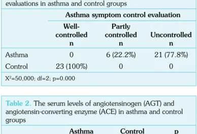
The frequencies of the level of asthma symptom control evaluations were found to be 22.2% for partially under control (n=6) and 77.8% for uncontrolled (n=21) in the asthma group and 100% for under
control (n=23) in the control group. The distribution of the asthma control evaluations were found to be significantly different between the groups (X2=50,000; df=2; p=0.000) (Table 1). The frequency
of the uncontrolled individuals was significantly higher in the patients than in the control (cohort analyses for uncontrolled=0.222; 95% CI=0.11–0.45; p=0.000).
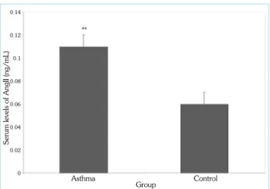
There were no statistically significant differences for the serum lev-els of AGT (15.94±1.9 ng/mL) and ACE (2.1±0.3 ng/mL) in the asthma group compared with the serum levels of AGT (13.24±1.4 ng/mL) and ACE (1.9±0.3 ng/mL) in the control groups, p=0.259 and p=0.763, respectively (Table 2). The serum levels of An-gII (0.11±0.01 ng/mL), ACE2 (0.1±0.01 ng/mL), and Ang1–7 (4.7±0.2 ng/mL) were significantly higher in the asthma groups compared with the serum level of AngII (0.06±0.01 ng/mL), ACE2 (0.08±0.03 ng/mL), and Ang1-7 (3.9±0.3 ng/mL) in the control group; p=0.002, p=0.01, and p=0.01, respectively (Figure 1-3).
DISCUSSION
RAS is the main regulatory system for water and salt homeostasis (1, 2). The counterbalanced action of this system occurs on different tissues via the tails of RAS called as ACE/AngII/AT1R and ACE2/ Ang1–7/Mas-receptor pathways (7, 8, 13). Activated RAS due to the elevated serum levels of renin and AngII have been established in acute asthma and acute severe asthma patients (17, 22). It has been also reported that the serum level of ACE is more elevated in acute severe asthma patients than that in control and mild and severe chronic asthma groups (17). This elevation results from even one-dose β-agonist treatment (22). The activation mechanisms of RAS in asthma have not been clarified yet. Up to now, we could not recognize any reports evaluating the serum level of AGT in chronic severe asthmatic patients. In our study, the serum levels of AGT and ACE were higher, although not significantly, in the chronic severe asthma group where β-agonist therapy was withheld compared with those of the control group. According to literature, this elevation can be affected by pharmacological agents such as β-agonists or the
88
Ayada et al. Serum Levels of RAS Components in Asthma Erciyes Med J 2015; 37(3): 87-90 Table 1. The frequencies of the asthma symptom control evaluations in asthma and control groupsAsthma symptom control evaluation Well- Partly
controlled controlled Uncontrolled n n n Asthma 0 6 (22.2%) 21 (77.8%) Control 23 (100%) 0 0
X2=50,000; df=2; p=0.000
Table 2. The serum levels of angiotensinogen (AGT) and angiotensin-converting enzyme (ACE) in asthma and control groups
Asthma Control p AGT (ng/mL) 15.94±1.9 13.24±1.4 0.259 ACE (ng/mL) 2.1±0.3 1.9±0.3 0.763
pathophysiological features of asthma (17, 22, 23). Because of the limitations of our study, we could not evaluate the exact reason for the elevated serum levels of AGT and ACE in the asthma group. On the other hand, it is known that the elevated plasma levels of AGT and ACE are related to their homozygote mutant variations (24). We
evaluated AGT and ACE polymorphisms in the asthma and control groups. We did not observe any statistically significant difference be-tween the groups for interested polymorphisms (data not shown). This can be one of the explanations for the elevated serum levels of AGT and ACE, which are not significant in the asthma group, com-pared with those of the control group. According to our results, we suppose that the elevation of the serum levels of AGT and ACE in chronic severe asthmatic patients lays the groundwork for the nega-tive effects of RAS such as the hypertrophy of bronchial smooth muscles and bronchoconstriction.
As mentioned above, the elevated serum level of AngII is deter-mined in acute asthma and acute severe asthma patients due to β-agonist therapy (17, 22). We could not observe any reports about serum level of AngII in chronic severe asthmatic patients. In our study, we found a significant increase for the serum level of An-gII in the chronic severe asthma group compared with that of the control group. According to our results, the elevated serum level of AngII in the asthma group is not affected by the serum level of ACE as its product. We could not evaluate the exact reason for the increased serum level of AngII in the asthma group. We suppose that the elevated serum level of AngII can cause adverse effects in asthma treatment such as the hypertrophy of bronchial smooth muscles and bronchoconstriction. We think that pharmacological treatments, which have antagonistic properties against AngII and inhibit AngII production or AT1R activation, provide benefits in addition to β-agonist treatment.
ACE2 is described as an ACE homolog and has carboxypeptidase activity. ACE2 converts AngII to Ang1–7, which regulates the counterbalanced activity of RAS (7, 25). To the best of our knowl-edge, there is no study indicating the serum levels of ACE2 and Ang1–7 in patients with asthma. Our results have revealed the ac-tivation of ACE2/Ang1-7 axis of RAS in chronic severe asthmatic patients. Increased ACE2 level can be induced via the elevated lev-el of AngII. The lev-elevation of both AngII and ACE2 naturally causes an increase in the level of Ang1–7. Unfortunately, we do not have a certain explanation for the mechanism of the increased level of ACE2 and Ang1–7. It has been supposed that the imbalanced ac-tion of ACE/AngII/AT1R and ACE2/Ang1-7/Mas-receptor axis of RAS can cause lung diseases. It has been thought that ACE inhibitors or AT1R blockers eliminate the negative effects of RAS during the treatment of lung diseases. However, these treatments could not provide a significant beneficial effect completely for some of them such as pulmonary fibrosis and pulmonary hypertension (3). It is believed that ACE2 activation or ACE2 gene therapies provide more beneficial effects during the treatment of pulmonary diseases besides RAS inhibitory strategies (26). We suppose that ACE2 elevation and Ang1–7 production, which may have occurred to maintain homeostatic balance in asthma patients in our study, by ACE2 activator therapeutics such as XNT, which is described as synthetic ACE2 activator, provides important clinical benefits.
CONCLUSION
We suppose that the activation of the two tails of RAS indicate that RAS acts as a homeostatic regulatory system even in treated asth-matic patients. We suppose that the inhibition of the ACE pathway of RAS and the homeostatic action of RAS, which is the activation
89
Ayada et al. Serum Levels of RAS Components in AsthmaErciyes Med J 2015; 37(3): 87-90
Figure 1. Serum levels of AngII in the asthma and control groups
**p<0.01 vs. control group (Mann–Whitney U test); AngII; angiotensin II Group
**
Asthma
Serum levels of AngII (ng/mL)
0.14 0.12 0.1 0.08 0.06 0.04 0.02 0 Control
Figure 3. Serum levels of Ang1-7 in the asthma and control groups
*p<0.05 vs. control group (Mann–Whitney U test); AngII: angiotensin II Group
Asthma
Serum levels of Ang1-7 (ng/mL)
6 5 4 3 2 1 0 Control *
Figure 2. Serum levels of ACE2 in the asthma and control groups
*p<0.05 vs. control group (Mann–Whitney U test); ACE2: angiotensin-converting enzyme 2 Group
Asthma
Serum levels of ACE2 (ng/mL)
0.12 0.1 0.08 0.06 0.04 0.02 0 Control *
of the ACE2 pathway, besides β-agonist treatment can provide more beneficial outcomes for asthma treatment. We think that get-ting more knowledge about RAS in asthma helps understand the pathophysiological processes in this disease. For this reason, we believe that further studies are needed to provide the effective com-bined therapeutic strategies, including the inhibition and/or activa-tion of specific components of RAS, in the treatment of asthma.
Ethics Committee Approval: Ethics committee approval was received for
this study.
Informed Consent: Written informed consent was obtained from patients
who participated in this study.
Authors’ Contributions: Conceived and designed the experiments or
case: CA, ÜT, OG. Performed the experiments or case: CA, ÜT, OG, SŞ. Analyzed the data: CA, ÜT, OG, SŞ, İB, ÖA, MA. Wrote the paper: CA. All authors have read and approved the final manuscript.
Conflict of Interest: No conflict of interest was declared by the authors.
Financial Disclosure: This study was supported by Dumlupınar University
Scientific Research Fund Commission (Project no: 2013/15).
REFERENCES
1. Goodfriend TL. Angiotensin receptors: history and mysteries. Am J Hypertens 2000; 13(4): 442-9. [CrossRef]
2. Der Sarkissian S, Huentelman MJ, Stewart J, Katovich MJ, Raizada MK. ACE2: A novel therapeutic target for cardiovascular diseases. Prog Biophys Mol Biol 2006; 91: 163-98. [CrossRef]
3. Kuba K, Imai Y, Penninger JM. Angiotensin-converting enzyme 2 in lung diseases. Curr Opin Pharmacol 2006; 6: 271-6. [CrossRef]
4. Weber MA, Giles TD. Inhibiting the renin-angiotensin system to pre-vent cardiovascular diseases: do we need a more comprehensive strat-egy? Rev Cardiovasc Med 2006; 7: 45-54.
5. Maibaum J, Feldman DL. Renin inhibitors as novel treatments for car-diovascular disease. Expert Opin Ther Patents 2003; 13(5): 589-603.
[CrossRef]
6. Griendling KK, Murphy TJ, Alexander RW. Molecular biology of the renin-angiotensin system. Circulation 1993; 87: 1816-28. [CrossRef]
7. Iwai M, Horiuchi M. Devil and angel in the renin-angiotensin system: ACE-angiotensin II-AT1 receptor axis vs. ACE2-angiotensin-(1-7)-Mas receptor axis. Hypertens Res 2009; 32(7): 533-6. [CrossRef]
8. Santos RA, Simoes e Silva AC, Maric C, Silva DM, Machado RP, de Buhr I, et al. Angiotensin-(1-7) is an endogenous ligand for the G protein-coupled receptor Mas. Proc Natl Acad Sci U S A. 2003; 100(14): 8258-63. [CrossRef]
9. Azizi M. Renin inhibition. Curr Opin Nephrol Hypertens 2006; 15(5): 505-10. [CrossRef]
10. Wright JW, Krebs LT, Stobb JW, Harding JW. The Angiotensin IV system: Functional implications. Front Neuroendocrinol 1995; 16(1): 23-52. [CrossRef]
11. Berry C, Touyz R, Dominiczak AF, Webb RC, Johns DG. Angiotensin receptors: signaling, vascular pathophysiology, and interactions with ceramide. Am J Physiol Heart Circ Physiol 2001; 281(6): 2337-65. 12. Gaddam RR, Chambers S, Bhatia M. ACE and ACE2 in
inflamma-tion: a tale of two enzymes. Inflamm Allergy Drug Targets 2014; 13(4): 224-34. [CrossRef]
13. Paul M, Poyan Mehr A, Kreutz R. Physiology of Local Renin-Angio-tensin Systems. Physiol Rev 2006; 86(3): 747-803. [CrossRef]
14. Barnes PJ. The role of inflammation and anti-inflammatory medication in asthma. Respir Med 2002; 96(supplement 1): 9-15. [CrossRef]
15. Bousquet J, Jeffery PK, Busse WW, Johnson M, Vignola AM. Asth-ma. From bronchoconstriction to airways inflammation and remodel-ing. Am J Respir Crit Care Med 2000; 161(5): 1720-45. [CrossRef]
16. Roche WR, Beasley R, Williams JH, Holgate ST. Subepithelial fibro-sis in the bronchi of asthmatics. Lancet 1989; 333(8637): 520-4.
[CrossRef]
17. Millar EA, Angus RM, Hulks G, Morton JJ, Connell JM, Thomson NC. Activity of the renin-angiotensin system in acute severe asthma and the effect of angiotensin II on lung function. Thorax 1994; 49(5): 492-5.
[CrossRef]
18. Nicholls MG, Richards AM, Agarwal M. The importance of the renin-angiotensin system in cardiovascular disease. J Hum Hypertens 1998; 12(5): 295-9. [CrossRef]
19. Hamming I, Timens W, Bulthuis ML, Lely AT, Navis G, van Goor H. Tissue distribution of ACE2 protein, the functional receptor for SARS coronavirus. A first step in understanding SARS pathogenesis. J Pathol 2004; 203(2): 631-7. [CrossRef]
20. Lambert DW, Hooper NM, Turner AJ. Angiotensin-converting en-zyme 2 and new insights into the renin-angiotensin system. Biochem Pharmacol 2008; 75(4): 781-6. [CrossRef]
21. Global Initiative for Asthma Guideline 2014. Available from: URL: http://www.ginasthma.org/local/uploads/files/GINA_Pock-et_2014_Jun11.pdf
22. Millar EA, Connell JM, Thomson NC. The effect of nebulized albuterol on the activity of the renin-angiotensin system in asthma. Chest 1997; 111(11): 71-4. [CrossRef]
23. Ramsay SG, Dagg KD, McKay IC, Lipworth BJ, McSharry C, Thom-son NC. Investigations on the renin-angiotensin system in acute severe asthma. Eur Respir J 1997; 10(12): 2766-71. [CrossRef]
24. Rigat B, Hubert C, Alhenc-Gelas F, Cambien F, Corvol P, Soubrier F. An insertion/deletion polymorphism in the angiotensin I-converting enzyme gene accounting for half the variance of serum enzyme levels. J Clin Invest 1990; 86(4): 1343-6. [CrossRef]
25. Donoghue M, Hsieh F, Baronas E, Godbout K, Gosselin M, Stagliano N, et al. A novel angiotensin-converting enzyme-related carboxypepti-dase (ACE2) converts angiotensin I to angiotensin 1-9. Circ Res 2000; 87: e1-9. [CrossRef]
26. Ferreira AJ, Murça TM, Fraga-Silva RA, Castro CH, Raizada MK, Santos RA. New cardiovascular and pulmonary therapeutic strategies based on the Angiotensin-converting enzyme 2/angiotensin-(1-7)/ mas receptor axis. Int J Hypertens 2012; 2012: 147825. [CrossRef]