Introduction
One of the best treatment methods for advanced arthrosis of the knee is total knee arthroplasty (TKA).[1]
Achieving correct postoperative alignment is one of the most impor-tant factors for success following TKA.[2]
Malalignment of the prosthesis can lead to complications such as wear and aseptic loosening that could require early revision. Thus, components should be implanted according to the mechanical axes during knee arthroplasty. However, the landmarks used for the femoral alignment are not always distinct enough to determine these axes due to its complex structure.[3,4]
Various axes and reference landmarks used during femoral implantation were mainly determined in the coro-nal plane since the prosthesis that provides a neutral align-ment in the coronal plane has been demonstrated to have a higher long-term survival success.[5,6]However, the situ-ation in sagittal alignment, which is as important as coro-nal alignment, was not demonstrated. The inability to achieve suitable alignment in the sagittal plane and place-ment of the femoral component in flexion lead to loss of extension and posterior polyethylene wear, whereas place-ment in extension with respect to the mechanical axis can result in anterior notching leading to periprosthetic frac-ture.[7–9]
Clinical significance of the relationship between
3D analysis of the distal femur and femoral
shaft anatomy in total knee arthroplasty
Mehmet Emin fiimflek1, Murat Bozkurt2 1
Department of Orthopedics and Traumatology, Lokman Hekim University, Ankara, Turkey
2
Department of Orthopedics and Traumatology, Ankara Y›ld›r›m Beyaz›t University, Ankara, Turkey
İD İD
Abstract
Objectives:A proper morphometric analysis of the anatomy of the distal femur is of utmost importance for providing cor-rect alignment for the survival of total knee arthroplasty (TKA). Herein, we aimed to conduct a detailed morphometric analy-sis of the distal femur, including the differences between men and women. We also aimed to determine landmarks in the sagittal and coronal planes for positioning of the femoral component during TKA and demonstrate the data that may affect clinical outcome.
Methods: Two-hundred adult femurs from the collection of anatomy department were enrolled in this study. Three-dimensional reconstruction of computed tomography scans were performed on these femurs. Differences between the reference axes and lines in the sagittal and coronal planes were obtained from the images, and correlation coefficients of the collected data were analyzed. All measurements were compared between men and women.
Results: The calculated mean angles between the sagittal mechanical axis, anterior cortical axis and distal medullary axis were found as 5.14±1.67° and 4.12±2.41°, respectively, and the mean angle difference between the posterior condylar line (PCL) and the epicondylar axis (EA) was 4.37±2.18°. The angle difference between PCL and EA was higher in females (p=0.047).
Conclusion:In addition to the gender-dependent anthropomorphic differences between the distal femurs of females and males, differences between the measurements used as reference in conventional TKA techniques may affect the post-oper-ative alignment.
Keywords: distal femur; femoral bowing; morphometry; total knee arthroplasty
Anatomy 2019;13(3):174–182 ©2019 Turkish Society of Anatomy and Clinical Anatomy (TSACA)
Received: July 15, 2019; Accepted: November 4, 2019
In addition to the sagittal morphometry of the distal femur, there are other factors that affect prosthesis vival and knee functions after primary and revision sur-geries. Rotation of the femoral component and one of its important determinants, i.e., posterior condylar struc-tures; femoral bowing; medullary canal diameters in the distal femur; and anteroposterior (AP) and mediolateral (ML) width of the distal femur are some of the fac-tors.[6,10]
Nearly two-thirds of the patients who undergo TKA are females. Moreover, studies have revealed variations in the morphometry of the distal femur based on factors such as gender and race. Thus, there is a need to develop dif-ferent designs considering all these variables to obtain desired results after TKA.[11]
Possible challenges that can be encountered by sur-geons in anatomic alignment of the mechanical axis of the lower extremity during TKA are now easier to over-come with the use of computer-assisted orthopedic sur-gery techniques. Identifying the reference points and angles to be used during femoral implantation and, fur-thermore, identifying the differences thereof according to an important variable such as gender would increase the success of surgery while using conventional knee prosthesis techniques, still being commonly used throughout the world.[12,13]
In this study, we aimed to conduct a detailed morpho-metric analysis of the anatomy of the distal femur, includ-ing many variables in the coronal and sagittal planes, to determine the differences among genders, and to demon-strate the data that could be associated with clinical out-comes.
Materials and Methods
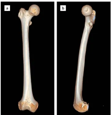
A total of 200 adult femurs were randomly selected from the collection of Department of Anatomy of Ankara University School of Medicine. The bones with deformi-ty, fracture, tumor, and other such changes were excluded. The bones were then assigned to two groups (100 females and 100 males) of unknown age. 115 bones were right side, and 85 were from the left. Each bone was assigned a num-ber for identification. Computed tomography (CT) scans were performed on each femur in the coronal, sagittal, and axial planes to encompass the entire femur, with a slice thickness of 0.6 mm (256-slice multidetector scanner; Siemens®, Erlangen, Germany). Each CT scan of the femurs was analyzed with the femur rotated fully in AP position and lateral position on three-dimensional (3D) reconstructions obtained by Leonardo Dr/Dsa Va30a soft-ware (Siemens®, Erlangen, Germany) in a digital environ-ment (Figure 1). Measureenviron-ments for each parameter were
carried out from the obtained images by three different observers (MES, SG, MA), with the intra- and inter-observer differences being determined. Measurements done for the morphometric analysis of the distal femur were as follows:
Measurements in the Sagittal Plane
• Femoral length: distance from the most superior point of the femoral head to the most distal point of the medi-al epicondyle (ME)
• Bowing angle: the angle between the vertical lines passing through the midpoint of the line drawn at the level of the flair point and the transverse line drawn below the lesser trochanter. The flair point is the point at which condyles start to expand in the distal part. This angle was determined as the tip of bowing and AP - ML medullary diameter measurements done at this level; the distance of the tip to the Blumensaat line (BL) was determined as the bowing tip distance (Figure 2).
• Anterior cortical axis (ACA): the line that connects the points drawn at 5 cm and 10 cm proximal to the dis-tal joint line (JL) on the anterior cortex[14]
(Figure 3). • Distal medullary axis (DMA): the line drawn between
1 cm anterior to the BL end point and the midpoint
Figure 1. Anteroposterior (a) and lateral (b) images of the femur after
3D reconstruction of the CT images obtained by Leonardo Dr/Dsa Va30a software (Siemens®, Erlangen, Germany). [Color figure can be
viewed in the online issue, which is available at www.anatomy.org.tr]
of the medullary canal at 20 cm proximal to the JL (Figure 3).
• Sagittal mechanical axis (sMA): the line that connects the center of the femoral head and the midpoint of the epicondylar axis (EA)[15]
(Figure 3).
Measurements in the Coronal and Axial Planes • Medial epicondyle (ME) and lateral epicondyle
(LE) distance: the distance from the most prominent
point of the medial and lateral epicondyles to the JL in the coronal plane (Figure 4).
• Epicondylar axis (EA): the line that connects the most prominent points of the medial and lateral epicondyle (Figure 4).
• Posterior condylar line (PCL): the line that connects the posterior borders of the condyles (Figure 4). • Medial and lateral posterior condylar offset (m-PCO
and l-PCO): the distance from the most prominent posterior point of the medial and lateral femoral condyles to the posterior femoral cortex in the sagittal plane measured from the 3D reconstruction of the PCO (Figure 4).
Figure 2. Determination of the tip of femoral bowing (a) and bowing
tip distance measurement (b). [Color figure can be viewed in the online issue, which is available at www.anatomy.org.tr]
Figure 3. Reference angles of the distal femur in the sagittal plane.
ACA: Anterior cortical axis; the line that connects the points drawn at 5 cm and 10 cm proximal to the distal joint line on the anterior cor-tex. DMA: distal medullary axis; the line drawn between 1 cm anteri-or of the BL and the midpoint of the medullary canal at 20 cm proxi-mal to the joint line. sMA: sagittal mechanical axis; the line that con-nects the center of the femoral head and the midpoint of the epi-condylar axis. [Color figure can be viewed in the online issue, which is available at www.anatomy.org.tr]
Figure 4. Medial epicondyle and lateral epicondyle distance (a), Measurement of posterior offset and reference axes (b). ME distance (MEd) and
LE distance (LEd); the distance from the most prominent point of the medial and lateral epicondyle distance to the joint line. EA: epicondylar axis; the line that connects the most prominent points of the MEd and LEd. PCL: posterior condylar line; the line that connects the posterior bor-ders of the condyles. m-PCO: medial posterior condylar offset; l-PCO: lateral posterior condylar offset; the distance from the most prominent posterior point of the medial and lateral femoral condyles to the posterior femoral cortex in the sagittal plane from the 3D reconstruction of the posterior condylar offset measurement (D: distal, L: lateral, M: medial, P: proximal, Po: posterior). [Color figure can be viewed in the online issue, which is available at www.anatomy.org.tr]
a a a b ISTHMUS Tip of bowing SB + 175 mm SB + 150 mm SB + 125 mm SB + 100 mm SB + 75 mm SB + 50 mm SB + 25 mm Subchondral bone ISTHMUS ACA DMA sMA Tip of bowing SB + 175 mm SB + 150 mm SB + 125 mm SB + 100 mm SB + 75 mm SB + 50 mm SB + 25 mm Subchondral bone
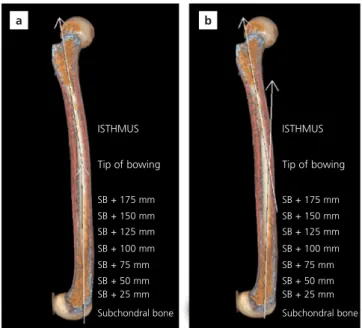
Additionally, AP and ML canal diameters at 25, 50, 75, 100, 125, 150, 175, and 200 mm were measured from distal to proximal starting from the BL (Figures 5 and 6).
In 5 mm slices starting from the BL, AP and ML diam-eters at the level with the narrowest medullary canal width was determined as the isthmus diameter and the distance of this level to the BL distal endpoint was determined as isth-mus distance.
The angle between EA and PCL was measured in the coronal plane. The angle difference between the axis meas-urements in the sagittal plane was calculated. The correla-tion of the measurements with bowing and each other in the sagittal plane was analyzed. All measurements were compared between men and women.
The variables with normal distribution were analyzed with the Shapiro–Wilk test. Descriptive statistics of the variables without normal distribution were expressed as median (minimum, maximum) values. In addition, mean±standard deviation values were also provided. Mann–Whitney U test was used to analyze the differences between variables according to gender. The relationship between the specified variables was studied with the Spearman’s rho correlation coefficient. In cases with sig-nificant relationships, the correlations were interpreted as “no correlation or negligible correlation” for correlation coefficients between 0.00–0.19, as “poor (low)” for 0.20–0.39, as “moderate” for 0.40–0.69, as “strong (high)”
for 0.70–0.89, and as “very strong” for those between 0.90–1.0.
IBM SPSS Statistics for Windows (Version 21, Armonk, NY, USA) and MS-Excel 2007 software were used for statistical analyses and calculations. The level of statistical significance was considered as p<0.05.
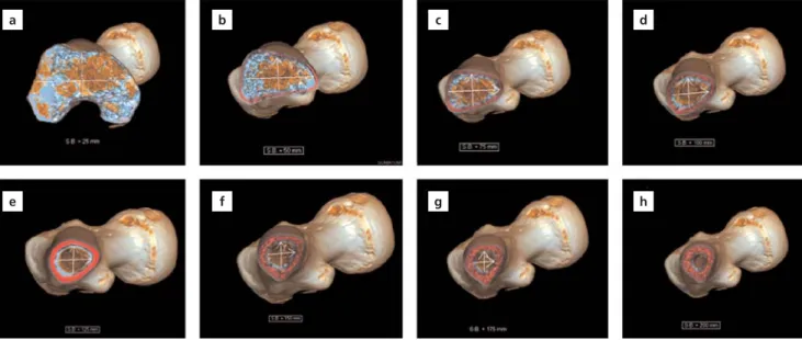
Figure 6. Examples of canal diameter measurements at different cross-section levels from distal end of the Blumensaat line level towards the
proximal part. Canal diameters at 25 mm (a), 50 mm (b), 75 mm (c), 100 mm (d), 125 mm (e), 150 mm (f), 175 mm (g), and 200 mm (h). [Color figure can be viewed in the online issue, which is available at www.anatomy.org.tr]
a b c d
e f g h
Figure 5. Measurement of canal diameters from distal end of the
Blumensaat line level towards the proximal part in the coronal (a) and sagittal (b) planes. [Color figure can be viewed in the online issue, which is available at www.anatomy.org.tr] a b ISTHMUS LT SB + 175 mm SB + 200 mm SB + 150 mm SB + 125 mm SB + 100 mm SB + 75 mm SB + 50 mm SB + 25 mm Subchondral bone ISTHMUS SB + 175 mm SB + 150 mm SB + 125 mm SB + 100 mm SB + 75 mm SB + 50 mm SB + 25 mm Subchondral bone
Results
No statistically significant difference was found between genders in terms of the values for femoral length, being 41.93±5.19 cm in males and 39.75±5.10 cm in females (p=0.475). However, the distance from the tip of bowing to the BL was 17.74±2.53 cm in females and 19.14±2.70 cm in males, the difference being statistically significant (p=0.017). Moreover, the variable bowing angle exhibit-ed a significant difference according to gender (p=0.013), with the mean bowing angle being 15.78± 4.47° and 13.25±4.42° in females and males, respectively.
For comparing the medullary canal diameters of the femurs by gender, AP and ML diameters were measured at 25 mm, 50 mm, 75 mm, 100 mm, 125 mm, 150 mm, 175 mm, and 200 mm levels with respect to the BL dis-tal endpoint. The two gender-groups had similar results in terms of the AP diameter at 200 mm and ML diame-ter at 25 mm; whereas, there was a statistically significant difference at all the other levels particularly being high-er in males: (AP 25 mm, p=0.05; AP 50 mm, p=0.036; AP 75 mm, p=0.041; AP 100 mm, p=0.049; AP 125 mm, p=0.045; AP 150 mm, p=0.037; AP 175 mm, p=0.05; AP 200 mm, p=0.051; ML 25 mm; p=0.052; ML 50 mm, p=0.043; ML 75 mm, p=0.032; ML 100 mm, p=0.04; ML 125 mm, p=0.038; ML 150 mm, p=0.024; ML 175 mm, p=0.049; and ML 200 mm, p=0.046). The comparison of
measured values and all variables according to gender are shown in Tables 1 and 2.
It was observed that there was a mean difference of 5.14±1.67° and 4.12±2.41° between sMA and the ACA and DMA, respectively. These two parameters are com-monly used in component positioning in the sagittal plane while using conventional total knee prosthesis techniques. And this difference was statistically signifi-cant in both male and female femurs (p=0.02 and p=0.031, respectively). The calculated mean angle between PCL and EA in the coronal plane was 4.37± 2.18°, and it this angle between the two lines showed a statistically significant difference according to gender (p=0.047). Moreover, it was observed that the m- and l-PCOs were longer in male femurs as compared to female femurs, with the difference being statistically significant (p=0.025 and p=0.037, respectively).
Correlation coefficients between the variables are shown in Table 3. There was no statistically significant relationship between the femoral bowing tip distance and isthmus distance from the distal part (p=0.837). A moderate correlation was observed between the medial offset and ME–JL distance (p<0.001) both in female and male femurs (p<0.001 and p<0.001, respectively). On evaluating statistical relationship between the femoral length and bowing angle, it was found that the bowing angle increased in parallel with increasing femoral length
Table 1
Angle and length measurements of the parameters used for morphometric analysis of the anatomy of distal femur and the descriptive statistics of these variables according to gender (mean±SD).
Gender
Male Female All
Variables Mean±SD Mean±SD Mean±SD p-value
Femoral length (cm) 41.93±5.19 39.75±5.10 40.84±5.23 0.475
Bowing angle 13.25±4.42° 15.78±4.47° 14.02±4.50° 0.013
Bowing tip distance (cm) 19.14±2.70 17.74±2.53 18.44±2.51 0.017
Bowing tip ML diameter (cm) 1.16±0.17 1.14±0.17 1.15±0.17 0.728
Bowing tip AP diameter (cm) 1.30±0.21 1.27±0.19 1.29±0.20 0.595
The angle between the anterior cortical axis and sagittal mechanical axis 4.10±1.80° 6.16±1.58° 5.14±1.67° 0.020
The angle between the distal medullary axis and sagittal mechanical axis 3.71±2.30° 4.82±2.36° 4.12±2.41° 0.031
Medial condylar offset (mm) 35.47±5.91 28.1±4.3 31.79±5.10 0.025
Lateral condylar offset (mm) 28.96±4.28 25.6±4.4 27.28±4.35 0.037
The angle between the posterior condylar line and epicondylar axis 3.60±2.10° 5.10±3.10° 4.37±2.18° 0.047
Lateral epicondyle distance (mm) 28.41±3.00 23.95±2.72 24.18±1.87 0.040
Medial epicondyle distance (mm) 37.1±2.8 30.88±4.14 32.97±3.49 0.003
Isthmus distance (cm) 25.41±2.89 23.31±2.04 24.36±2.96 0.022
Isthmus diameter ML (cm) 1.37±0.43 0.97±0.15 1.17±0.44 0.456
in both male and female femurs (p<0.05 and p<0.001, respectively). Bowing angle with ACA/sMA and bowing angle with DMA/sMA were also correlated (p<0.01 and p=0.043, respectively), and this correlation was found to be higher in female femurs.
The inter- and intra-observer differences in the meas-urements were not statistically significant (p>0.05).
Discussion
This study showed that significant difference between genders in terms of various parameters for the distal and medullary canal structures of males and females, with a discussion on the improvements in the femoral compo-nent and stem designs and positioning that these differ-ences may require. In addition to the gender-dependent
Table 2
Measurement of anteroposterior and mediolateral canal diameters at 25, 50, 75, 100, 125, 150, 175, and 200 mm from the Blumensaat line distal end point towards the proximal part and the descriptive statistics of these variables according to gender.
Gender
Male Female All
Variables Mean±SD Mean±SD Mean±SD p-value
ML at 25 mm (cm) 6.18±0.84 6.08±0.79 6.13±0.81 0.052 AP at 25 mm (cm) 3.87±0.46 3.51±0.30 3.76±0.28 0.050 ML at 50 mm (cm) 3.61±0.69 3.47±0.70 3.59±1.70 0.043 AP at 50 mm (cm) 2.72±2.45 2.16±0.40 2.48±1.40 0.036 ML at 75 mm (cm) 2.75±1.50 2.42±0.58 2.68±0.90 0.032 AP at 75 mm (cm) 2.31±0.25 1.98±0.29 2.24±0.27 0.041 ML at 100 mm (cm) 2.21±0.38 2.06±0.39 2.18±0.88 0.040 AP at 100 mm (cm) 1.86±0.25 1.66±0.97 1.71±0.26 0.049 ML at 125 mm (cm) 1.77±0.29 1.62±0.29 1.69±0.79 0.038 AP at 125 mm (cm) 1.65±0.22 1.52±0.24 1.59±0.29 0.045 ML at 150 mm (cm) 1.57±0.37 1.37±0.25 1.41±0.23 0.024 AP at 150 mm (cm) 1.53±0.25 1.28±0.29 1.49±0.35 0.037 ML at 175 mm (cm) 1.37±0.76 1.19±0.17 1.24±0.16 0.049 AP at 175 mm (cm) 1.39±0.21 1.23±0.22 1.33±0.49 0.050 ML at 200 mm (cm) 1.23±0.67 1.13±0.16 1.18±0.46 0.046 AP at 200 mm (cm) 1.22±0.18 1.14±0.19 1.18±0.19 0.051 ML at Isthmus (cm) 0.97±0.15 1.37±0.43 1.07±0.44 0.784 AP at Isthmus (cm) 1.07±0.13 1.06±0.12 1.07±0.13 0.646
AP: anteroposterior; ML: mediolateral.
Table 3
Correlation coefficients between the variables in general and according to gender. Gender
Male (n=100) Female (n=100) All (n=200)
Compared parameters rho p-value rho p-value rho p-value
Isthmus distance - Femoral bowing tip distance 0.134 0.514 0.118 0.748 0.015 0.837
Medial epicondylar distance -Medial condylar offset 0.716 <0.001 0.633 <0.001 0.682 <0.001
Lateral epicondylar distance - Lateral condylar offset 0.089 0.379 0.065 0.522 0.071 0.262
Femoral length - Bowing angle 0.715 <0.05 0.878 0.001 0.799 <0.001
ACA/sMA - Bowing angle 0.413 <0.05 0.876 <0.001 0.517 <0.01
DMA/sMA - Bowing angle 0.489 <0.05 0.817 <0.001 0.615 0.043
variables, differences between the angular parameters that are commonly used in knee prosthesis during femoral implantation were also analyzed.
Previous studies have provided different suggestions for femoral component design due to the gender-dependent morphometric differences in the anatomy of the distal femur between males and females.[16,17] Especially in revision surgeries that involve the use of long stems, not paying attention to the femoral width and bowing could lead to excessive stress and, in turn, to fractures during surgery, rapid wear and also early failure in the postoperative period. One of the important find-ings of this study was that the isthmus and tip of bowing measured from the distal part were at different levels in male and female femurs, and the tip of femoral bowing was closer to the joint than the isthmus. Moreover, we observed that the bowing angle was higher and that the tip of bowing was closer to the JL in female femurs as compared to the male ones. It was also found that the isthmus was closer to the joint in female femurs as com-pared to male femurs. It was observed that increased bowing resulted in increased flexion in the distal part of the femur, thereby causing an increase in the DMA in female femurs. Therefore, it is possible to say that using shorter femoral stems would be necessary in females with the same bowing angle, as the tip of bowing would be closer to the JL.
Today, many authors emphasize that correct compo-nent alignment in primary and revision surgeries have important effects on the survivorship and clinical success of arthroplasty.[18]
Apart from the planning in the coronal plane, planning in the sagittal plane to align the femoral component in accordance with the mechanical axis is of utmost importance for the survival of the component. The mentioned positioning can be achieved more suc-cessfully by using knee prosthesis techniques that involve navigation rather than conventional methods.[19,20] Studies have shown that the femoral component position achieved by using DMA and ACA references with con-ventional techniques could be in flexion or extension with respect to the mechanical axis.[14]Placement of the femoral component in flexion or extension can lead to many complications such as early polyethylene wear, limitation of movement, and periprosthetic fracture. In this study, ACA, DMA, and sMA axes described in the literature were measured in all the bones to reveal the differences between these axes, and it was confirmed that the mentioned differences were statistically significant. Our study showed that there was a strong correlation between the femoral bowing and ACA, DMA, and sMA.
The angles between ACA and sMA and DMA and sMA were significantly increased in parallel with increased femoral bowing in male and female femurs (p<0.01 and p=0.043, respectively). In a similar study, Chung et al.[21] found that the difference between DMA and sMA according to the measurement method was highly affect-ed by anterior bowing. The same study showaffect-ed that each 1-degree increase in anterior bowing resulted in a 0.15 degree increase in DMA with respect to sMA. Our study has shown that the mentioned increment exhibited a higher variation in female femurs as compared to male femurs. A distal bowing in addition to the mentioned bowing can be relevant in females since the deviation between DMA, ACA, and the sagittal mechanical axis exhibits a higher increment with the change in bowing angle in females as compared to the males who have the same bowing angle. Knowing the differences between these reference points used during femoral component implantation in systems that do not involve navigation would ensure that the intramedullary and extramedullary guides are correctly routed and interpreted, thereby helping the positioning of the component closer to the desired mechanical axis.
Suitable rotation of the femoral component is one of the most important parameters for the survival of the prosthesis and functional outcomes. Malrotation of the femoral component is associated with many complica-tions such as patellofemoral mal-tracking, limitation of movement and stiffness, and early loosening.[22,23]
In the presence of normal condylar anatomy, PCL is at 3–4° internal rotation with respect to EA, and this angle dif-ference is the main parameter that determines the amount of resection. In our study, m- and l-PCO values were smaller in female femurs as compared to male femurs (p=0.025 and p=0.037, respectively). Moreover, in female femurs, the l-PCO value exhibited a higher decrease with respect to the medial value, in comparison to male femurs, and the angle between PCL and EA was higher in female femurs (p=0.047). Therefore, having a shorter resection in the lateral aspect as compared to the medial aspect in femoral posterior chamfer and posteri-or offset resections would provide a femposteri-oral component rotation more consistent with the EA in comparison to the knee replacement implant systems that use a stan-dard resection with 3°.
Bellemans et al.[24]were the first to describe the con-cept of PCO. They suggested that the maximum flexion following TKA was limited to the angle from the poste-rior edge of the tibia to its contact point on the posteri-or edge of the femur. In our study, considering the
rela-tionship between ME and LE–JL distance and l- and m-PCOs, the only linear correlation observed was for the medial aspect. Therefore, we suggest that l- and m-PCOs should be evaluated separately to adjust the ideal offset. Although it is thought that increasing PCO with TKA would increase flexion, according to a study by Mitsuyasu et al.,[25]
posterior tissue tension increases due to enlarged posterior femoral component, thereby tight-ening the extension gap. To prevent this instability, m-and l-PCO measurements should be conducted separate-ly using CT, considering the triangular structure of the distal femur, and it should be kept in mind that conven-tional x-rays could lead to faulty measurement results.
Conclusion
Since there is a wide range of gender-dependent anthro-pomorphic differences between the distal femurs of males and females, as well as a wide range of variables from the measurement of each morphological structure around the knee; it is necessary to have various implants with different designs. Size and morphologic measure-ments exhibit differences not only between genders but also within the same gender. Therefore, there is no stan-dard value. Further studies are necessary to evaluate the effect of using designs based on the relevant variables on the clinical outcomes in individuals who undergo TKA.
Acknowledgement
The authors gratefully acknowledge those who donated their bodies to medical research and their families.
References
1. Garriga C, Murphy J, Leal J, Price A, Prieto-Alhambra D, Carr A, Arden NK, Rangan A, Cooper C, Peat G, Fitzpatrick R, Barker K, Judge A. Impact of a national enhanced recovery after surgery pro-gramme on patient outcomes of primary total knee replacement: an interrupted time series analysis from “The National Joint Registry of England, Wales, Northern Ireland and the Isle of Man”. Osteoarthritis Cartilage 2019;27:1280–93.
2. Ritter MA, Faris PM, Keating EM, Meding JB. Postoperative align-ment of total knee replacealign-ment. Its effect on survival. Clin Orthop Relat Res 1994;(299):153–6.
3. Bargren JH, Blaha JD, Freeman MA. Alignment in total knee arthro-plasty. Correlated biomechanical and clinical observations. Clin Orthop Relat Res 1983;(173):178–83.
4. Berend ME, Ritter MA, Meding JB, Faris PM, Keating EM, Redelman R, Faris GW, Davis KE. Tibial component failure mech-anisms in total knee arthroplasty. Clin Orthop Relat Res 2004;(428): 26–34.
5. Larose G, Fuentes A, Lavoie F, Aissaoui R, de Guise J, Hagemeister N. Can total knee arthroplasty restore the correlation between radi-ographic mechanical axis angle and dynamic coronal plane alignment during gait? Knee 2019;26:586–94.
6. Kim CW, Lee CR. Effects of femoral lateral bowing on coronal alignment and component position after total knee arthroplasty: a comparison of conventional and navigation-assisted surgery. Knee Surg Relat Res 2018;30:64–73.
7. O’Rourke MR, Callaghan JJ, Goetz DD, Sullivan PM, Johnston RC. Osteolysis associated with a cemented modular posterior-cruciate-substituting total knee design : five to eight-year follow-up. J Bone Joint Surg Am 2002;84:1362–71.
8. Yehyawi TM, Callaghan JJ, Pedersen DR, O’Rourke MR, Liu SS. Variances in sagittal femoral shaft bowing in patients undergoing TKA. Clin Orthop Relat Res 2007;(464):99–104.
9. Jethanandani R, Patwary MB, Shellito AD, Meehan JP, Amanatullah DF. Biomechanical consequences of anterior femoral notching in cruciate-retaining versus posterior-stabilized total knee arthroplasty. Am J Orthop 2016;45:E268–72.
10. Nagamine R, Inoue S, Miura H, Matsuda S, Iwamoto Y. Femoral shaft bowing influences the correction angle for high tibial osteoto-my. J Orthop Sci 2007;12:214–8.
11. Vaidya SV, Ranawat CS, Aroojis A, Laud NS. Anthropometric measurements to design total knee prostheses for the Indian popula-tion. J Arthroplasty 2000;15:79–85.
12. Schiffner E, Wild M, Regenbrecht B, Schek A, Hakimi M, Thelen S, Jungbluth P, Schneppendahl J. Neutral or natural? Functional impact of the coronal alignment in total. Knee Arthroplasty. J Knee Surg 2019;32:820–4.
13. Okamoto Y, Otsuki S, Nakajima M, Jotoku T, Wakama H, Neo M. Sagittal alignment of the femoral component and patient height are associated with persisting flexion contracture after primary total knee arthroplasty. J Arthroplasty 2019;34:1476–82.
14. Asada S, Mori S, Matsushita T, Hashimoto K, Inoue S, Akagi M. Influence of the sagittal reference axis on the femoral component size. J Arthroplasty 2013;28:943–9.
15. Lustig S, Lavoie F, Selmi TA, Servien E, Neyret P. Relationship between the surgical epicondylar axis and the articular surface of the distal femur: an anatomic study. Knee Surg Sports Traumatol Arthrosc 2008;16:674–82.
16. Booth RE Jr. Sex and the total knee: gender-sensitive designs. Orthopedics 2006;29:836–8.
17. Ritter MA, Thong AE, Keating EM, Faris PM, Meding JB, Berend ME, Pierson JL, Davis KE. The effect of femoral notching during total knee arthroplasty on the prevalence of postoperative femoral fractures and on clinical outcome. J Bone Joint Surg Am 2005;87: 2411–4.
18. Rand JA, Coventry MB. Ten-year evaluation of geometric total knee arthroplasty. Clin Orthop Relat Res 1988;(232):168–73.
19. Huang TW, Kuo LT, Peng KT, Lee MS, Hsu RW. Computed tomography evaluation in total knee arthroplasty: computer-assisted navigation versus conventional instrumentation in patients with advanced valgus arthritic knees. J Arthroplasty 2014;29:2363–8. 20. Mullaji A, Kanna R, Marawar S, Kohli A, Sharma A. Comparison of
limb and component alignment using computer-assisted navigation versus image intensifier-guided conventional total knee arthroplasty: a prospective, randomized, single-surgeon study of 467 knees. J Arthroplasty 2007;22:953–9.
21. Kuriyama S, Hyakuna K, Inoue S, Kawai Y, Tamaki Y, Ito H, Matsuda S. Bone-femoral component interface gap after sagittal mechanical axis alignment is filled with new bone after cementless
total knee arthroplasty. Knee Surg Sports Traumatol Arthrosc 2018;26:1478–84.
22. Tao K, Cai M, Li SH. The anteroposterior axis of the tibia in total knee arthroplasty for chinese knees. Orthopedics 2010;33:799. 23. Boldt JG, Stiehl JB, Hodler J, Zanetti M, Munzinger U. Femoral
component rotation and arthrofibrosis following mobile-bearing total knee arthroplasty. Int Orthop 2006;30:420–5.
24. Bellemans J, Banks S, Victor J, Vandenneucker H, Moemans A. Fluoroscopic analysis of the kinematics of deep flexion in total knee arthroplasty. Influence of posterior condylar offset. J Bone Joint Surg Br 2002;84:50–3.
25. Mitsuyasu H, Matsuda S, Fukagawa S, Okazaki K, Tashiro Y, Kawahara S, Nakahara H, Iwamoto Y. Enlarged post-operative pos-terior condyle tightens extension gap in total knee arthroplasty. J Bone Joint Surg Br 2011;93:1210–6.
Correspondence to: Mehmet Emin fiimflek, MD Department of Orthopedics and Traumatology, Lokman Hekim University, Ankara, Turkey Phone: +90 506 632 74 13
e-mail: [email protected]
Conflict of interest statement: No conflicts declared.
This is an open access article distributed under the terms of the Creative Commons Attribution-NonCommercial-NoDerivs 3.0 Unported (CC BY-NC-ND3.0) Licence (http://creativecommons.org/licenses/by-nc-nd/3.0/) which permits unrestricted noncommercial use, distribution, and reproduction in any medium, provided the original work is properly cited. Please cite this article as: fiimflek ME, Bozkurt M. Clinical significance of the relationship between 3D analysis of the distal femur and femoral shaft anatomy in total knee arthroplasty. Anatomy 2019;13(3):174–182.
ORCID ID: M. E. fiimflek 0000-0002-8081-0550; M. Bozkurt 0000-0001-8160-5375