Experimental and ab initio Computational Studies on
Dimethyl-(4-{4-{3-methyl-3-phenyl-cyclobutyl)-thiazol-2-yl]-hydrazonomethyl}-phenyl)-amine
Çiğdem Yüksektepe,* Hanife Saraçoğlu,† Nezihe Çalışkan,‡ Ibrahim Yilmaz,§ and Alaaddin Cukurovali# Department of Physics, Faculty of Science, Cankiri Karatekin University, 18100-Ballica, Cankiri, Turkey
*E-mail: [email protected], [email protected]
†Department of Physics Education, Faculty of Education, Ondokuz Mayis University, 55139- Kurupelit, Samsun, Turkey ‡Department of Physics, Faculty of Arts and Sciences, Ondokuz Mayis University, 55139- Kurupelit, Samsun, Turkey
§Department of Chemistry, Faculty of Science, Karamanoglu Mehmetbey University, 70100-Karaman, Turkey #Department of Chemistry, Faculty of Science, Firat University, 23119-Elazig, Turkey
Received June 14, 2010, Accepted September 24, 2010
A new hydrazone derivative compound has been synthesized and characterized by IR, 1H-NMR, 13C-NMR and UV-vis. spectroscopy techniques, elemental analysis and single-crystal X-ray diffraction (XRD). The new compound crystallizes in monoclinic space group C2/c. In addition to the crystal structure from X-ray experiment, the molecular geometry, vib -rational frequencies and frontier molecular orbitals analysis of the title compound in the ground state have been calculat -ed by using the HF/6-31G(d, p), B3LYP/6-311G(d, p) and B3LYP/6-31G(d, p) methods. The comput-ed vibrational fre -quencies are used to determine the types of molecular motions associated with each of the observed experimental bands. To determine conformational flexibility, molecular energy profile of (1) was obtained by semi-empirical (AM1) calcula -tion with respect to a selected degree of torsional freedom, which was varied from ‒180o to +180o in steps of 10o. Mole -cular electrostatic potential of the compound was also performed by the theoretical method.
Key Words: Synthesis, Crystal structure, Vibrational frequency, DFT
Introduction
Benzothiazolium groups have been used in organic dyes as either electron-withdrawing or electron-donating substituents, depending on whether the N atom is cationic or not.1 Various thiazole derivatives show herbicidal, anti-inflammatory, antimi-crobial and antiparasite activity and also liquid crystal properti-es.2,4 The thiazole ring is known to be a part of vitamin B1, cocar-boxylase, and the cyclic system of penicillin.5 Thiazole itself and its derivatives are of importance in biological systems as anti-inflammatory, analgesic agents and inhibitors on lipoxy-genase activities.6,7 Schiff bases constitute an interesting class of chelating agents, capable of coordination with one or more metal ions to form mononuclear as well as polynuclear metal complexes.8,9 Some of these applications could be found in an-alytical chemistry and serve as biochemical models.10,11 Most Schiff bases have antibacterial, anticancer, anti-inflammatory and antitoxic activities and the sulfur-containing Schiff bases are particularly effective.12 Hydrazone derivatives have been synthesized in order to investigate the relationship between structure and biological activity.13-15 Hydrazine has been report-ed to methylate DNA and interfere in the urea cycle, with the result that citrulline levels are raised in the livers of experimental animals.16-18 Substituted hydrazines have also found many scien-tific and commercial applications.19,20 Hydrazones have been utilized for the determination of carbonyl compounds.21,22 Tak-ing into account the above observations this compound has been synthesized in a similar manner of our ongoing biological active compounds research program.23
A number of papers have recently appeared in the literature
concerning the calculation of vibrational assignments by quan
-tum-chemistry methods.24-28 These papers indicate that geo
-metry optimization is a crucial factor in an accurate determina
-tion of computed vibra-tional frequencies. Moreover, it is known that the density functional theory (DFT) adequately takes into account electron correlation contributions, which are especially important in systems containing extensive electron conjugation
and/or electron lone pairs. However, considering that as mole
-cular size increases, computing-time also increases. To optimize computing-time the DFT level was used. It was proposed that the single-point calculation of magnetic shielding by DFT
methods was combined with a fast and reliable geometry-op
-timization procedure at the molecular mechanics level.29 In most cases, in order to take into account correlation effects, post-
Hartree-Fock calculations of organic molecules have been per
-formed using (i) Møller-Plesset perturbation methods, which are very time consuming and hence applicable only to small
molecular systems, and (ii) density functional theory (DFT) me
-thods, which usually provide significant results at a relatively low computational cost. In this regard, DFT methods have been preferred in the study of large organic molecules, metal complexes and organometallic compounds in all those cases in
which the electron correlation contributions were not negligi
-ble.30-33
In this study, we present results of a detailed investigation of the synthesis and structural characterization of dimethyl-(4- {4-{3-methyl-3-phenyl-cyclobutyl)-thiazol-2-yl]-hydrazono- methyl}-phenyl)-amine by using single crystal X-ray, 1H-NMR, 13C-NMR and UV-vis.and quantum chemical methods. The
vibrational assignments, and frontier molecular orbitals (FMO) analysis of the title compound in the ground state have been
N S NH N H3C CH N CH3 CH3 H2N SCNH N CH N CH3 CH3 C H3C O H2C Cl + 5% NH3 Ethanol A
Scheme 1. Synthetic pathway for the synthesis of the target compound
(a)
(b)
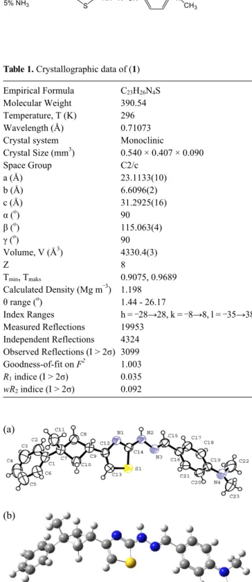
Figure 1. (a) ORTEP drawing of the basic crystallographic unit of the title compound (1), showing the atom-numbering scheme. Displace-ment ellipsoids are drawn at the 50% probability level and all H atoms are shown as small spheres of arbitrary radii. (b) Gaussian03View drawing of the title compound (1).
Table 1. Crystallographic data of (1) Empirical Formula Molecular Weight Temperature, T (K) Wavelength (Å) Crystal system Crystal Size (mm3) Space Group a (Å) b (Å) c (Å) α (o) β (o) γ (o) Volume, V (Å3) Z Tmin, Tmaks Calculated Density (Mg m‒3) θ range (o) Index Ranges Measured Reflections Independent Reflections Observed Reflections (I > 2σ) Goodness-of-fit on F2 R1 indice (I > 2σ) wR2indice (I > 2σ) C23H26N4S 390.54 296 0.71073 Monoclinic 0.540 × 0.407 × 0.090 C2/c 23.1133(10) 6.6096(2) 31.2925(16) 90 115.063(4) 90 4330.4(3) 8 0.9075, 0.9689 1.198 1.44 - 26.17 h = ‒28→28, k = ‒8→8, l = ‒35→38 19953 4324 3099 1.003 0.035 0.092 (B3LYP) methods with 6-311G(d, p) and 6-31G(d, p) basis sets.
A comparison of the experimental and theoretical spectra can be very useful in making correct assignments and understanding the molecular structure relationship. And so, these calculations are valuable for providing insight into molecular analysis.
Experimental and Theoretical Methods
General method. IR spectra were recorded on a Mattson 1000 (Unicam Ltd., Cambridge, UK) Fourier transform-infrared (FT- IR) Spectrometer using KBr pellets in the range 4000 - 400 cm‒1. The UV spectra of the compound were performed on a Schi-madzu UV-1700 spectrometer in CHCl3 solvent. The 1H and 13C-NMR spectra were recorded on a Brucker 400 MHz spec-trometer using TMS as internal standard. Microanalyses were performed on a LECO CHNSO-932 auto elemental analysis apparatus. Melting points were determined on a Gallenkamp melting point apparatus and checked by differential scanning calorimeter (DSC) and are uncorrected.
Synthesis. The compound was synthesized as in Scheme 1 by the following procedure. To a solution of hydrazone com-pound A (2.2231 g, 10 mmol) in 50 mL of ethanol, a solution of 1-methyl-1-phenyl-3-(2-chloro-1-oxoethyl) cyclobutane (2.2271 g, 10 mmol) in 20 mL of absolute ethanol was added. After the addition of the α-haloketone, the temperature was raised to 323 - 328 K and kept at this temperature for 2 h. The solution was cooled to room temperature and then made alkaline with an aqueous solution of NH3 (5%), and dark brown precipi-tate separated by suction, washed with aqueous NH3 solution several times and dried in air. Suitable single crystal for crystal structure determination was obtained by slow evaporation of its ethanol solution. Yield: 74%. mp 437 K. IR (KBr, υ cm‒1) 3166 (-NH-), 3078 - 3020 (aromatics), 2960 - 2802 (aliphatics), 1606 (C=N azomethine), 1575 (C=N thiazole), 703 (C-S-C). 1H- NMR (CDCl3, TMS, δ ppm) 1.58 (s, 3H,-CH3, on cyclobutane ring), 2.54 (d, j = 8.9 Hz, 4H, -CH2-, in cyclobutane ring), 3.03 (s, 6H, -CH3, on aniyine N), 3.67 (quint, j = 8.4 Hz, 1H, >CH-, in cyclobutane ring), 6.21 (d, j = 0.9 Hz, 1H, = CH-S, in thiazole ring), 6.71 (d, j = 8.95 Hz, 2H, on aniline ring), 7.14-7.22 (m, 3H aromatics), 7.27-7.35 (m, 2H, aromatics), 7.55 (d, j = 8.95 Hz, 2H, on aniline ring), 7.71 (s, 1H, -N=CH-, azomethine), 10.56 (s, 1H, -NH-, D2O exchangeable). 13C-NMR (CDCl3, TMS, δ ppm) 168.69, 155.99, 152.35, 151.33, 142.60, 128.20, 128.11, 125.08, 124.77, 122.01, 111.92, 101.58, 40.26, 40.20, 38.86, 30.84, 30.14. Anal. calc. for C23H26N4S (390.54); C: 70.73, H: 6.71, N: 14.35, S: 8.21; found; C: 70.57, H: 6.85, N: 13.93, S: 8.34.
Crystal structure determination. The data collection was per-formed at 293 K on a Stoe-IPDS-2 diffractometer equipped with a graphite monochromated Mo Kα radiation (λ = 0.71073
Table 3. Hydrogen bond interaction of the title compound (1) (Å, o)
Hydrogen bond(Å,o) D-H H...A D...A D-H...A
N2-H2A...N1i 0.86 2.29 3.037(2) 145
Symmetry code: (i): 1-x, 2-y, -z
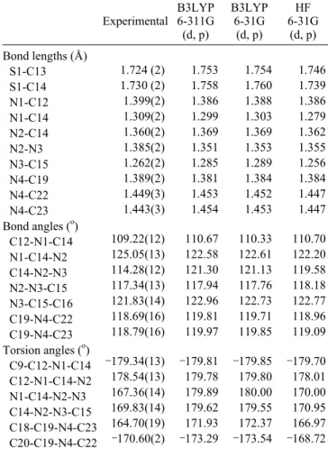
Figure 2. A partial packing diagram of the title compound (1). N-H…N interaction has been shown as broken lines. Hydrogen atoms not in-volved in hydrogen bonding have been omitted [Symmetry code: (i): 1-x, 2-y, -z].
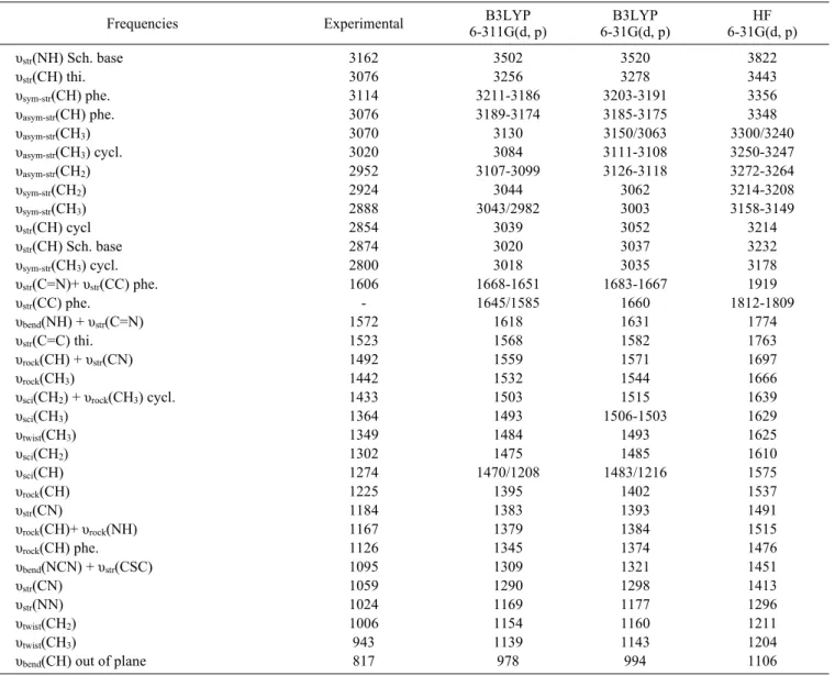
Table 2. Selected geometrical parameters of the title compound (1) with X-ray structure, DFT and HF methods
Experimental B3LYP6-311G (d, p) B3LYP 6-31G (d, p) HF 6-31G (d, p) Bond lengths (Å) S1-C13 S1-C14 N1-C12 N1-C14 N2-C14 N2-N3 N3-C15 N4-C19 N4-C22 N4-C23 Bond angles (o) C12-N1-C14 N1-C14-N2 C14-N2-N3 N2-N3-C15 N3-C15-C16 C19-N4-C22 C19-N4-C23 Torsion angles (o) C9-C12-N1-C14 C12-N1-C14-N2 N1-C14-N2-N3 C14-N2-N3-C15 C18-C19-N4-C23 C20-C19-N4-C22 1.724 (2) 1.730 (2) 1.399(2) 1.309(2) 1.360(2) 1.385(2) 1.262(2) 1.389(2) 1.449(3) 1.443(3) 109.22(12) 125.05(13) 114.28(12) 117.34(13) 121.83(14) 118.69(16) 118.79(16) ‒179.34(13) 178.54(13) 167.36(14) 169.83(14) 164.70(19) ‒170.60(2) 1.753 1.758 1.386 1.299 1.369 1.351 1.285 1.381 1.453 1.454 110.67 122.58 121.30 117.94 122.96 119.81 119.97 ‒179.81 179.78 179.89 179.62 171.93 ‒173.29 1.754 1.760 1.388 1.303 1.369 1.353 1.289 1.384 1.452 1.453 110.33 122.61 121.13 117.76 122.73 119.71 119.85 ‒179.85 179.80 180.00 179.55 172.37 ‒173.54 1.746 1.739 1.386 1.279 1.362 1.355 1.256 1.384 1.447 1.447 110.70 122.20 119.58 118.18 122.77 118.96 119.09 ‒179.70 178.01 170.00 170.95 166.97 ‒168.72
Å). The structure was solved by direct methods using SHELXS- 97 and refined by a full-matrix least-squares procedure using the program SHELXL-97.34 All non-hydrogen atoms were easi-ly found from the difference Fourier map and refined aniso-tropically. All hydrogen atoms were included using a riding model and refined isotropically with C-H = 0.93 - 0.97 Å and N-H = 0.86 Å. Uiso (H) = 1.2 Ueq (C, N), Uiso (H) = 1.5 Ueq (for methyl group).
Theoretical methods. Theoretical calculations were carried
out using semi-empirical AM1 and ab initio HF/6-31G(d, p) and density functional B3LYP/6-311G(d, p) and B3LYP/6-31G (d, p) quantum chemical methods.35-37 For modeling, the experi-mental data were supplemented by using semi-emprical and ab initio quantum chemical calculations. Molecular geometry is restricted and all the calculations are performed without speci
-fying any symmetry for the title molecule by using Gaussian
03 Program package on a personal computer.38 In addition to
these, vibrational frequencies for optimized molecular structures
have been also calculated. No scale factor was used in the
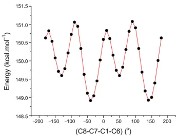
calculated frequencies. To identify low energy conformations, a selected degree of torsional freedom, T(C8-C7-C1-C6), was varied from ‒180o to +180o in steps of 10o, and the molecular energy profile was obtained at the semi-empirical AM1 level.
Result and Discussions
Description of the crystal structure. The title compound con-tains thiazole, hydrazone, phenyl and cyclobutane moieties. De-tails of crystal parameters, data collection, structure solution and refinement are given in Table 1 and the crystal structure with the formula, C23H26N4S, (I) shown in Figure 1. The central five-member thiazole ring is essentially planar, to within 0.0006 Å. The dihedral angles between the phenyl ring A(C1 through C6), cyclobutane ring B(C7 through C10), thiazole ring C(C12- C13-S1-C14-N1) and the other phenyl ring D(C16 through C21) are 38.69(6)o (A/B), 49.30(7)o (B/C) and 41.49(5)o (C/D). Only some of bond distances in thiazole ring show partial double bond character so that C12-N1, S1-C13 and S1-C14 bond dis-tances of 1.399(2), 1.724(2), 1.730(2) Å are showing the values of single bond character (see Table 2). The S-C bond distances are shorter than the accepted value for an S-C sp2 single bond with 1.76 Å.39 It is worth noting that the C14-N1 bond distance value of 1.309(2) Å is falling in to the C=N double bond distance region, and is shorter than the C=N double bond distance found in related thiazole ring structure,40 in thiazole ring the C12-C13 bond distance of 1.344(2) Å is also showing the value of C=C double bond character.
C and D rings are linked by the strictly planar N2-N3 = C15-C16 fragment and its torsion angle is 175.88(14)o. In hy-drazone group, C15-N3 double bond distance of 1.262(2) Å is shorter than the C=N bond distance found related hydrazone structures, i.e. 1.2810(19) Å41 and 1.272(2) Å.42 The steric inter-action between the substituent groups on the cyclobutane ring means that this ring derivates significantly from planarity. Literature values for the puckering of the cyclobutane ring are 29.03(13)o 43 and 26.8(2)o.44 In this paper, the C8/C7/C10 plane forms a dihedral angle of 25.74(15)o with the C8/C9/C10 plane. The crystal structure does not exhibit intramolecular
4000 3500 3000 2500 2000 1500 1000 500 Wavenumbers 70 60 50 40 30 20 10 T %
Figure 3. The experimental FT-IR spectrum of the title compound (1).
Table 4. Vibrational frequencies of the title compound (1) with X-ray structure, DFT and HF methods
Frequencies Experimental 6-311G(d, p)B3LYP 6-31G(d, p)B3LYP 6-31G(d, p)HF
υstr(NH) Sch. base υstr(CH) thi. υsym-str(CH) phe. υasym-str(CH) phe. υasym-str(CH3) υasym-str(CH3) cycl. υasym-str(CH2) υsym-str(CH2) υsym-str(CH3) υstr(CH) cycl υstr(CH) Sch. base υsym-str(CH3) cycl. υstr(C=N)+ υstr(CC) phe. υstr(CC) phe. υbend(NH) + υstr(C=N) υstr(C=C) thi. υrock(CH) + υstr(CN) υrock(CH3)
υsci(CH2) + υrock(CH3) cycl.
υsci(CH3) υtwist(CH3) υsci(CH2) υsci(CH) υrock(CH) υstr(CN) υrock(CH)+ υrock(NH) υrock(CH) phe. υbend(NCN) + υstr(CSC) υstr(CN) υstr(NN) υtwist(CH2) υtwist(CH3)
υbend(CH) out of plane
3162 3076 3114 3076 3070 3020 2952 2924 2888 2854 2874 2800 1606 -1572 1523 1492 1442 1433 1364 1349 1302 1274 1225 1184 1167 1126 1095 1059 1024 1006 943 817 3502 3256 3211-3186 3189-3174 3130 3084 3107-3099 3044 3043/2982 3039 3020 3018 1668-1651 1645/1585 1618 1568 1559 1532 1503 1493 1484 1475 1470/1208 1395 1383 1379 1345 1309 1290 1169 1154 1139 978 3520 3278 3203-3191 3185-3175 3150/3063 3111-3108 3126-3118 3062 3003 3052 3037 3035 1683-1667 1660 1631 1582 1571 1544 1515 1506-1503 1493 1485 1483/1216 1402 1393 1384 1374 1321 1298 1177 1160 1143 994 3822 3443 3356 3348 3300/3240 3250-3247 3272-3264 3214-3208 3158-3149 3214 3232 3178 1919 1812-1809 1774 1763 1697 1666 1639 1629 1625 1610 1575 1537 1491 1515 1476 1451 1413 1296 1211 1204 1106 Sch. base: schiff base, thi: thiazole, phe: phenyl, cycl: cyclobutane, str: stretching, bend: bending, rock: rocking, twist: twisting, sci: scissorsing
action. There is, however, an intermolecular N-H…N (head to head) hydrogen bonding, details of which are given in Table 3. In this hydrogen bonding, atom N2 of the molecule at (x, y, z) acts as hydrogen-bond donor, via atom H2A, to atom N1 of the molecule at (1-x, 2-y, -z), so generating by inversion a dimer, centered at (1/2, 1, 0) and characterized by an R22(8) motif (see
Figure 2). The R22(8) rings formed by hydrogen bonds are
centered at [n/2, m/2, k] and [n/2, m/2, 1/2+k] (n, m and k are zero or integer). Beside of these dimers, the weak π-π and π...ring interactions also stabilize to crystal packing.
Vibrational frequency. As mentioned in the Experimental section, the experimental FT-IR spectra was obtained in KBr discs using a Mattson 1000 FT-IR spectrometer, and shown in
Figure 3. Vibrational frequencies calculated at B3LYP/6-311G
(d, p), B3LYP/6-31G(d, p) and HF/6-31G(d, p) levels. Some
primary calculated harmonic frequencies are listed in Table 4 and compared with the experimental data. The descriptions con-cerning the assignment have also been indicated in the Table 4.
Gauss-View Molecular Visualization program was used to
assign the calculated harmonic frequencies.45 As seen from Table 4, the predicted IR spectra by using B3LYP/6-311G(d,
Figure 4. Atom-by-atom superimposition of the structures calculated (red)(B3LYP/6–311G(d, p)) over the X-ray structure (black) for the title compound. Hydrogen atoms have been omitted for clarity. The RMS overlay error of 0.601 Å does not include hydrogen atoms.
‒200 ‒150 ‒100 ‒50 0 50 100 150 200 (C8-C7-C1-C6) (o) 151.5 151.0 150.5 150.0 149.5 149.0 148.5 Energy (kcal.mol –1 )
Figure 5. Molecular energy profile against the selected torsional degree
T(C8-C7-C1-C6) of freedom for AM1 method. p) and B3LYP/6-31G(d, p) methods are consistent with those
by HF/6-31G(d, p) method. It is well known that the calculated HF and DFT ‘raw’ or ‘non-scale’ harmonic frequencies could significantly overestimate experimental values due to lack of electron correlation, insufficient basis sets and anharmonicity. Much effort has been devoted to accurately reproducing ex-perimental frequencies in theoretical calculations. The Hartree- Fock calculated results are usually more overestimated than
the corresponding DFT ones.46 To compare these, we calculated
the theoretical vibrational spectra of the title compound by using both HF/6-31G(d, p) and B3LYP/6-311G(d, p) with B3LYP/ 6-31G(d, p) methods and compared with the experimental results.
The characteristic υ(CH) stretching vibrations of heteroaro-matic structures are expected to appear in 3000 - 3100 cm‒1 fre-quency ranges.47,48 In the present study, experimentally four υ (CH) stretching vibrations of the title compound (I) are observed at 3114, 3076, 2874 and 2854 cm‒1. The highest normal mode of vibration corresponds to the υ(CH) stretching in the thiazole ring, which is calculated at 3443 cm‒1 for HF/6-31G(d, p) me-thod. On the other hand, the CH3 asymmetric and symmetric stretching frequencies are established at 3070, 3020, 2888 and 2800 cm‒1 in infrared spectra. All this results are in good agree-ment with the literature values.47,49,50 As shown in Table 4, the experimentally determined vibrational bands of the compound were found to be significantly lower than calculated values; however, υ(NH) stretching vibration is observed at 3162 cm‒1 and bending vibration is observed at 1572 cm‒1 for experimental values. Due to N-H…N intermolecular hydrogen bonding, it can be said that experimental υ(NH) bending vibration increases while υ(NH) stretching vibration decreases.51Other essential
characteristic vibrations of the title compound such as rocking, bending, scissorsing, out of plane bending, twisting are compar-ed in Table 4. As a result, experimental frequency values are in good agreement with the calculated frequencies with HF and B3LYP methods.
Theoretical structures. Some selected geometric parameters
experimentally obtained and theoretically calculated by HF/6-
31G(d, p), B3LYP/6-311G(d, p) and B3LYP/6-31G(d, p)me
-thods are listed in Table 2. When the X-ray structure of the title
compound is compared with B3LYP/6-311G(d, p) optimized
counterpart (see Figure 4), conformational discrepancy is ob-served between its. The dihedral angles between A, B, C and D planes are calculated at 49.73o (A/B), 47.55o (B/C) and 33.42o (C/D) for AM1, at 36.79o (A/B), 46.05o (B/C) and 0.93o (C/D)
for B3LYP/6-311G(d, p), at 28.36o (A/B), 37.96o (B/C) and 38.17o (C/D) for B3LYP/6-31G(d, p) and at 36.18o (A/B), 43.71o (B/C) and 8.27o (C/D) for 36.79o (A/B), 46.05o (B/C) and 0.93o
(C/D) for HF/6-31G(d, p).
For the optimized geometric parameters, various methods in
-cluding HF method estimates some bond lengths well to some
extent.52-54 We noted that the experimental results belong to
solid phase and theoretical calculations belong to gaseous phase. In the solid state, the existence of the crystal field along with the intermolecular interactions have connected the molecules together, which result in the differences of bond parameters between the calculated and experimental values. It is well known that DFT optimized bond lengths are usually longer and more
accurate than HF, due to inclusion of electron correlation. How
-ever, according to our calculations, HF method correlates well for both bond length, bond angle and torsion angle compared with the other method (Table 2). The largest differences between experimental and calculated HF and B3LYP bond lengths are
about 0.030 Å and 0.034 Å, respectively. The bond angles pro
-vided by HF method is the closest to the experimental values (see Table 2). The largest difference is about 5.30o in the case of
HF method, while this difference is 7.02o for B3LYP method.
The same trend was also observed in torsion angles. The largest
differences are 12.64o and 2.64o for B3LYP and HF methods,
respectively. As a result, the optimized bond lengths, bond an
-gles and torsion an-gles obtained by HF method show the best agreement with the experimental values.
Conformational analysis. In order to define the preferential position of the phenyl ring system with respect to the cyclo-butane group, a preliminary search of low-energy structures was performed by using AM1 computation as a function of the se-lected torsion angle, T(C8-C7-C1-C6). The respective value of the selected torsion angle is 151.74(15)o in the X-ray struc-ture, whereas the corresponding values in optimized geometries are 142.686o for AM1, 143.251o for HF/6-31G(d, p), 142.169o for B3LYP/6-31G(d, p) and 142.295o for B3LYP/6-311G(d, p). The molecular energy profile with respect to rotations about the selected torsion angle is presented in Figure 5. According to the results, the low-energy domains for T(C8-C7-C1-C6) are
‒0.030 0.030 0
Figure 6. Molecular electrostatic potential map (in a.u.) of the title com-pound with the B3LYP/6-311G(d, p) method (with an isodensity value of 0.0004 a.u.).
Table 5. Molecular energy profile against the selected torsional degree
T(C8-C7-C1-C6) of freedom for AM1 method
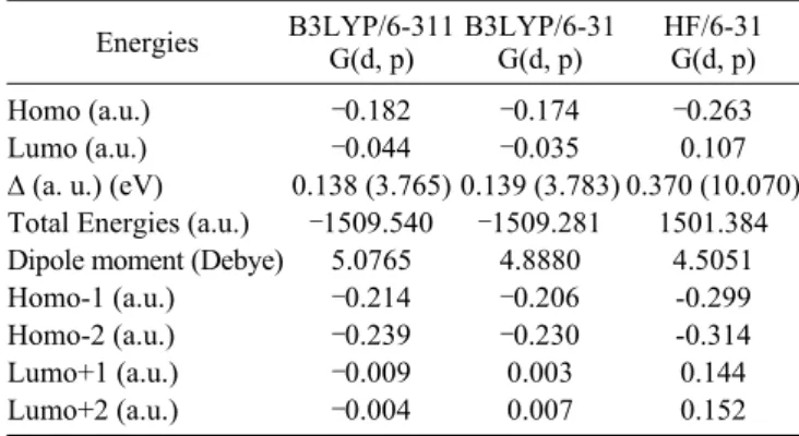
Energies B3LYP/6-311G(d, p) B3LYP/6-31G(d, p) HF/6-31G(d, p) Homo (a.u.)
Lumo (a.u.) ∆ (a. u.) (eV) Total Energies (a.u.) Dipole moment (Debye) Homo-1 (a.u.) Homo-2 (a.u.) Lumo+1 (a.u.) Lumo+2 (a.u.) ‒0.182 ‒0.044 0.138 (3.765) ‒1509.540 5.0765 ‒0.214 ‒0.239 ‒0.009 ‒0.004 ‒0.174 ‒0.035 0.139 (3.783) ‒1509.281 4.8880 ‒0.206 ‒0.230 0.003 0.007 ‒0.263 0.107 0.370 (10.070) 1501.384 4.5051 -0.299 -0.314 0.144 0.152 1 a.u.= 27.2116 eV 200 400 600 800 1000 1200 Wavelength (nm) Absor bance 3 2.5 2 1.5 1 0.5 0
Figure 7. The experimental UV-vis absorption spectra of the title compound (1).
located at ‒40 and 140o with energies of 148.913 and 148.924 kcal mol‒1. As shown in Figure 5, the energy difference between the most favourable and most unfavourable conformers, which arises from the rotational potential barrier calculated with re-spect to the selected torsion angle, is calculated to be 1.920 kcal mol‒1.
The molecular energy can be divided into bonded and non-
bonded contributions. The bonded energy is considered to be independent of torsional angle changes and therefore vanished
when relative conformer energies are calculated. The non-bond
-ed energy is further separat-ed into torsional steric and electro
-static terms.55 Since the title compound contains no intramole
-cular hydrogen bond, it can be deduced from the computational results that the most stable conformer of the title compound is principally determined by the non-bonded torsional energy term affected by packing of the molecules.
Molecular electrostatic potential. The molecular electrostatic potential (MEP) is related to the electronic density and is a very useful descriptor for determining sites for electrophilic attack and nucleophilic reactions as well as hydrogen-bonding interactions.56-58 To predict reactive sites for electrophilic and nucleophilic attack for the title molecule, MEP was calculated at the B3LYP/6-311G(d, p) optimized geometry. The negative (red) regions of MEP were related to electrophilic reactivity and the positive (blue) regions to nucleophilic reactivity shown in Figure 6. As can be seen from the figure, there is possible site on the title compound for electrophilic attack. The negative region is localized on the unprotonated nitrogen atom of the thiazole ring, N1, with a maximum value of ‒0.030 a.u. How-ever, maximum positive region is localized on atom N2 probably due to the hydrogen, with a maximum value of 0.030 a.u. These results provide information concerning the region where the compound can interact intermolecularly and bond metallically. Therefore, Figure 6 confirms the existence of an intermolecular N-H...N interaction between the protonated and unprotonated N atoms of the thiazole ring and hydrazone group.
Frontier molecular orbital analysis. The calculations indicate that the compound has 104 occupied molecular orbitals (MOs). The highest occupied molecular orbital (HOMO) energies, the lowest unoccupied molecular orbital (LUMO) energies, the energy gap for mentioned molecule in above have calculated and given in Table 5. The frontier molecular orbitals play an
im-portant role in the electric and optical properties, as well as in UV-vis spectra and chemical reactions.59Based on B3LYP/6-
311G(d, p), B3LYP/6-31G(d, p) and HF/6-31G(d, p)optimized
geometry, the total energy of the title compound has been cal
-culated by this three methods, which are ‒1509.540,‒1509.281
and ‒1501.384 a.u., respectively. An electronic system with a larger HOMO-LUMO gap should be less reactive than one hav-ing a smaller gap.60 In the present study, the HOMO-LUMO gap values of the molecule are at 3.765, 3.783 and 10.070 eV for B3LYP/6-311G(d, p), B3LYP/6-31G(d, p) and for HF/6-
31G(d, p) methods, respectively, as seen in Table 5.
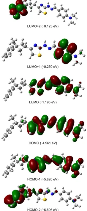
The UV-vis absorption spectra of the title compound were recorded in the CHCl3 solutions. The compound exhibits absorp-tion peaks in the UV-visible region. The UV-vis absorpabsorp-tion spectra of the title compound is shown in Figure 7. The experi-mentally absorption peaks are observed at 349, 239 nm for the title compound. It can be seem that these peaks equal to n → π* and π → π*transitions.3D plots of the HOMO-2, HOMO-1, HOMO, LUMO, LUMO+1, LUMO+2 and the corresponding energy levels for the title compound are shown in Figure 8. The theoretically electronic transfers (ET) for the compound B3LYP/6-311G(d, p) and B3LYP/6-31G(d, p) basis sets are at 330, 243 and 329, 234 nm to correspond to the UV-vis spectral
LUMO+2 (‒0.123 eV) LUMO+1 (‒0.250 eV) LUMO (‒1.195 eV) HOMO (‒4.961 eV) HOMO-1 (‒5.820 eV) HOMO-2 (‒6.506 eV)
Figure 8. Molecular orbital surfaces and energy levels given in paren-theses for the HOMO-2, HOMO-1, HOMO, LUMO, LUMO+1 and LUMO+2 of the title compound have been computed with the B3LYP/ 6-311G(d, p) method. The positive phase is red, and the negative phase is green.
absorption peaks, and the corresponding electronic transfers happened between HOMO and LUMO, HOMO and LUMO+1, respectively. The bigger theoretical absorption wavelengths of the compound have slight blue-shifts comparing with the corr-esponding experimental ones.
Natural population analysis indicates that the electronic tran-sitions are mainly derived from the contributions of bands π → π*as reported literature.61 As shown from Figure 7, the elect-ron clouds of the HOMO and HOMO-1 are delocalized on the thiazole, hydrazone bridge and phenyl ring but HOMO-2 is
delocalized on the other phenyl ring. These orbitals are seem to be π-bonding type orbital. LUMO is found mainly delocalized on thiazole, hydrazone bridge and phenyl ring, but LUMO+1 and LUMO+2 are found mainly delocalized on different parts of the title molecule. In all cases, LUMOs are π*-anti-bonding orbitals.
Conclusions
In this study, we have synthesized a novel hydrazone com-pound, C23H26N4S, and characterized by using spectroscopic (IR, 1H-NMR, 13C-NMR, UV-vis) and structural (XRD) tech-niques. The X-ray structure is found to be very slightly different from its optimized counterparts, and the crystal structure is stabilized by N-H...N-type hydrogen bond between the pro-tonated N atom of hydrazone group and unpropro-tonated N atom of adjacent thiazole ring as well as by van der Waals forces. The results of the HF method show a better fit to experimental values than B3LYP in evaluating geometrical parameters. It is noted here that the experimental results are for the solid phase and the theoretical calculations are for the gaseous phase. In the solid state, the existence of the crystal field together with the intermolecular interactions holds the molecules together, which results in differences between the calculated and experi-mental values for the bond parameters. Despite the differences observed in the geometric parameters, the general agreement is good and the theoretical calculations support the solid-state structures. However, it can be seen from the theoretical results that the B3LYP method is more appropriate than the HF method for the calculation of vibrational frequencies. The calculated MEP map agrees well with the solid-state interactions. As men-tioned in the introduction, this compound has been synthesized as a part of our ongoing research program. According to the ob-served results, there is a good relationship between the results of the present study and expected functionalities from the structure of the molecule.
Supplementary Data. Crystallographic data for the structural analysis have been deposited with the Cambridge Crystallo-graphic Data Centre, CCDC No 748348 Copies of this informa-tion may be obtained free of charge from the Director, CCDC, 12 Union Road, Cambridge CB2 1EZ, UK (fax: +44-1223- 336033; e-mail: [email protected] or www: http://www. ccdc.cam.ac.uk).
Acknowledgments. The authors wish to acknowledge the Faculty of Arts and Sciences, Ondokuz Mayis University, Turk-ey, for the use of the STOE IPDS-II diffractometer (purchased under grant F.279 of the University Research Fund).
References
1. Zollinger, H. Colour Chemistry Syntheses Properties and
Applica-tions of Organic Dyes and Pigments, 2nd ed.; Weinheim: VCH,
1991.
2. Koparır, M.; Cansız, A.; Ahmedzade, M.; Çetin, A. Heteroat.
Chem. 2004, 15, 26.
2003, 25, 51.
4. Coghi, L.; Lanfredi, A. M. M.; Tripicchio, A. J. Chem. Soc. Perkin.
Trans. 1976, 2, 1808.
5. Saprykina,V. A.; Vinogradova, V. I.; Ambartsumova, R. F.; Ibragi-mov, T. F.; Shakhidoyatov, Kh. M. Chem. Nat. Compd. 2006, 42, 4470.
6. Hadjipavlou-Litina, J. D.; Geronikaki, A. Arzneim Forsch/ Drug.
Res. 1996, 46, 805.
7. Holla, B. S.; Malini, K. V.; Rao, B. S.; Sarojini, B. K.; Kunari, N. S. Eur. J. Med. Chem. 2003, 38, 313.
8. Tarafder, M. T. H.; Jin, K. T.; Crouse, K. A.; Ali, A. M.; Yamin, B. M.; Fun, H.-K. Polyhedron 2002, 21, 2547.
9. Guerriero, P.; Tamburini, S.; Vigato, P. A. Coord. Chem. Rev. 1995, 139, 17.
10. Sreekala, R.; Yusuff, K. K. M. React. Kinet. Catal. Lett. 1992, 48, 575.
11. Das, N. N.; Dash, A. C. Polyhedron 1995, 14, 1221. 12. Williams, D. R. Chem. Rev. 1972, 72, 203.
13. Tsapkov, V. I. Russ. J. Gen. Chem. 2002, 72, 276.
14. Ghosh, S.; Bandyopadhyay, T. K. Trans. Met. Chem. 1985, 10, 57. 15. Shan, S.; Xu, D.-J.; Wu, J.-Y.; Chiang, M. Y. Acta Crystallogr.
2002, E58, o1444.
16. Bosan, W. S.; Shank, R. C.; MacEwen, J. D.; Gaworski, C. L.; Newberne, P. M. Carcinogenesis 1987, 8, 439.
17. Roberge, A.; Gosslin, C.; Charbonneau, R. Biochem. Pharmacol. 1971, 20, 2231.
18. Waterfield, C. J.; Asker, D. S.; Timbrell, J. A. Chem. Biol. Int. 1997, 107, 157.
19. Rothgery, E. F. Kirk-Othmer Encyclopedia of Chemical
Techno-logy, 5th ed.; Wiley: Hoboken, N. J. 2005; 27, pp 22957.
20. Schmidt, E.W. Hydrazine and its Derivatives: Preparation,
Pro-perties, Applications, 2nd ed.; Wiley: Hoboken, N. J. 2001.
21. Tezcan, H.; Tunç, T.; Şahin, E.; Yagbasan, R. Analy. Sci. 2004, 20, x137.
22. Townshend, A.; Wheatly, R. A. Analy. 1998, 123, 1041. 23. Cukurovali, A.; Yilmaz, I.; Gur, S.; Kazaz, C. Eur. J. Med. Chem.
2006, 41, 201.
24. Dinçer, M.; Avcı, D.; Şekerci, M.; Atalay, Y. J. Mol. Model. 2008,
14, 823.
25. Özdemir, N.; Dinçer, M.; Çukurovalı, A.; Büyükgüngör, O. J. Mol.
Model. 2009, 15, 1435.
26. Misra, N.; Prasad, O.; Sinha, L.; Pandey, A. J. Mol. Struct.
(Theo-chem.) 2007, 822, 45.
27. Jian, F.; Zhao, P.; Guo, H.; Li, Y. Spectrochim. Acta 2008, A69, 647.
28. Choo, J.; Yoo, S.; Moon, S.; Kwon, Y.; Chung, H. Vibr. Spectrosc. 1998, 17, 173.
29.Forsyth, D. A.; Sebag, A. B. J. Am. Chem. Soc. 1997, 119, 9483. 30. Cimino, P.; Gomez-Paloma, L.; Duca, D.; Riccio, R.; Bifulco, G.
Magn. Reson. Chem. 2004, 42, 26.
31. Friesner, R. A.; Murphy, R. B.; Beachy, M. D.; Ringnalda, M. N.; Pollard, W. T.; Dunietz, B. D.; Cao, Y. J. Phys. Chem. 1999, A103
(13), 1913.
32. Rulìsek, L.; Havlas, Z. Int. J. Quantum Chem. 2003, 91, 504. 33. Ziegler, T. Chem. Mater. Sci. 1997, 69.
34. Sheldrick, G. M. SHELXS-97 and SHELXL-97, Univ. Gottingen, Germany, 1997.
35. Lee, C.; Yang, W.; Parr, R. G. Phys. Rev. 1988, B37, 785. 36. Becke, A. D. J. Chem. Phys. 1993, 98, 5648.
37. Ditchfield, R.; Hehre, W. J.; Pople, J. A. J. Chem. Phys. 1971, 54,
724.
38. Frisch, M. J.; Trucks, G. W.; Schlegel, H. B.; Scuseria, G. E.; Robb, M. A.; Cheeseman, J. R.; Montgomery J. A.; Jr., Vreven, T.; Kudin, K. N.; Burant, J. C.; Millam, J. M.; Iyengar, S. S.; Tomasi, J.; Bar
-one, V.; Mennucci, B.; Cossi, M.; Scalmani, G.; Rega, N.; Peters
-son, G. A.; Nakatsuji, H.; Hada, M.; Ehara, M.; Toyota, K.; Fukuda, R.; Hasegawa, J.; Ishida, M.; Nakajima, T.; Honda, Y.; Kitao, O.; Nakai, H.; Klene, M.; Li, X.; Knox, J. E.; Hratchian, H. P.; Cross, J. B.; Bakken, V.; Adamo, C.; Jaramillo, J.; Gomperts, R.; Stratmann, R. E.; Yazyev, O.; Austin, A. J.; Cammi, R.; Pomelli, C.; Ochterski, J. W.; Ayala, P. Y.; Morokuma, K.; Voth, G. A.; Salvador, P.; Dan
-nenberg, J. J.; Zakrzewski, V. G.; Dapprich, S.; Daniels, A. D.; Strain, M. C.; Farkas, O.; Malick, D. K.; Rabuck, A. D.; Rag
-havachari, K.; Foresman, J. B.; Ortiz, J. V.; Cui, Q.; Baboul, A. G.; Clifford, S.; Cioslowski, J.; Stefanov, B. B.; Liu, G.; Liashenko, A.; Piskorz, P.; Komaromi, I.; Martin, R. L.; Fox, D. J.; Keith, T.; Al- Laham, M. A.; Peng, C. Y.; Nanayakkara, A.; Challacombe, M.; Gill, P. M. W.; Johnson, B.; Chen, W.; Wong, M. W.; Gonzalez, C.; Pople, J. A. Gaussian 03. Revision E.01. Gaussian Inc., Walling
-ford, CT, 2004.
39. Allen, F. H. Acta Crystallogr. 1984, B40, 64.
40. Yüksektepe, Ç.; Çalışkan, N.; Yılmaz, I.; Çukurovali, A. Acta
Cry-stallogr. 2006, E62, o2762.
41. Liu, G.; Liu, L.; Jia, D.; Yu, K. J. Chem. Crystallogr. 2005, 35, 497. 42. Ma, Q.; Lu, L.-P.; Zhu, M.-L. Acta Crystallogr. 2008, E64, o2026. 43. Yüksektepe, Ç.; Saraçoğlu, H.; Koca, M.; Çukurovali, A.;
Çalış-kan, N. Acta Crystallogr. 2004, C60, o509.
44. Yüksektepe, Ç.; Soylu, M. S.; Saraçoğlu, H.; Yılmaz, I.; Çukuro-vali, A.; Çalışkan, N.; Acta Crystallogr. 2005, E61, o1158. 45.Dennington, R. I. I.; Keith, T.; Millam, J. GaussView, Version
4.1.2. Semichem Inc, Shawnee Mission, KS, 2007.
46. Zhou, W.; Lu, J.; Zhang, Z.; Zhang, Y.; Cao, Y.; Lu, L.; Yang, X. Vibr. Spectrosc. 2004, 34, 199.
47.Arslan, H.; Algül, O. Int. J. Mol. Sci. 2007, 8, 760.
48. Siddiqui, S. A.; Dwivedi, A.; Singh, P. K.; Hasan, T.; Jain, S.; Pra-sad, O.; Misra, N. J. Struc. Chem. 2009, 50, 411.
49. Sundaraganesan, N.; Kumar, K. S.; Meganathan, C.; Joshua, B. D.
Spectrochim. Acta 2006, A65, 1186.
50. Krishnakumar, V.; Ramasamy, R. Spectrochim. Acta 2005, A62, 570.
51. Silverstein, R. M.; Bassler, G. C.; Morrill, T. C. Spectrometric
Identification of Organic Compounds; John Wiley & Sons: New
York, 1963.
52.Wheeless, C. J. M.; Zou, X.; Liu, R. J. Phys. Chem. 1995, 99(33),
12488.
53. Lee, S. Y.; Boo, B. H. J. Phys. Chem. 1996, 100, 15073. 54. Atalay, Y.; Avcı, D.; Başoğlu, A.; Okur, I. J. Mol. Struct. (Theo
-chem.) 2005, 713, 21.
55.Weiqun, Z.; Baolong, L.; Yang, C.; Yong, Z.; Xujie, L.L.Y. J. Mol. Struct. (Theochem.) 2005, 715, 117.
56. Scrocco, E.; Tomasi, J. Adv. Quant. Chem. 1979, 11, 115. 57. Luque, F. J.; Lopez, J. M.; Orozco, M. Theor. Chem. Acc. 2000,
103, 343.
58. Okulik, N.; Jubert, A. H. Internet Electron. J. Mol. Des. 2005, 4, 17. 59. Fleming, I. Frontier Orbitals and Organic Chemical Reactions;
Wiley: London, 1976.
60. Kurtaran, R.; Odabaşoğlu, S.; Azizoğlu, A.; Kara, H.; Atakol, O.
Polyhedron 2007, 26, 5069.
61. Sun, Y.-X.; Wei, W.-X.; Hao, Q.-L.; Lu, L.-D.; Wang, X.