Abstract
The purpose of this study was to determine the structural and functional properties of the distal muscles of front and hind legs of Malakan Horses. Thus, totally 10 Malakan horses (5 females and 5 males) were used in the study. From front and hind legs of the horses, the findings of totally 28 muscles were received. Accordingly, it is determined that the longest muscle in the distal of the front leg was m. flexor carpi ulnaris, the heaviest one was m. extensor carpi radialis and the muscle with the highest value in terms of parameters of tendon length and total muscle tendon length was m. extensor digitorum communis. From the distal muscles of the hind leg; it is observed that the longest one was m. tibialis cranialis, the heaviest one was m. gastrocnemius and the muscle with the highest value in terms of parameters of tendon length and total muscle tendon length was m. flexor digitorum superficialis. It was observed that while the difference determined between some of the values in the front and hind leg muscles had a statistical significance in the comparison performed between the genders (P<0.05), the differences had no statistical significance in the comparison performed based on the direction (right-left).
Keywords: Muscle, Malakan horse, Tendon, Front and hind leg
Malakan Atlarında (Equus caballus) Ön ve Arka Bacağın
Distal’indeki Kasların Yapısal ve Fonksiyonel Özellikleri
Özet
Bu çalışma, Malakan Atlarında ön ve arka bacağın distal’inde bulunan kasların yapısal ve fonksiyonel özelliklerini belirlemek amacıyla yapıldı. Bu amaçla 5 dişi, 5 erkek olmak üzere toplam 10 Malakan Atı kullanıldı. Atların ön ve arka bacağından toplam 28 kasın bulgusu alındı. Buna göre; ön bacağın distal’inde bulunan kaslardan en uzununun m. flexor carpi ulnaris, en ağırının m. extensor carpi radialis, tendo uzunluğu ve toplam kas tendo uzunluğu parametreleri bakımından ise en yüksek değere sahip olan kasın m. extensor digitorum communis olduğu belirlendi. Arka bacağın distal kaslarından en uzun olanın m. tibialis cranialis, en ağır olanın m. gastrocnemius, tendo uzunluğu ve toplam kas-tendo uzunluğu bakımından ise en yüksek değere sahip olan kasın m. flexor digitorum superficialis olduğu görüldü. Cinsiyetler arası yapılan karşılaştırmada ön ve arka bacakta kasların bazı değerleri arasında belirlenen farkın istatistiksel önem taşıdığı belirlenirken (P<0.05), yöne (sağ-sol) göre yapılan kıyaslamada farkların istatistiksel anlam taşımadığı tespit edildi.
Anahtar sözcükler: Kas, Malakan atı, Tendo, Ön ve arka bacak
Structural and Functional Properties of the Distal Muscles of
Front and Hind Legs of Malakan Horses (Equus caballus)
[1]Yasin DEMİRASLAN
1
İftar GÜRBÜZ
2Mustafa Orhun DAYAN
3Yalçın AKBULUT
4Kadir ASLAN
2Sami ÖZCAN
2Derviş ÖZDEMİR
5[1] 1 2 3 4 5
This study has been financially supported by Kafkas University Scientific Research Projects Coordination Unit. Project No: 2014-VF-20
Department of Anatomy, Faculty of Veterinary Medicine, Mehmet Akif Ersoy University, TR-15000 Burdur - TURKEY Department of Anatomy, Faculty of Veterinary Medicine, Kafkas University, TR-36000 Kars - TURKEY
Department of Anatomy, Faculty of Veterinary Medicine, Selçuk University, TR-42100 Konya - TURKEY Health School, Kafkas University, TR-36000 Kars - TURKEY
Department of Anatomy, Faculty of Veterinary Medicine, Atatürk University, TR-25000 Erzurum - TURKEY
INTRODUCTION
Malakan Horse is an endemic horse breed which is raised in the north-eastern regions of Eastern Anatolia, can resist to extreme winter conditions and is used in carriage,
draught, and horse riding. Its wide body and thick bone structure are remarkable. Also, the commonly seen types are the ones with black, gray, bay, and red coats [1-3].
There has been a serious literature deficit regarding this horse race in terms of basic sciences, clinical sciences
İletişim (Correspondence)
+90 542 7681365
[email protected]
and raising, husbandry and feeding in veterinary medicine. Morphologic structure of the muscle determines its function [4-6]. While the muscle structure may vary between
different muscles of the individual [7,8], the same muscles
in different individuals can be different in terms of structure [9,10]. Also it is known that exercise [11], genetic
structure and gender [12] are effective on the characteristics
of the muscle structure. Burkholder [13] also add the muscle
fiber volume, physiological characteristics, number of the fibers and their sequence to the factors affecting the structure of the muscle. Regarding the structural and functional properties of the muscles in Equidae family, Brown et al.[7] and Payne et al.[8] conducted a study on horses
and Fayed [14] and Demiraslan and Özcan [15] conducted a
study on donkeys. Also, the studies conducted on the muscle-tendon structure of four-leg animals is rather important in order to reveal the differences between the functions of front and hind legs [7,8].
The purpose of the study was to determine structural and functional properties of the distal muscles of front and hind legs of Malakan horses which are a native horse breed.
MATERIAL and METHODS
Totally 10 Malakan horses (5 males and 5 females) aged between 5 and 7 years were used in the study. The Malakan horses used had gray (3 males, 1 female), isabella (1 female), bay (2 males, 1 female), and red (2 females) coats. Average live weights of male and female Malakan horses were 314.00±5.47 kg and 287.80±7.69 kg, average cidago height was 142.40±3.04 cm and 138.40±1.14 cm, and average ridge height was 146.20±2.94 cm and 142.40±0.89 cm.
In this study, approval of the ethics committee was taken upon the decision no. 31 dated 20.02.2014 of Atatürk University Animal Experiments Local Ethics Committee regarding the usage of Malakan horses. The animals were taken to deep anesthesia according to the cadaver preparation techniques. Xylazine HCl (8 ml/100 kg, intravenous) and cloralhydrate (20 mg/kg intraperitoneal) were applied for this process [16]. After the blood of the
animals taken to deep anesthesia was drained through arteria carotis communis, the front legs were cut from the art. humeri level and the hind legs were cut from the art. coxae level and separated as right and left. Then, in order to reduce the effect of rigor mortis that could occur in the muscles, the legs were kept in extension position in the refrigerator of Kafkas University Faculty of Veterinary Medicine Department of Anatomy at 4°C for 24 h and within 48 h, the distal muscles of the front and hind legs were dissected and required results were taken. In order to determine the study limits, only the distal muscles of the front and hind legs were preferred.
The dissected muscles were subject to a set of measurements and calculations in order to determine the structural and functional properties. For these measurements, Payne et al.[8] and Fayed [14] were taken as
a reference. While the structural parameters in the study were determined as muscle length (KU cm), tendon length (TU cm), total muscle-tendon length (TKTU cm), muscle weight (KA g), tendon weight (TA g), total muscle-tendon weight (TKTA g), muscle volume (KH cm3), tendon
volume (TH cm3), total muscle-tendon volume (TKTH cm3),
pennation angle (PA°), and muscle bundle length (KDU cm), the functional parameters were determined as the physiological cross-sectional area of the muscle (PCSA cm2),
maximum isometric force (FmaxMPa), structural index (AI), tendon cross-sectional area (TCSA cm2), tendon pressure
on the maximum isometric force (TSfmaxMPa) and the per-centage of the distention ratio on the tendon pressure (TS%). Functional parameters were calculated by using the following parameters.
a. PCSA = KH/KDU d. TCSA = TH/TU b. Fmax = PCSA x 0.3 MPa e. TSfmax = Fmax/TCSA c. AI = KDU/KU f. TS% = TSfmax/1.5 GPa
While the distal front leg muscles evaluated in the study was examined as m. extensor carpi radialis (ECR), m. extensor digitalis lateralis (EDlat), m. extensor carpi obliquus (OEM), m. flexor carpi radialis (FCR), m. flexor carpi ulnaris (FCU), m. extensor carpi ulnaris (ECU), m. flexor digitorum superficialis (FDS) and mm. interossei (MIO); m. extensor digitorum communis (EDC) was investigated in two parts as superficial head, Thierness (Th) and deep head, Philips (Ph) and mm. flexores digitorum profundii (FDP) was observed in three parts as caput humerale (FDPH), ulnare (FDPU), and radiale (FDPR). While the distal hind leg muscles were examined as m. extensor digitorum longus (EDL), m. extensor digitorum lateralis (EDlat), m. peroneus tertius (PT), m. tibialis cranialis (Tcr), m. soleus (S), m. flexor digitorum superficialis (FDS), m. popliteus (P), m. extensor digitorum brevis (EDB), m. interosseus medius (lig. suspensorium - MIO), m. flexores digitorum profundi (FDP) was examined in three parts as m. flexor digitorum medialis (FDM), m. flexor digitorum lateralis (FDL), m. tibialis caudalis (Tcd) and m. gastrocnemius (G) was examined in two parts as caput lateralis (GL) and caput medialis (GM).
Data obtained from the study were standardized because the horses were at different ages (5-7) and different live weights (CA) [17]. Geometric similarity method was used in
the study for standardization [18-20]. According to this method,
data were calculated by using the following formulas. a. KA/CA b. TA/CA c. TU/CA d.KDU/CA e. PCSA/CA
For the analysis of the descriptive values of the measurements and the calculations obtained from the study according to gender and direction (right/left),
2-t test (P≤0.05), a parametric test, was performed in SPSS statistical package software (16.0 version). In this study, terms in Nomina Anatomica Veterinaria [21] were
taken as a basis.
RESULTS
Table 1, 2, 3, and 4 illustrates mean and standard
deviation data of structural and functional properties obtained in the study.
Accordingly, it was observed that the muscle with maximum KU value in the front leg distal of Malakan horse was FCU and the muscle with minimum KU value was FDPR. When considered in terms of TU and TKTU para-meters, it was observed that EDC had the maximum value. When KDU parameter was observed, it was determined that maximum and minimum values belonged to Th and FDS. ECR had the maximum KA and TKTA values and FDPH
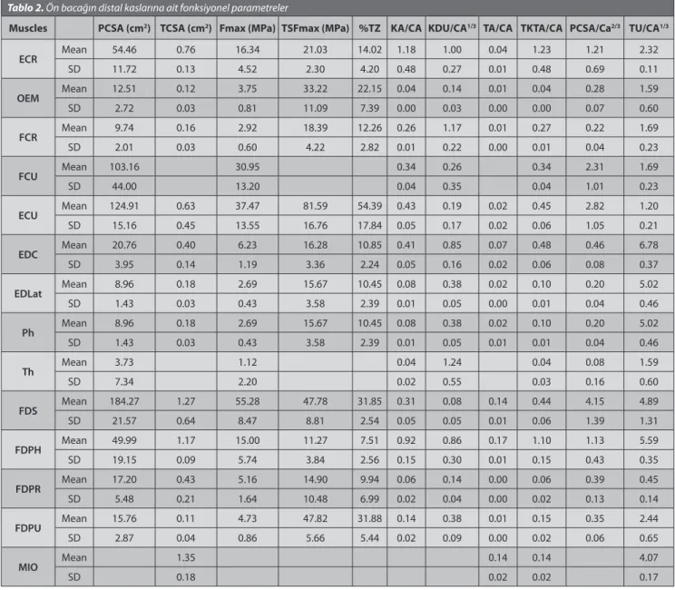
had the maximum TA value in the distal muscles of the front leg. When functional muscle properties were considered, it was observed that FDS and Th had the maximum and minimum PCSA and Fmax values in the distal of the front leg. In terms of TCSA, it was considered that the maximum value belonged to MIO. The muscles having the maximum and minimum values in terms of PA were determined as FCU and Th, respectively.
When the structural parameters of the distal muscles of front leg according to the gender were examined, it was found that FCR (12.50-10.13) and ECU (9.25-6.63) in terms of TU, FCU (117.27-85.97), ECU (147.94-113.22) and FDS (109.74-73.92) in terms of KA (g), ECR (15.14-11.36), FCR (2.62-1.55) and FDS (45.99-36.49) in terms of TA (g), FCR (83.69-76.72) in terms of TKTA (g), FCU (110.63-81.11), ECU (139.57-106.81) and FDS (103.53-69.74) in terms of KH (cm3), ECR (13.52-10.15), FCR (2.34-1.38) and FDS
(41.06-32.58) in terms of TH (cm3), FCR (78.82-72.29) in
Table 1. Structural parameters of distal muscle of front leg Tablo 1. Ön bacağın distal kaslarına ait yapısal parametreler
Muscles KU (cm) TU (cm) TKTU (cm) KDU (cm) AI KA (g) TA (g) TKTA (g) PA (°) KH (cm3) TH (cm3) TKTH (cm3)
ECR Mean 29.64 15.46 45.10 6.70 0.15 354.18 13.25 367.43 35.80 334.13 11.83 345.96 SD 1.57 0.82 1.06 1.80 0.04 42.95 2.43 44.47 5.50 34.86 2.17 36.21 OEM Mean 15.93 10.63 26.55 0.94 0.04 11.93 1.34 13.27 31.30 11.26 1.20 12.45 SD 2.34 4.08 5.32 0.22 0.01 1.10 0.43 1.21 7.00 1.04 0.39 1.13 FCR Mean 26.28 11.31 34.21 7.82 0.23 78.12 2.09 80.20 22.00 73.69 1.86 75.55 SD 1.04 1.67 8.70 1.44 0.13 4.58 0.66 5.09 4.00 4.32 0.59 4.77 FCU Mean 34.18 34.18 1.69 0.05 101.62 101.62 47.90 95.87 95.87 SD 1.10 1.10 2.29 0.06 18.40 18.40 2.00 17.36 17.36 ECU Mean 33.02 8.00 41.02 1.30 0.03 128.18 6.23 134.41 45.40 120.92 5.57 126.49 SD 1.11 1.55 1.10 1.15 0.03 22.31 5.56 24.98 5.00 21.05 4.96 23.41 EDC Mean 25.53 45.21 70.74 5.67 0.08 122.04 20.15 142.18 25.80 115.13 17.99 133.11 SD 1.77 2.40 2.17 1.02 0.02 17.55 6.35 22.35 3.00 16.55 5.67 20.83 EDLat Mean 23.24 33.50 56.74 2.56 0.04 23.89 6.60 30.49 21.20 22.54 5.89 28.43 SD 1.83 2.99 2.33 0.36 0.01 2.34 1.27 2.63 2.00 2.20 1.13 2.46 Ph Mean 23.24 33.50 56.74 2.56 0.04 23.89 6.60 30.49 21.20 22.54 5.89 28.43 SD 1.83 2.99 2.33 0.36 0.01 2.34 1.27 2.63 2.50 2.20 1.13 2.46 Th Mean 19.20 19.20 8.25 0.41 10.77 11.31 20.40 10.16 10.64 SD 5.15 5.15 3.64 0.20 7.89 9.11 1.11 7.45 8.53 FDS Mean 30.33 32.59 62.91 0.54 0.01 91.83 41.24 133.07 30.10 86.63 36.82 123.45 SD 1.11 8.87 9.11 0.32 0.00 10.06 5.38 15.01 2.00 8.92 4.80 13.34 FDPH Mean 31.90 37.20 69.10 5.76 0.08 272.63 50.87 323.50 39.70 257.20 45.42 302.62 SD 1.16 2.46 2.25 2.06 0.03 50.51 3.52 52.57 3.00 47.65 3.14 49.48 FDPR Mean 15.68 2.94 18.61 0.94 0.05 16.51 1.40 17.91 27.98 15.57 1.25 16.82 SD 2.68 1.05 3.19 0.29 0.02 5.12 0.86 5.63 4.74 4.83 0.77 5.28 FDPU Mean 20.08 16.21 36.29 2.54 0.07 41.55 1.90 43.45 29.20 39.20 1.70 40.89 SD 2.11 4.09 3.43 0.63 0.02 7.98 0.73 7.84 2.00 7.52 0.65 7.40 MIO Mean 27.15 27.15 41.01 41.01 36.62 36.62 SD 0.95 0.95 4.52 4.52 4.04 4.04
terms of TKTH (cm3) were higher in males compared to
females and the difference was statistically significant (P<0.05). It was determined that FDS (29.53-31.13) in terms of KU (cm) and Th (0.29-0.60) in terms of Yİ were higher in females compared to males and the difference was statistically significant (P<0.05).
When the functional parameters of the distal muscles of front leg according to the gender were examined, it was found that ECR (0.86-0.67) and FCR (0.19-0.14) in terms of TCSA (cm2), FCU (0.37-0.31), ECU (0.47-0.40), and
FDS (0.35-0.26) in terms of KA/CA, ECU (0.50-0.41) in terms of TKTA/CA, FCR (1.84-1.55) and ECU (1.36-1.01) in terms of TU/CA1/3 were higher in males compared to females
and the difference was statistically significant (P<0.05). It was determined that ECU (37.65-126.50) in terms of TSFmax (MPa) and FDS (4.81-4.96) in terms of TU/CA1/3
had a higher value in females compared to males and the difference was statistically significant (P<0.05).
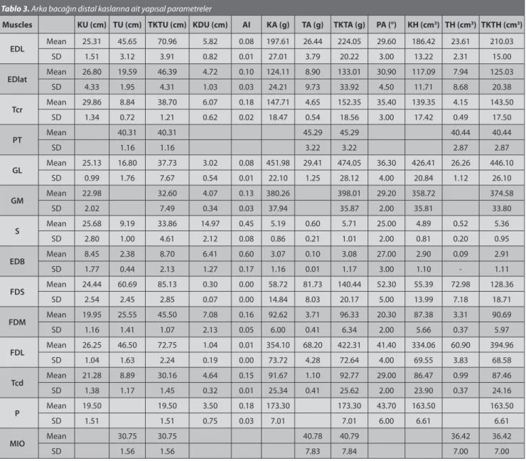
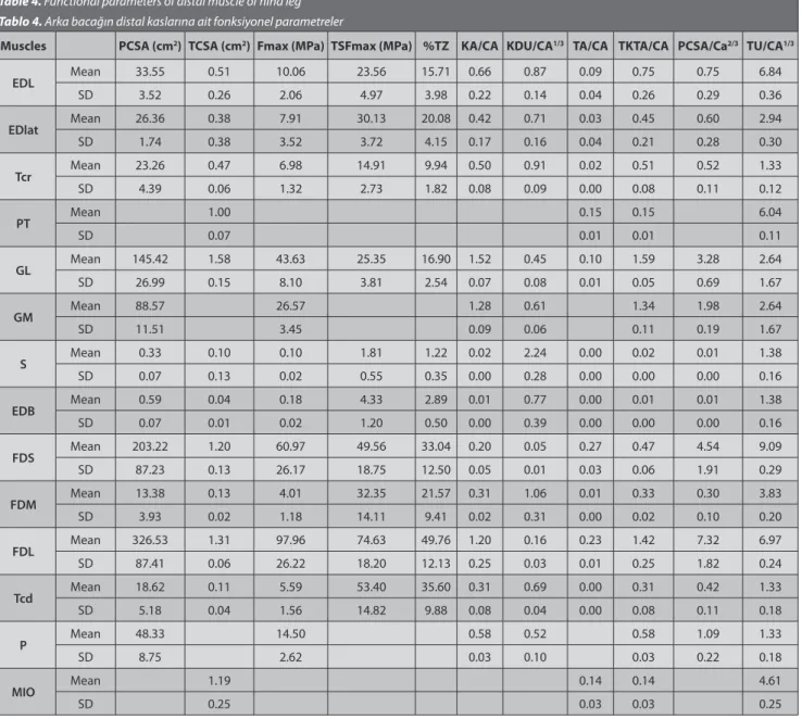
When the results regarding the distal muscles of the hind leg were considered, the muscles with maximum and minimum KU values were determined as Tcr and EDB. In terms of TU and TKTU, the muscles with high and low values were specified as FDS and EBD. It was observed that in the distal of the hind leg, maximum value in terms of the KDU parameter belonged to S and minimum value belonged to FDS. G had the maximum value in terms of KA and TKTA, and FDS had the maximum value in terms of TA. When the functional properties of the muscles were considered, it was observed that maximum and minimum values of PCSA and Fmax parameters belonged to FDL and S, maximum TCSA parameter belonged to G, maximum value in terms of PA belonged to FDS and minimum value belonged to FDM.
When the structural parameters of the distal muscles of hind leg according to the gender were observed, it was found that Tcr (30.88-28.85), GM (24.45-21.50), FDL
Table 2. Functional parameters of distal muscle of front leg Tablo 2. Ön bacağın distal kaslarına ait fonksiyonel parametreler
Muscles PCSA (cm2) TCSA (cm2) Fmax (MPa) TSFmax (MPa) %TZ KA/CA KDU/CA1/3 TA/CA TKTA/CA PCSA/Ca2/3 TU/CA1/3
ECR Mean 54.46 0.76 16.34 21.03 14.02 1.18 1.00 0.04 1.23 1.21 2.32 SD 11.72 0.13 4.52 2.30 4.20 0.48 0.27 0.01 0.48 0.69 0.11 OEM Mean 12.51 0.12 3.75 33.22 22.15 0.04 0.14 0.01 0.04 0.28 1.59 SD 2.72 0.03 0.81 11.09 7.39 0.00 0.03 0.00 0.00 0.07 0.60 FCR Mean 9.74 0.16 2.92 18.39 12.26 0.26 1.17 0.01 0.27 0.22 1.69 SD 2.01 0.03 0.60 4.22 2.82 0.01 0.22 0.00 0.01 0.04 0.23 FCU Mean 103.16 30.95 0.34 0.26 0.34 2.31 1.69 SD 44.00 13.20 0.04 0.35 0.04 1.01 0.23 ECU Mean 124.91 0.63 37.47 81.59 54.39 0.43 0.19 0.02 0.45 2.82 1.20 SD 15.16 0.45 13.55 16.76 17.84 0.05 0.17 0.02 0.06 1.05 0.21 EDC Mean 20.76 0.40 6.23 16.28 10.85 0.41 0.85 0.07 0.48 0.46 6.78 SD 3.95 0.14 1.19 3.36 2.24 0.05 0.16 0.02 0.06 0.08 0.37 EDLat Mean 8.96 0.18 2.69 15.67 10.45 0.08 0.38 0.02 0.10 0.20 5.02 SD 1.43 0.03 0.43 3.58 2.39 0.01 0.05 0.00 0.01 0.04 0.46 Ph Mean 8.96 0.18 2.69 15.67 10.45 0.08 0.38 0.02 0.10 0.20 5.02 SD 1.43 0.03 0.43 3.58 2.39 0.01 0.05 0.01 0.01 0.04 0.46 Th Mean 3.73 1.12 0.04 1.24 0.04 0.08 1.59 SD 7.34 2.20 0.02 0.55 0.03 0.16 0.60 FDS Mean 184.27 1.27 55.28 47.78 31.85 0.31 0.08 0.14 0.44 4.15 4.89 SD 21.57 0.64 8.47 8.81 2.54 0.05 0.05 0.01 0.06 1.39 1.31 FDPH Mean 49.99 1.17 15.00 11.27 7.51 0.92 0.86 0.17 1.10 1.13 5.59 SD 19.15 0.09 5.74 3.84 2.56 0.15 0.30 0.01 0.15 0.43 0.35 FDPR Mean 17.20 0.43 5.16 14.90 9.94 0.06 0.14 0.00 0.06 0.39 0.45 SD 5.48 0.21 1.64 10.48 6.99 0.02 0.04 0.00 0.02 0.13 0.14 FDPU Mean 15.76 0.11 4.73 47.82 31.88 0.14 0.38 0.01 0.15 0.35 2.44 SD 2.87 0.04 0.86 5.66 5.44 0.02 0.09 0.00 0.02 0.06 0.65 MIO Mean 1.35 0.14 0.14 4.07 SD 0.18 0.02 0.02 0.17
(27.00-25.50), and Tcd (22.30-20.25) in terms of KU (cm), EDL (48.05-43.25) and PT (41.13-39.50) in terms of TU (cm), FDM (46.30-44.70) in terms of TKTU (cm), Tcr (0.19-0.16) in terms of Yİ, GM (405.90-354.62) in terms of KA (g), GL (30.07-28.10) and FDM (4.05-3.37) in terms of TA (g), GL(496.58-451.52) in terms of TKTA (g), GM (382.93-334.52) in terms of KH (cm3), GL (26.85-25.08) and FDM (3.62-3.00)
in terms of TH (cm3), GL (466.95-425.25) in terms of TKTH
(cm3) were higher in males compared to females and the
difference was statistically significant (P<0.05). It was observed that EDB (5.22-7.60) in terms of KDU (cm) and EDL (0.08-0.09) and EDB (0.44-0.76) in terms of Yİ were higher in females compared to males and the difference was statistically significant (P<0.05).
When the functional parameters of the distal muscles of hind leg according to the gender were observed, it was found that GM (98.26-78.88) and EDB (0.65-0.52) in terms of PCSA (cm2), GM (29.48-23.67) and EDB (0.19-0.16) in terms
of Fmax (Mpa), Edlat (39.60 20.66) in terms of TSFmax (MPa), Edlat (26.40 13.77) in terms of %TZ, GM (2.13-18.4) in terms of PCSA/Ca2/3 were higher in males compared to females
and the difference was statistically significant (P<0.05). EDL (0.78-0.97) in terms of KDU/Ca1/3, S (1.31-1.45) in terms
of TU/CA1/3 were higher in females compared to males and
the difference was statistically significant (P<0.05).
In the analysis performed according to the direction (right-left), no statistically significant difference was determined.
DISCUSSION
In the study, structural and functional properties of the distal muscles of front and hind legs of Malakan Horses and the possible differences of these properties between the right-left legs and females-males were determined. Since the obtained results have distinguishing characteristics
Table 3. Structural parameters of distal muscle of hind leg Tablo 3. Arka bacağın distal kaslarına ait yapısal parametreler
Muscles KU (cm) TU (cm) TKTU (cm) KDU (cm) AI KA (g) TA (g) TKTA (g) PA (°) KH (cm3) TH (cm3) TKTH (cm3)
EDL Mean 25.31 45.65 70.96 5.82 0.08 197.61 26.44 224.05 29.60 186.42 23.61 210.03 SD 1.51 3.12 3.91 0.82 0.01 27.01 3.79 20.22 3.00 13.22 2.31 15.00 EDlat Mean 26.80 19.59 46.39 4.72 0.10 124.11 8.90 133.01 30.90 117.09 7.94 125.03 SD 4.33 1.95 4.31 1.03 0.03 24.21 9.73 33.92 4.50 11.71 8.68 20.38 Tcr Mean 29.86 8.84 38.70 6.07 0.18 147.71 4.65 152.35 35.40 139.35 4.15 143.50 SD 1.34 0.72 1.21 0.62 0.02 18.47 0.54 18.56 3.00 17.42 0.49 17.50 PT Mean 40.31 40.31 45.29 45.29 40.44 40.44 SD 1.16 1.16 3.22 3.22 2.87 2.87 GL Mean 25.13 16.80 37.73 3.02 0.08 451.98 29.41 474.05 36.30 426.41 26.26 446.10 SD 0.99 1.76 7.67 0.54 0.01 22.10 1.25 28.12 4.00 20.84 1.12 26.10 GM Mean 22.98 32.60 4.07 0.13 380.26 398.01 29.20 358.72 374.58 SD 2.02 7.49 0.34 0.03 37.94 35.87 2.00 35.81 33.80 S Mean 25.68 9.19 33.86 14.97 0.45 5.19 0.60 5.71 25.00 4.89 0.52 5.36 SD 2.80 1.00 4.61 2.12 0.08 0.86 0.21 1.01 2.00 0.81 0.20 0.95 EDB Mean 8.45 2.38 8.70 6.41 0.60 3.07 0.10 3.08 27.00 2.90 0.09 2.91 SD 1.77 0.44 2.13 1.27 0.17 1.16 0.01 1.17 3.00 1.10 - 1.11 FDS Mean 24.44 60.69 85.13 0.30 0.00 58.72 81.73 140.44 52.30 55.39 72.98 128.36 SD 2.54 2.45 2.85 0.07 0.00 14.84 8.03 20.17 5.00 13.99 7.18 18.71 FDM Mean 19.95 25.55 45.50 7.08 0.16 92.62 3.71 96.33 20.30 87.38 3.31 90.69 SD 1.16 1.41 1.07 2.13 0.05 6.00 0.41 6.34 2.00 5.66 0.37 5.97 FDL Mean 26.25 46.50 72.75 1.04 0.01 354.10 68.20 422.31 41.40 334.06 60.90 394.96 SD 1.04 1.63 2.24 0.19 0.00 73.72 4.28 72.64 4.00 69.55 3.83 68.58 Tcd Mean 21.28 8.89 30.16 4.64 0.15 91.67 1.10 92.77 29.00 86.47 0.99 87.46 SD 1.38 1.17 1.45 0.32 0.01 25.34 0.41 25.62 2.00 23.90 0.37 24.16 P Mean 19.50 19.50 3.50 0.18 173.30 173.30 43.70 163.50 163.50 SD 1.51 1.51 0.75 0.03 7.01 7.01 6.00 6.61 6.61 MIO Mean 30.75 30.75 40.78 40.79 36.42 36.42 SD 1.56 1.56 7.83 7.84 7.00 7.00
compared to other animals (especially other horse breeds and donkeys), this study plays a key role in terms of making comparison and determining the locomotor activity.
Alexander et al.[22] argued that the decrease in the
bundle length of the distal muscles in the legs of big animals can be tolerated by the tendons in this region which have a long and elastic structure. Shortening in the muscle bundles specified that the bundles can fit into a narrower area and number of fiber per unit area may increase. Thus, this situation caused an increase in PCSA and the force generated (see equipment and method). As a result, the short muscle fibers which can be packaged in a narrow area will be able to provide higher power generation by the increase of PCSA and able to increase the elastic energy quantity level which can be stored and released on the tendon via sufficient muscle-tendon movement [8]. Thus, the fact that muscles have small
volume, short fibers and large PCSA shows that they have
a high power generation capacity. Consequently, the force required for the muscle to be dynamic or static is produced by the muscles with a small volume and long tendons [7,8,23,24]. In the study, it was observed that the
muscles in the distal of front and hind legs of Malakan horses generally had small volume, short fibers and high PCSA. The most significant muscles were determined as EDC, FDPH, FDS in the front leg and FDS and FDL in the hind leg.
Determination of the scaling and standardization of the findings obtained through the evaluation of muscle structure provides opportunity in the comparison of the animal types of results [22]. Alexander et al.[22] standardized
various values (KA, TA, KDU, PCSA, TU) with the live body weight in their studies. Another standardization value in revealing the structural properties of the muscles is the structural index (KDU/KU). Structural index provides opportunity for the relatively comparison of the muscle
Table 4. Functional parameters of distal muscle of hind leg Tablo 4. Arka bacağın distal kaslarına ait fonksiyonel parametreler
Muscles PCSA (cm2) TCSA (cm2) Fmax (MPa) TSFmax (MPa) %TZ KA/CA KDU/CA1/3 TA/CA TKTA/CA PCSA/Ca2/3 TU/CA1/3
EDL Mean 33.55 0.51 10.06 23.56 15.71 0.66 0.87 0.09 0.75 0.75 6.84 SD 3.52 0.26 2.06 4.97 3.98 0.22 0.14 0.04 0.26 0.29 0.36 EDlat Mean 26.36 0.38 7.91 30.13 20.08 0.42 0.71 0.03 0.45 0.60 2.94 SD 1.74 0.38 3.52 3.72 4.15 0.17 0.16 0.04 0.21 0.28 0.30 Tcr Mean 23.26 0.47 6.98 14.91 9.94 0.50 0.91 0.02 0.51 0.52 1.33 SD 4.39 0.06 1.32 2.73 1.82 0.08 0.09 0.00 0.08 0.11 0.12 PT Mean 1.00 0.15 0.15 6.04 SD 0.07 0.01 0.01 0.11 GL Mean 145.42 1.58 43.63 25.35 16.90 1.52 0.45 0.10 1.59 3.28 2.64 SD 26.99 0.15 8.10 3.81 2.54 0.07 0.08 0.01 0.05 0.69 1.67 GM Mean 88.57 26.57 1.28 0.61 1.34 1.98 2.64 SD 11.51 3.45 0.09 0.06 0.11 0.19 1.67 S Mean 0.33 0.10 0.10 1.81 1.22 0.02 2.24 0.00 0.02 0.01 1.38 SD 0.07 0.13 0.02 0.55 0.35 0.00 0.28 0.00 0.00 0.00 0.16 EDB Mean 0.59 0.04 0.18 4.33 2.89 0.01 0.77 0.00 0.01 0.01 1.38 SD 0.07 0.01 0.02 1.20 0.50 0.00 0.39 0.00 0.00 0.00 0.16 FDS Mean 203.22 1.20 60.97 49.56 33.04 0.20 0.05 0.27 0.47 4.54 9.09 SD 87.23 0.13 26.17 18.75 12.50 0.05 0.01 0.03 0.06 1.91 0.29 FDM Mean 13.38 0.13 4.01 32.35 21.57 0.31 1.06 0.01 0.33 0.30 3.83 SD 3.93 0.02 1.18 14.11 9.41 0.02 0.31 0.00 0.02 0.10 0.20 FDL Mean 326.53 1.31 97.96 74.63 49.76 1.20 0.16 0.23 1.42 7.32 6.97 SD 87.41 0.06 26.22 18.20 12.13 0.25 0.03 0.01 0.25 1.82 0.24 Tcd Mean 18.62 0.11 5.59 53.40 35.60 0.31 0.69 0.00 0.31 0.42 1.33 SD 5.18 0.04 1.56 14.82 9.88 0.08 0.04 0.00 0.08 0.11 0.18 P Mean 48.33 14.50 0.58 0.52 0.58 1.09 1.33 SD 8.75 2.62 0.03 0.10 0.03 0.22 0.18 MIO Mean 1.19 0.14 0.14 4.61 SD 0.25 0.03 0.03 0.25
bundle length among the muscles. Fayed [14] specified that
AI value in the distal muscles of front leg of the donkey varied between 0.024 and 0.759 and Demiraslan and Özcan [15] stated that AI value in the distal muscles of the
hind leg of the donkey varied between 0.02 and 0.34. According to the results obtained in the study, it was observed that the structural index values of the muscles varied between 0.01 and 0.23 for the front leg and between 0.01 and 0.60 for the hind leg.
Tendons provide the muscles and bones engage each other and also increase the mobility of the leg bones in the distal of the leg [25]. However, it is notified that tendons
act as an elastic energy storage [7]. The muscles evaluated
in the study with maximum TSfmax and TZ% values were determined as ECU for the front leg and as FDL for the hind leg. In the study of Fayed [14], he evaluated the front leg distal
muscles of the donkeys and the muscle with maximum TSfmax value belonged to the caput accessorium of EDC (Thiernesse muscle-Th). Fayed [14] specified that maximum
TS% value belonged to FDPU which is one of the digital flexor muscles in the front leg.
In a study of Payne et al.[8], they remarked that the
volumes of the distal leg muscles and mean KDU values between each other were similar. In the study, this similarity was also determined in the distal leg muscles of Malakan horses. Payne et al.[8] specified in the same study
that m. gastrocnemius was volumetrically the largest distal muscle in hind leg of the horse and it ended with a durable and a common tendon. The results obtained in the study revealed that in parallel to the literature, the most volumetric muscle among the distal muscles of the hind leg was m. gastrocnemius and it reached to insertion point with a durable and common tendon.
While Fayed [14] specified that TA/CA value of m.
inter-osseus medius was 0.65, the same value was specified as 0.14 in this study. According to these values, it can be asserted that m. interosseus medius rendered more volume in the front leg of donkey compared to the front leg of Malakan horse.
Energy storage capability or number of the tendon depends on the size of the tendon and the pressure applied on that tendon. In this case, the energy storage capacity of the tendon can be estimated by the elongation amount of the tendon and weight of the tendon and the length in dysfunctional (at rest) position [20]. When TU
and TA were considered in the study, it was determined that the muscles having the longest tendon were EDC, FDS, and FDPH in the front leg and EDL, FDS, and FDL in the hind leg.
In the study of Fayed [14] regarding the front leg distal
muscles of the donkeys, he determined the muscles with maximum value in terms of KDU, KU, TU, KA, TA para-meters were FCR, FCU, FDPH, ECR, and FDPH respectively. In the study, this sequence was specified as Th, FDPH, EDC, ECR, and FDPH. In this case, it is remarkable that sequence of high muscle length is different in donkeys and Malakan horses.
In the study conducted by Brown et al.[7], they evaluated
the structural and functional properties of front leg distal muscles of the horses and specified that high values for KU, TU, KH, PCSA, and KDU parameters belonged to FDPH, FCU, ECR, FDS, and FCU muscles. In our study, these muscles were determined as FDPH, EDC, ECR, FDS, and TH.
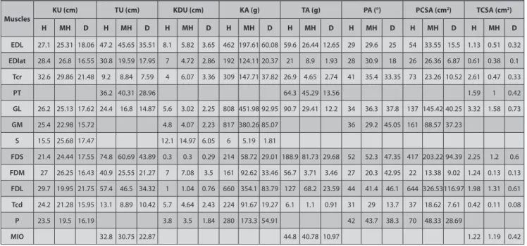
Table 5 illustrates comparatively the results obtained
Table 5. Some characteristics of hind leg distal muscle of horse, H: Horse (Payne et al.[8]), MH: Malakan Horse (Study), D: Donkey (Demiraslan, [26])
Tablo 5. At, Malakan Atı, ve merkep arka bacağı distal kaslarının bazı özellikleri, H: At (Payne et al.[8]), MH: Malakan Atı (Çalışma), D: Merkep (Demiraslan, [26])
Muscles KU (cm) TU (cm) KDU (cm) KA (g) TA (g) PA (°) PCSA (cm
2) TCSA (cm2) H MH D H MH D H MH D H MH D H MH D H MH D H MH D H MH D EDL 27.1 25.31 18.06 47.2 45.65 35.51 8.1 5.82 3.65 462 197.61 60.08 59.6 26.44 12.65 29 29.6 25 54 33.55 15.5 1.13 0.51 0.32 EDlat 28.4 26.8 16.55 30.8 19.59 17.95 7 4.72 2.86 192 124.11 20.37 21 8.9 1.93 28 30.9 18 26 26.36 6.87 0.61 0.38 0.1 Tcr 32.6 29.86 21.48 9.2 8.84 7.59 4 6.07 3.36 309 147.71 37.82 26.9 4.65 2.74 41 35.4 33.35 73 23.26 10.52 2.61 0.47 0.33 PT 36.2 40.31 28.96 64.3 45.29 13.56 1.59 1 0.42 GL 26.2 25.13 17.62 24.4 16.8 14.87 5.6 3.02 2.25 808 451.98 92.95 90.7 29.41 12.2 34 36.3 37.8 137 145.42 40.25 3.32 1.58 0.73 GM 25.4 22.98 15.72 4.8 4.07 2.23 817 380.26 85.07 36 29.2 45.05 161 88.57 37.23 S 15.5 25.68 17.47 12.1 14.97 6.05 6 5.19 1.81 FDS 21.4 24.44 17.55 74.8 60.69 43.89 0.3 0.3 0.29 214 58.72 29.01 188.9 81.73 29.68 52 52.3 47.35 417 203.22 94.39 2.25 1.2 0.6 FDM 27 26.25 16.43 40.9 25.55 21.27 7 7.08 3.5 161 92.62 33.46 56.7 3.71 3.46 27 20.3 42.95 22 13.38 9.02 1.24 0.13 0.13 FDL 29.7 19.95 21.75 57.4 46.5 34.32 1 1.04 0.76 660 354.1 83.79 127 68.2 23.59 44 41.4 46.1 644 326.53116.97 1.98 1.31 0.61 Tcd 24.2 21.28 15.95 13.1 8.89 10.42 5.7 4.64 2.43 224 91.67 19.27 6.1 1.1 0.91 31 29 13.7 37 18.62 7.61 0.42 0.11 0.08 P 23.5 19.5 16.19 3.8 3.5 1.84 280 173.3 54.91 42 43.7 38.3 70 48.33 28.69 MIO 32.8 30.75 22.87 44.8 40.78 10.97 1.22 1.19 0.42
from the hind leg distal muscles for the horses from the study of Payne et al.[8], for the donkeys from the study of
Demiraslan [15] and for Malakan Horses from this study.
When the muscles are generally observed according to the table, it is seen that high muscle structural and functional parameters are similar in horses and donkeys.
In the comparison performed between the genders; while it was specified that the difference observed between some values of the muscles in the front and hind legs had a statistical significance (P<0.05), the differences in the comparison performed according to the direction (right-left) had no statistically significance. Abe et al.[12]
reported that gender is effective upon properties of the muscle structure. While the data obtained from our study support literature in terms of the differences between the genders, it also reveals that gender should not be neglected during the preparation of the anatomical or biomechanical muscle models in equidae.
In parallel to the results of the studies previously performed in the species of equidae family, it was determined for the Malakan horse that the regional muscles evaluated in the study had generally long tendons, pennate, and short muscle bundles. This situation is accepted as an indicator for the leg distal that acts as an elastic energy storage. Especially the extent of the effect of the differences between the species on structural and functional properties of the muscles are better understood at the end of the study. Thus, the thesis specifying that the differences between the species can be effective in developing a leg muscle model is supported by this study.
REFERENCES
1. Güleç E: Türk At Irkları. Anadolu At Irklarını Yaşatma ve Geliştirme
Derneği, Ankara, 1995.
2. Güleç E: Ardahan Atı (Malakan Atı). Anadolu At IrklarınıYaşatma ve
Geliştirme Derneği, Ankara, 1997.
3. Hendricks BL: International Encyclopedia of Horse Breeds. University
of Oklahoma Press, Oklahoma State, 1995.
4. Sacks RD, Roy RR: Architecture of the hind limb of muscle of cats:
Functional significance. J Morphol, 173, 185-195, 1982. DOI: 10.1002/ jmor.1051730206
5. Lieber RL, Shoemaker SD: Muscle, joint, and tendon contributions to
the torque profile of frog hip joint. Am J Physiol, 263, 586-590, 1992.
6. Lieber RL: Skeletal muscle anatomy. In, Lieber RL (Ed): Skeletal Muscle
Structure and Function. 1-43, Williams and Wilkins, Baltimore, 1992.
7. Brown NA, Kawcak CE, McIlwraith CW, Pandy M: Architectural
properties of distal forelimb muscles in horses, Equus caballus. J Morph, 258, 106-114, 2003. DOI: 10.1002/jmor.10113
8. Payne RC, Hutchinson JR, Robilliard JJ, Smith NC, Wilson AM:
Functional specialisation of pelvic limb anatomy in horses (Equus caballus). J Anat, 206, 557-574, 2005. DOI: 10.1111/j.1469-7580.2005.00420.x
9. Abe T, Kumagai K, Brechue WF: Fascicle length of leg muscles is
greater in sprinters than distance runners. Med Sci Sports Exerc, 32, 1125-1129, 2000.
10. Blazevich AJ, Gill ND, Zhou S: Intra- and intermuscular variation in
human quadriceps femor is architecture assessed in vivo. J Anat, 209, 289-310, 2006. DOI: 10.1111/j.1469-7580.2006.00619.x
11. Blazevich AJ, Gill ND, Bronks R, Newton RU: Training specific muscle
architecture adaptation after 5-wk training in athletes. Med Sci Sports Exerc, 35, 2013-2022, 2003. DOI: 10.1249/01.MSS.0000099092.83611.20
12. Abe T, Brechue WF, Fujita S, Brown B: Gender differences in FFM
accumulation and architectural characteristics of muscle. Med Sci Sports Exerc, 30, 1066-1070, 1998. DOI: 10.1097/00005768-199807000-00007
13. Burkholder TJ, Fingado B, Baron S, Lieber RL: Relationship between
muscle fiber types and sizes and muscle architectural properties in the mouse hind limb. J Morph, 22, 177-190, 1994.
14. Fayed MH: Architecture and functional specifications of the muscles
of the antibrachium and manus regions of the African Ass (Equus asinus). Adv Biol Res, 4 (1): 45-64, 2010.
15. Demiraslan Y, Özcan S: Merkepte regio cruris ve pedis’i çevreleyen
kasların yapısal ve fonksiyonel özellikleri. Eurasian J Vet Sci, 31, 1-7, 2015. DOI: 10.15312/EurasianJVetSci.201518470
16. Uçar Y: Yerli merkep’in (Equus asinus) gövde ve ard bacak iskelet
kasları üzerinde makro-anatomik araştırmalar. Ank Üniv Vet Fak Derg, 27, 103-124, 1980.
17. Biewener AA: Biomechanical consequences of scaling. J Exp Biol, 208,
1665-1676, 2005. DOI: 10.1242/ jeb.01520
18. Pollock CM, Shadwick RE: Allometry of muscle, tendon, and elastic
energy storage capacity in mammals. Am J Physiol, 266, 1022-1031, 1994.
19. Bullimore S, Burn J: Scaling of elastic energy storage in mammalian
limb tendons: Do small mammals really lose out? Biol Lett, 1, 57-62, 2005. DOI: 10.1098/rsbl.2004.0243
20. Payne RC, Crompton RH, Isler K,Savage R, Vereecke EE, Gunther MM, Thorpe SKS, Aount KD: Morphological analysis of the hind
limb in apes and humans. II. Moment arms. J Anat, 208, 725-742, 2006. DOI: 10.1111/j.1469-7580.2006.00564.x
21. International Committee on Veterinary Gross Anatomical Nomenclature: General Assembly of the World Association of Veterinary
Anatomists. Nomina Anatomica Veterinaria. 5th ed., Gent, 2012.
22. Alexander RM, Jayes AS, Maloiy GM, Wathuta EM: Allometry of the
leg muscles of mammals. J Zool, 194, 539-552, 1981. DOI: 10.1111/j.1469-7998.1981.tb04600.x
23. Pasi BM, Carrier DR: Functional trade-offs in the limb muscles
of dogs selected for running vs. fighting. J Evol Biol, 16, 324-332, 2003. DOI: 10.1046/j.1420-9101.2003.00512.x
24. Smith NC, Wilson AM, Jespers K, Payne RC: Muscle architecture and
functional anatomy of the pelvic limb of the ostrich (Struthio camelus). J Anat, 209, 765-780, 2006. DOI: 10.1111/j.1469-7580.2006.00658.x
25. Williams SB, Payne RC, Wilson AM: Functional specialization of
the pelvic limb of the hare (Lepus europeus). J Anat, 210, 472-490, 2007. DOI: 10.1111/j.1469-7580.2007.00703.x
26. Demiraslan Y: Yerli merkeplerde (Equus asinus) crus ve regio pedis’i
çevreleyen kasların yapısal ve fonksiyonel özellikleri. Doktora Tezi, Kafkas Üniv. Sağlık. Bil. Enst., 2013.