The changes in biomarkers for necrotising enterocolitis
in premature calves with respiratory distress syndrome
Ramazan Yildiz
1*, Mahmut Ok
2,
Merve Ider
2, Aybars Akar
1, Amir Naseri
2,
Erman Koral
21Department of Internal Medicine, Faculty of Veterinary Medicine, Mehmet Akif University, Burdur, Turkey
2Department of Internal Medicine, Faculty of Veterinary Medicine, Selcuk University, Konya, Turkey
*Corresponding author: [email protected]
Citation: Yildiz R, Ok M, Ider M, Akar A, Naseri A, Koral E (2019): The changes in biomarkers for necrotising
entero-colitis in premature calves with respiratory distress syndrome. Veterinarni Medicina 64, 440–447.
Abstract: The aim of this study was to determine the changes of the biomarkers used for the diagnosis of necrotising
enterocolitis of human neonates in premature calves with respiratory distress syndrome (RDS). Novel biomarkers including the intestinal fatty acid binding protein (IFABP), the liver-type FABP (LFABP), trefoil factor-3 (TFF3), actin gamma 2 smooth muscle (ACTG2), and Claudin-3 were investigated using bovine specific ELISA kits. Thirty premature calves with respiratory distress syndrome (the RDS group), seven premature calves without RDS (the non-RDS group), and seven healthy calves (control) were included in the study. Blood samples from all the groups were taken at 0 and 48 h for the blood gas and biomarker measurement. It was determined that IFABP (P < 0.05), LFABP (P < 0.05), TFF3 (P < 0.05), ACTG2 (P < 0.05), and Claudin-3 (P < 0.05) in the control group were significantly higher than those in the RDS and non-RDS groups at 0 hour. The LFABP and Claudin-3 con-centrations in the control group were statistically higher (P < 0.05) than those in the RDS and non-RDS groups at 48 h, whereas the ACTG2 and TFF3 contents were significantly higher (P < 0.05) than the non-RDS group. A significant increase in the contents of IFABP (P ≤ 0.01), LFABP (P < 0.05), TFF3 (P < 0.05), ACTG2 (P < 0.05) at 48 h was detected in the RDS group only. In conclusion, the changes in the biomarkers support the suspicion of intestinal damage such as necrotising enterocolitis (NEC) after enteral feeding in premature calves with RDS. Intestinal damage biomarkers such as IFABP, LFABP, TFF3, and ACTG2 may be useful in the diagnosis of intestinal damage in premature calves. These results also indicate that the plasma concentrations of the intestinal biomarkers change in new born calves with their gestational age.
Keywords: actin; claudin; FABP; hypoxia; trefoil factor
In prematurity, because of a surfactant deficiency, the sufficient gas exchange in the lungs is disrupted and it leads one to develop a respiratory distress syn-drome (RDS). RDS and the associated complications are the major causes of death in premature calves. One of these complications is necrotising
enterocol-itis (NEC), which is related to immature intestinal interaction with aggressive enteral feeding, and in-testinal epithelial damage due to hypoxia or altered intestinal blood flow. Some possible consequences of intestinal immaturity are an immature physiochemi-cal luminal environment, decreased gut motility, ab-Supported by the Selcuk University, Scientific Research Project Office, Konya (Project No. 16401150).
normal microbiota, immature barrier function and, imbalance in the inflammatory responses to vari-ous agents (Neu 2005; Patole 2007; Neu et al. 2008; Gephart et al. 2012). Novel biomarkers such as the intestinal fatty acid binding protein (IFABP), the liv-er-type FABP (LFABP), trefoil factor-3 (TFF3), actin gamma 2, smooth muscle (ACTG2), and Claudin-3 are now being used to determine intestinal damage in infants with NEC (Kanda et al. 2011; Benkoe et al. 2014; Srivastava et al. 2015; Hong et al. 2017).
The fatty acid binding protein (FABP) family con-tains a number of cytoplasmic small size proteins with high organ specificity. The IFABP expression is reported to be the highest in the jejunum, where-as the LFABP expression is the highest in the colon of healthy cattle (Hayashi et al. 2013).The levels of LFABP and IFABP have been reported to be signifi-cantly higher in necrotising enterocolitis in infants (Ng et al. 2013). TFF3, secreted by the goblet cells of the small and large intestine, protects the in-testinal mucosa against damage. Srivastava et al. (2015) have shown that the elevated levels of TFF3 are associated with a gastrointestinal tract injury, such as those in an inflammatory bowel syndrome. Smooth muscle actin-α (SMA-α) (ACTG2) arises from smooth muscle of the intestine (Fatigati and Murphy 1984; Hong et al. 2017) and its contents in the blood may increase when the smooth muscle layer of the gut is damaged, such as in severe NEC (Evennett et al. 2014). The loss of paracellular bar-riers and tight junctions is a sign of compromised intestinal integrity. These tight junctions consist of a large complex of intra- and extracellular proteins, including Claudin-3, an important sealing protein (Rahner et al. 2001; Turksen and Troy 2004; Thuijls et al. 2010), which disappears rapidly from the tight junctions following a haemorrhagic shock and in-flammatory bowel disease (Zeissig et al. 2007). In previous human infant studies, IFABP, LFABP, TFF3, ACTG2, and Claudin-3 have been reported as intestine-specific markers (Thuijls et al. 2010; Ng et al. 2013; Evennett et al. 2014).
We hypothesised that necrotising enterocolitis, which is commonly observed in human preterm infants, may develop in premature calves with RDS and can be detected by intestinal-specific biomark-ers used in cases of NEC.
The aim of this study was to determine the pres-ence of an intestinal injury in premature calves using serum biomarkers that define the loss of integrity in the intestinal wall and inflammation. Furthermore,
the usefulness of the serum IFABP, LFABP, TFF3, ACTG2, and Claudin-3 in detecting enterocolitis in premature calves with RDS and screening the presence of NEC gastrointestinal symptoms like in human preterm infants are evaluated.
MATERIAL AND METHODS
Animals. The research procedure was carried out by the approval of the Institutional Ethics Com-mittee of the Faculty of Veterinary Medicine, Selcuk University (No. 2015/16). Thirty sponta-neous premature calves with RDS (group RDS) and 7 premature calves without RDS (non-RDS group) that were admitted to the Large Animal Hospital of the Faculty of Veterinary Medicine of Selcuk University for treatment were included in the study. Seven healthy calves (control) that had completed the gestational age were supplied from Selcuk University Veterinary Faculty Research and Application Farm. The gestational age range for the premature and healthy calves (according to the artificial insemination records) was 240–250 days and > 280 days, respectively. All the groups includ-ed Holsteins breinclud-ed calves.
Clinical examination. The criteria for prematu-rity, such as short silky hair coat, low body weight, soft hooves, the incomplete eruption of incisors, weakness, lateral recumbency, and weak or no suckling reflex were recorded (Ok et al. 2000; Guzelbektes et al. 2012; Yildiz and Ok 2017). The respiratory rate, body temperature, and peripheral capillary oxygen saturation (SpO2) of the calves were instantly monitored using a Compact 7 sys-tem(Medical Econet, Oberhausen, Germany) and the parameters were recorded at 0 and 48 hour.
Hypoxemia (arterial partial oxygen (PaO2) less than 60 mmHg), hypercapnia (arterial partial carbon dioxide (PaCO2) greater than 45 mmHg), tachypnoea (respiratory rate equal or greater than 45 breaths/min), and expiration accentuated by an abdominal lift and expiratory grunt were used as the criteria for grouping into the RDS group (Bleul 2009; Yildiz and Ok 2017). The premature calves which did not meet the RDS criteria were in-cluded in the non-RSD group. The healthy termed calves were evaluated during the first hours after birth and before the first feeding.
Standard treatment protocol for premature calves. The premature calves in all the groups
were administered standard support and an oxy-gen therapy protocol (Yildiz and Ok 2017). The premature calves received fresh or frozen colos-trum (10% of the body weight, divided into four feedings) during the first 48 h from the birth. The calves with a weak suckling reflex received colos-trum via a stomach tube instead of a nipple bottle (Yildiz and Ok 2017).
Sample collection and blood analyses. The blood samples were taken from all the groups at 0 and 48 hour. The arterial blood samples were taken anaerobically using a plastic insulin syringe containing sodium heparin from the arteria
au-ricularis (Yildiz and Ok 2017). The arterial blood
gases including the pH, the partial pressure of car-bon dioxide (PaCO2), the partial pressure of oxy-gen (PaO2), and the oxygen saturation (SaO2) was measured using an automated blood gas analyser (GEM Premier 3000, Inst. Laboratory, Lexington, USA) within 5 min after collection. Ten millilitres of blood were collected by jugular venepuncture and divided into two fractions. The first fraction was placed in tubes containing potassium ethylene diamine tetra-acetic acid (EDTA) for a complete blood count (CBC) using an automated cell coun-ter (MS4e, Melet Schloesing Laboratories, Osny, France) and was measured within 5 min; the sec-ond was placed in tubes without an anticoagulant for the measurement of the intestinal biomark-ers, allowed to clot and centrifuged at 2500 g for 15 min at 4 °C. The serum samples were stored at −80 °C until further analysis and defrosted im-mediately before the batch analysis. The sample collection phase of this study was completed within 12 months.
The serum concentrations of the biomarkers (IFABP, LFABP, TFF3, Claudin-3, and ACTG2) were determined using commercially available bovine specific enzyme-linked immunosorbent assay kits(ELISA kits, MyBioSource, San Diego, USA) following the manufacturer’s instructions and the absorbance was measured with a Universal Microplate Reader (ELX800, BIOTEK®, USA). The
manufacturer reported the intra-assay and inter-assay coefficients of the variations (CV) and the minimum detectable concentrations (MDC) for the biomarkers are presented in Table 1.
Statistical analysis. The mean values and stand-ard error of the mean (mean ± SEM) was calculated for each parameter. The Kolmogorov-Smirnov test was used to evaluate the data for normality. All the
parameters were normally distributed. A one-way analysis of variance (ANOVA) (with post-hoc Duncan’s test) was used for comparing the differ-ence between the groups. A paired t-test was used for comparing the difference within the groups. The P-value of < 0.05 was accepted as the level of statistical significance. All statistical analyses were performed using SPSS software for Windows, version 21.0.
RESULTS
Clinical findings
In the RDS group, twenty of 30 premature calves and all the non-RDS calves survived. In the non-surviving calves, hypothermia was recorded throughout the treatment process and these calves developed an abdominal distention after the oral feeding, they could not stand in the sternal position without support and had a prominent depressive appearance. All the recorded clinical indicators are presented in Table 2.
Blood gas and complete blood count
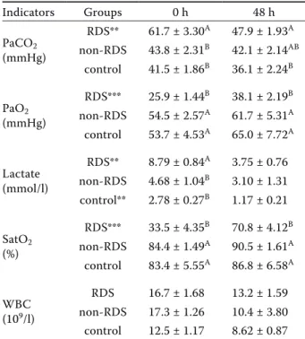
The measurements of the white blood cell (WBC) counts and arterial blood gas variables of the groups are presented in Table 3. The arterial blood gas analysis showed that, at the time of admission, the PaCO2 of the RDS group was significantly higher
Table 1. The manufacturer reported intra-assay and inter-assay coefficients of variations and minimum detectable concentrations for the biomarkers
Biomarkers Intra-assay CV Inter-assay CV MDC
IFABP ≤ 8% ≤ 12% 0.05 ng/ml
LFABP ≤ 8% ≤ 12% 0.06 ng/ml
TFF3 5.9% ≤ 7.1% < 0.26 ng/ml
ACTG2 ≤ 15% ≤ 15% 0.1 ng/ml
Claudin-3 ≤ 15% ≤ 15% 0.1 ng/ml ACTG2 = Actin Gamma 2, smooth muscle; CV = coef-ficients of variations; IFABP = intestinal fatty acid bind-ing protein; LFABP = liver-type fatty acid bindbind-ing protein; MDC = minimum detectable concentrations; TFF3 = trefoil factor-3
(P < 0.05) than those in the non-RDS and control groups and remained significantly higher (P < 0.05) than in the control group at 48 hour. Also, it was apparent that the PaCO2 levels in the RDS group at 48 h in comparison to the time of admission were significantly higher (P < 0.05) than those in the con-trol and non-RDS groups, but this difference was not statistically significant. When the within-group PaCO2 levels were evaluated at 0 and 48 h, the val-ues of PaCO2 decreased (P ≤ 0.01) in the RDS group at 48 h only (Table 3). The PaO2 concentration in the RDS group was lower (P < 0.05) than those in the non-RDS and control groups at 0 and 48 hour. A significant increase in PaO2 at 48 h (P ≤ 0.001) was determined in the RDS group. When the blood lactate levels of all the groups were compared, the blood lactate levels in the RDS group were higher at 0 hour. However, compared to the 0 h sampling time, a statistically significant decrease in the blood lactate (P ≤ 0.01) was observed in the RDS and control groups at 48 h (Table 3). The blood SatO2 contents in the RDS group were found to be signifi-cantly lower (P < 0.05) than those in the non-RDS and control groups at 0 and 48 hour. A significant within-group increase in SatO2 at 48 h (P ≤ 0.01) was determined for the RDS group.
Intestinal damage biomarkers
The findings of the intestinal damage biomarkers are presented in Table 4. It was determined that, at the time of admission, (0 hour) IFABP, LFABP, TFF3, ACTG2, and Claudin-3 levels in the control group were significantly higher (P < 0.05) than in the RDS and non-RDS groups. There was not any significant differentiation between the RDS and non-RDS groups at the time of admission. The LFABP and Claudin-3 levels of the control group were significantly higher (P < 0.05) than the RDS and non-RDS groups, whereas the ACTG2 and TFF3 levels of the control group were significant-ly higher (P < 0.05) than the non-RDS group at 48 hour. The results of the intestinal damage bio-markers within the groups shown that a significant increase in the levels of IFABP (P ≤ 0.01), LFABP (P < 0.05), TFF3 (P < 0.05), and ACTG2 (P < 0.05) from time of admission to 48 h were detected in the RDS group only. The RDS group also showed higher Claudin-3 levels at 48 h, but without any statistical significance.
Table 3. Changes in the arterial blood gas indicators and the WBC value in the three groups (Mean ± SEM)
Indicators Groups 0 h 48 h PaCO2 (mmHg) RDS** 61.7 ± 3.30A 47.9 ± 1.93A non-RDS 43.8 ± 2.31B 42.1 ± 2.14AB control 41.5 ± 1.86B 36.1 ± 2.24B PaO2 (mmHg) RDS*** 25.9 ± 1.44B 38.1 ± 2.19B non-RDS 54.5 ± 2.57A 61.7 ± 5.31A control 53.7 ± 4.53A 65.0 ± 7.72A Lactate (mmol/l) RDS** 8.79 ± 0.84A 3.75 ± 0.76 non-RDS 4.68 ± 1.04B 3.10 ± 1.31 control** 2.78 ± 0.27B 1.17 ± 0.21 SatO2 (%) RDS*** 33.5 ± 4.35B 70.8 ± 4.12B non-RDS 84.4 ± 1.49A 90.5 ± 1.61A control 83.4 ± 5.55A 86.8 ± 6.58A WBC (109/l) RDS 16.7 ± 1.68 13.2 ± 1.59 non-RDS 17.3 ± 1.26 10.4 ± 3.80 control 12.5 ± 1.17 8.62 ± 0.87 PaCO2 = partial pressure of the arterial carbon dioxide; PaO2 = partial pressure of the arterial oxygen; SatO2 = oxy-gen saturation; SEM = standard error of the mean; RDS = respiratory distress syndrome; WBC = white blood cell The different letters (A,B) in the columns are statistically significant (P < 0.05)
The within-group differences between 0 and 48 h are ex-pressed as *P ≤ 0.05, **P ≤ 0.01, ***P ≤ 0.001
Table 2. Changes in the clinical indicators in the three groups (Mean ± SEM)
Indicator Groups 0 h 48 h Temperature (°C) RDS 35.7 ± 0.36B 37.7 ± 0.19B non-RDS 37.2 ± 0.54AB 38.1 ± 0.14AB control 38.4 ± 0.08A 38.5 ± 0.07A Respiratory rate breaths/min RDS 60.9 ± 3.16A 48.5 ± 4.04 non-RDS 46.6 ± 7.10AB 41.6 ± 5.01 control 37.0 ± 1.19B 36.7 ± 1.10 SpO2 (%) RDS 77.2 ± 1.73B 85.2 ± 1.65B non-RDS 91.7 ± 1.55A 94.8 ± 0.82A control 95.5 ± 1.13A 97.2 ± 0.68A SEM = standard error of the mean; SpO2 = peripheral capil-lary oxygen saturation
The different letters (A,B) in the column are statistically significant (P < 0.05)
DISCUSSION
To our knowledge, this is the first explanatory study about the changes in the diagnostic biomark-ers for necrotising enterocolitis in premature calves with RDS. We believe that these findings may pro-vide basic knowledge for further studies about NEC in the veterinary field.
It has been reported that the ideal indicators to evaluate the hypoxia caused by lung function insufficiency are the arterial PaO2, SatO2, SpO2, lactate, PaCO2 and the respiratory rate (Yildiz and Ok 2017) in premature calves with RDS. In the RDS group, the arterial PaO2 (< mean 38 mmHg) was lower than the critical value (60 mmHg) (Pickel et al. 1989; Bleul et al. 2008; Yildiz and Ok 2017), while the arterial SatO2 (mean 70.8%) and SpO2 (mean 85%) were below the desired level (88–95%)
(Yildiz and Ok 2017); the arterial PaCO2 value was above the critical value (> 45 mmHg) as reported in the previous studies (Bleul et al. 2008; Aydogdu et al. 2016; Yildiz and Ok 2017; Yildiz et al. 2017). Furthermore, the lactate level (mean 3.75 mmol/l) was close to critical value (4 mmol/l) (Yildiz et al. 2017) and the respiratory rate was above the criti-cal value (> 45 breaths/min) after the treatment (48 h) in the RDS group. These results suggested that persistent hypoxia in the RDS group might cause a decrease in the intestinal oxygenation and result in regional ischemia. Yildiz and Ok (2017) reported that premature calves with hypoxia fail to pass the meconium combined with a progressive abdominal distention after feeding. In addition, the autopsy findings of non-surviving premature calves revealed bleeding and oedema in both the abomasum and intestines (Yildiz and Ok 2017). The previous studies (Bleul et al. 2008; Guzelbektes et al. 2012; Canikli Engin et al. 2017; Yildiz and Ok 2017) have also reported that an abnormal pulmonary function is not the only critical factor in the premature calves. Therefore, the inflamma-tion of the mucosa of the forestomach and aboma-sum should also be taken into consideration when treating these animals. Enteral feeding plays a key role in the pathogenesis of NEC. There are many physical barriers against bacteria in the mature in-testine, such as gastric acid, proteolytic enzymes, peristalsis and intestinal mucus; tight junctions and cell surface glycoconjugates provide more advan-tages for the adherence and translocation for the pathogenic organisms (Berseth 1994; Claud 2009). Earlier studies have identified that the introduc-tion of enteral feeding causes a disrupintroduc-tion to the mucosal integrity, blood flow, and motility, and it has a pivotal role in the development of NEC (Pietz et al. 2007; Gregory et al. 2011). Yildiz and Ok (2017) observed that premature calves with hy-poxia develop an abdominal distension after the oral feeding. Guzelbektes et al. (2012) reported re-laxation in the lower oesophageal sphincter, sug-gesting a gastroesophageal reflux. The reason for the abomasal fluid escaping into the oesophagus may be related to an abdominal distension and a decrease in the intestinal motility. Canikli Engin et al. (2017) reported that premature calves have abomasal hypomotility and the administration of erythromycin before the oral feeding may be useful for emptying the abomasal contents. Normal mo-tility is crucial for the prevention of the stasis and
Table 4. Changes in the intestinal damage biomarkers in the three groups (Mean ± SEM)
Indicators Groups 0 h 48 h IFABP (ng/ml) RDS** 0.42 ± 0.02B 0.61 ± 0.06 non-RDS 0.38 ± 0.01B 0.43 ± 0.03 control 0.71 ± 0.10A 0.64 ± 0.08 LFABP (ng/ml) RDS* 1.22 ± 0.03B 1.47 ± 0.10B non-RDS 1.17 ± 0.05B 1.23 ± 0.05B control 1.59 ± 0.06A 1.85 ± 0.09A TFF3 (ng/ml) RDS* 0.90 ± 0.10B 1.96 ± 0.40AB non-RDS 0.63 ± 0.11B 0.77 ± 0.13B control 2.21 ± 0.38A 2.4 ± 0.43A ACTG2 (ng/ml) RDS* 1.34 ± 0.05B 1.90 ± 0.20AB non-RDS 1.33 ± 0.07B 1.35 ± 0.06B control 2.57 ± 0.62A 2.62 ± 0.49A Claudin-3 (ng/ml) RDS 6.14 ± 0.15B 6.64 ± 0.51B non-RDS 5.82 ± 0.19B 5.43 ± 0.18B control 9.40 ± 2.99A 10.13 ± 1.77A ACTG2 = Actin Gamma 2, smooth muscle; IFABP = intes-tinal fatty acid binding protein; LFABP = liver-type fatty acid binding protein; RDS = respiratory distress syndrome; SEM = standard error of the mean; TFF3 = trefoil factor-3 The different letters (A,B) in the columns are statistically significant (P < 0.05)
The within-group differences between 0 and 48 h are ex-pressed as *P ≤ 0.05, **P ≤ 0.01, ***P ≤ 0.001
subsequent diseases in the gastrointestinal tract and for the digestion and absorption of nutrients for the development of healthy organs and systems (Gregory et al. 2011). An abnormal peristaltic ac-tivity in preterm infants may increase the bacte-rial adhesion and allow bactebacte-rial overgrowth that increases the endotoxin exposure and risk of NEC (Berseth 1994; Claud 2009).
Kanda et al. (2011) determined a cut-off value (3.1 ng/ml) of IFABP in the diagnosis of an intesti-nal ischaemia, which is a reliable and non-invasive method for evaluating the risk of ischemia in the small intestine of the patients with acute abdominal complaints. It has been stated that the evaluation of the concentrations of IFABP in the abdominal fluids and plasma may be useful for predicting sur-vival in horses and the need for abdominal surgery in horses with colic (Nieto et al. 2005). Niewold et al. (2004) reported that the plasma IFABP levels were significantly increased in the intestinal dam-age of pigs and it is a reliable parameter that could be used to detect intestinal damage and monitor the health of the intestines. In human infants with necrotising enterocolitis, the LFABP concentration was found to be high in the initial phase of the disease, whereas the IFABP level was high in the advanced phase (Guthmann et al. 2002). Ng et al. (2013) reported 45% mortality in necrotising en-terocolitis in infants and that the LFABP, IFABP and TFF3 levels of the infants were significantly higher than the surviving infants. It has been determined that the level of TFF3 increases significantly dur-ing ulcerative colitis (Srivastava et al. 2015). IFABP and Claudin-3 significantly increased in new-borns suspected of NEC. They promise to be new non-invasive markers for the early detection of NEC (Thuijls et al. 2010).Evennett et al. (2014) has re-ported that SMA is a clinically useful marker of transmural intestinal necrosis. Plasma SMA has been reported to be able to provide information about the need for surgical intervention in neo-nates with elevated plasma IFABP concentrations, but further research is needed to examine its use in conditions that may cause intestinal necrosis (Evennett et al. 2014).
In this study, we assessed certain intestinal dam-age biomarkers in healthy mature calves and pre-mature calves with and without RDS. The contents of IFABP, LFABP, TFF3, ACTG2, and Claudin-3 in the healthy calf group were significantly (P < 0.05) higher than both the premature calves with and
without RDS (Table 4). In a study, investigating IFABP and brain-caspase-3 responses in preterm and term mice in which an experimental asphyxi-ated model was established, it was reported that the expression of these markers in the term mice was higher than the preterm ones. It was indicated that the expression of the markers by the gestational age may be related to the different levels of the expres-sion (Figueira et al. 2016).It has been reported that a significant portion of TFF3 in rats is expressed late in term pregnancy (Lin et al. 1999). It was also reported that the expression of TFF3 was relatively deficient in immature rats and an insufficient TFF3 expression may play a role in the development of NEC in premature infants (Lin et al. 1999). The results from the present study indicate that the plasma level of intestinal markers changes in the new-born calves with the gestational age. However, the intestinal tissue expressions of these markers still need to be investigated in calves.
In the present study, a significant elevation was found in the contents of IFABP, LFABP, TFF3, and ACTG2, while there was a non-significant increase in the level of Claudin-3 in premature calves with RDS only at 48 hour. The absence of an increase in the intestinal biomarkers in non-the hypoxic premature and control calves at 48 h suggests that this may be a condition like NEC in human infants triggered by intestinal hypoxia after oral feeding in premature calves with RDS. As previous studies report (Berseth 1994; Kleigman 2003; Caplan et al. 2005; Claud 2009; Henry and Moss 2009; Gregory et al. 2011), an immature intestinal system, the lack of a protective tight junction protein expression, hypomotility and hypoxia-induced microcircula-tion degradamicrocircula-tion in premature calves with RDS trigger pathogenic bacterial growth and abdominal distention after enteral feeding and cause further intestinal damage.
In preterm calves, it was seen that intestinal dam-age may occur after enteral feeding in cases when RDS persists. Intestinal damage biomarkers such as IFABP, LFABP, TFF3, and ACTG2 may be useful in the diagnosis of intestinal damage in premature calves. The results indicate that the serum con-tents of the intestinal biomarkers change in the new born calves with their gestational age. When the clinical signs and intestinal damage markers were evaluated in premature calves with RDS, a disease similar to necrotising enterocolitis in human in-fants can be identified. However, there are several
limitations of this study. A histopathology was not performed in order to correlate the amount of intestinal damage with the contents of IFABP, LFABP, TFF3, ACTG2, and Claudin-3 in order to gauge how well they indicate in vivo necrotising enterocolitis. No samples could be taken from the non-surviving calves because their death times could not be foreseen. Therefore, the significance of the specified biomarkers as mortality indicators could not be demonstrated. In the premature calves with RDS, a low survival rate is predicted to be associated with complications, such as in the gas-trointestinal tract. It has been observed that oral nutrition management should be considered in pre-mature calves. There is need for further research to improve the treatments for the increase in the intestinal microcirculation, motility and reduction of the intestinal tissue damage due to pathogenic bacterial colonisation.
REFERENCES
Aydogdu U, Yildiz R, Guzelbektes H, Coskun A, Sen I (2016): Cardiac biomarkers in premature calves with respiratory distress syndrome. Acta Veterinaria Hungarica 64, 38–46. Benkoe TM, Mechtler TP, Weninger M, Pones M, Rebhandl
W, Kasper DC (2014): Serum levels of interleukin-8 and gut-associated biomarkers in diagnosing necrotizing en-terocolitis in preterm infants. Journal of Pediatric Surgery 49, 1446–1451.
Berseth CL (1994): Gut motility and the pathogenesis of ne-crotizing enterocolitis. Clinics in Perinatology 21, 263–270. Bleul UT, Bircher BM, Kahn WK (2008): Effect of intrana-sal oxygen administration on blood gas variables and outcome in neonatal calves with respiratory distress syn-drome: 20 cases (2004–2006). Journal of the American Veterinary Medical Association 233, 289–293.
Bleul U (2009): Respiratory distress syndrome in calves. Veterinary Clinics of North America: Food Animal Prac-tice 25, 179–193.
Canikli Engin S, Sevinc M, Guzelbektes H (2017): The ef-fects on abomasal emptying rate of erythromycin and bethanechol in healthy, premature and diarrheic calves. Eurasian Journal of Veterinary Sciences 33, 228–234. Caplan MS, Simon D, Jilling T (2005): The role of PAF, TLR,
and the inflammatory response in neonatal necrotizing enterocolitis. Seminars in Pediatric Surgery 14, 145–151. Claud EC (2009): Neonatal necrotizing enterocolitis – inflam-mation and intestinal immaturity. Anti-Inflammatory & Anti-Allergy Agents in Medicinal Chemistry 8, 248–259.
Evennett N, Cerigioni E, Hall NJ, Pierro A, Eaton S (2014): Smooth muscle actin as a novel serologic marker of severe intestinal damage in rat intestinal ischemia-reperfusion and human necrotising enterocolitis. The Journal of Sur-gical Research 191, 323–330.
Fatigati V, Murphy RA (1984): Actin and tropomyosin variants in smooth muscles. Dependence on tissue type. The Journal of Biological Chemistry 259, 14383–14388. Figueira RL, Goncalves FL, Simoes AL, Bernardino CA,
Lopes LS, Castro E Silva O, Sbragia L (2016): Brain cas-pase-3 and intestinal FABP responses in preterm and term rats submitted to birth asphyxia. Brazilian Journal of Medical and Biological Research 49. doi: 10.1590/1414-431X20165258.
Gephart SM, McGrath JM, Effken JA, Halpern MD (2012): Necrotizing enterocolitis risk: state of the science. Ad-vances in Neonatal Care 12, 77–87.
Gregory KE, Deforge CE, Natale KM, Phillips M, Van Mar-ter LJ (2011): Necrotizing enMar-terocolitis in the premature infant: neonatal nursing assessment, disease pathogen-esis, and clinical presentation. Advances in Neonatal Care 11, 155–164.
Guthmann F, Borchers T, Wolform C, Wustrack T, Bartho-lomaus S, Spener F (2002): Plasma concentration of in-testinal- and liver FABP in neonates suffering from necrotizing enterocolitis and in healthy preterm neo-nates. Molecular and Cellular Biochemistry 239, 227–234. Guzelbektes H, Coskun A, Ok M, Aydogdu U, Sen I (2012):
Prevalence of gastroesophageal reflux disease in prema-ture calves. Journal of Veterinary Internal Medicine 26, 1051–1055.
Hayashi H, Maruyama S, Fukuoka M, Kozakai T, Nakajima K, Onaga T, Kato S (2013): Fatty acid-binding protein expression in the gastrointestinal tract of calves and cows. Animal Science Journal 84, 35–41.
Henry MC, Moss RL (2009): Necrotizing enterocolitis. An-nual Review of Medicine 60, 111–124.
Hong J, Gilder E, Blenkiron C, Jiang Y, Evennett NJ, Petrov MS, Phillips ARJ, Windsor JA, Gillham M (2017): Nonoc-clusive mesenteric infarction after cardiac surgery: poten-tial biomarkers. Journal of Surgical Research 211, 21–29. Kanda T, Tsukahara A, Ueki K, Sakai Y, Tani T, Nishimura A, Yamazaki T, Tamiya Y, Tada T, Hirota M, Hasegawa J, Funaoka H, Fujii H, Hatakeyama K (2011): Diagnosis of ischemic small bowel disease by measurement of serum intestinal fatty acid-binding protein in patients with acute abdomen: a multicenter, observer-blinded validation study. Journal of Gastroenterology 46, 492–500. Kleigman RM (2003): The relationship of neonatal feeding
practices and the pathogenesis and prevention of ne-crotizing enterocolitis. Pediatrics 111, 671–672.
Lin J, Holzman IR, Jiang P, Babyatsky MW (1999): Expres-sion of intestinal trefoil factor in developing rat intestine. Biology of the Neonate 76, 92–97.
Neu J (2005): Neonatal necrotizing enterocolitis: an update. Acta Paediatrica 94, 100–105.
Neu J, Mshvildadze M, Mai V (2008): A roadmap for un-derstanding and preventing necrotizing enterocolitis. Current Gastroenterology Reports 10, 450–457. Ng EW, Poon TC, Lam HS, Cheung HM, Ma TP, Chan KY,
Wong RP, Leung KT, Lam MM, Li K, Ng PC (2013): Gut-associated biomarkers LFABP, IFABP, and TFF3 and LIT score for diagnosis of surgical necrotizing enterocolitis in preterm infants. Annals of Surgery 258, 1111–1118. Nieto JE, Aldridge BM, Beldomenico PM, Aleman M, Snyder
JR (2005): Characterization of equine intestinal fatty acid binding protein and its use in managing horses with colic. American Journal of Veterinary Research 66, 223–232. Niewold TA, Meinen M, Van der Meulen J (2004): Plasma
intestinal fatty acid binding protein (IFABP) concentra-tions increase following intestinal ischemia in pigs. Re-search in Veterinary Science 77, 89–91.
Ok M, Birdane FM, Sen I, Guzelbektes H (2000): Study on some blood biochemical parameters in premature calves. Indian Veterinary Journal 77, 859–861.
Patole S (2007): Prevention and treatment of necrotising enterocolitis in preterm neonates. Early Human Develop-ment 83, 635–642.
Pickel M, Zaremba W, Grunert E (1989): Comparison of arterial and venous blood gas and acid-base values in prematurely born healthy calves or calves with a late as-phyxia. Zentralblatt fuer Veterinarmedizin A 36, 653–663. Pietz J, Achanti B, Lilien L, Stepka EC, Mehta SK (2007): Prevention of necrotizing enterocolitis in preterm infants: a 20-year experience. Pediatrics 119, e164–e170.
Rahner C, Mitic LL, Anderson JM (2001): Heterogeneity in expression and subcellular localization of claudins 2, 3, 4, and 5 in the rat liver, pancreas, and gut. Gastroenterol-ogy 120, 411–422.
Srivastava S, Kedia S, Kumar S, Pratap Mouli V, Dhingra R, Sachdev V, Tiwari V, Kurrey L, Pradhan R, Ahuja V (2015): Serum human trefoil factor 3 is a biomarker for mucosal healing in ulcerative colitis patients with minimal disease activity. Journal of Crohn’s and Colitis 9, 575–579. Thuijls G, Derikx JP, Van Wijck K, Zimmermann LJ,
De-graeuwe PL, Mulder TL, Van der Zee DC, Brouwers HA, Verhoeven BH, Van Heurn LW, Kramer BW, Buurman WA, Heineman E (2010): Non-invasive markers for early diagnosis and determination of the severity of necrotiz-ing enterocolitis. Annals of Surgery 251, 1174–1180. Turksen K, Troy TC (2004): Barriers built on claudins.
Jour-nal of Cell Science 117, 2435–2447.
Yildiz R, Aydogdu U, Guzelbektes H, Coskun A, Sen I (2017): Venous lactate, pH and partial pressure of carbon dioxide levels as prognostic indicators in 110 premature calves with respiratory distress syndrome. Veterinary Record 180. doi: 10.1136/vr.103730.
Yildiz R, Ok M (2017): Clinical efficacy of combinations of nebulised fluticasone, salbutamol and furosemide on lung function in premature calves with respiratory distress syndrome. Veterinary Medicine 62, 541–552.
Zeissig S, Burgel N, Gunzel D, Richter J, Mankertz J, Wahn-schaffe U, Kroesen AJ, Zeitz M, Fromm M, Schulzke JD (2007): Changes in expression and distribution of claudin 2, 5 and 8 lead to discontinuous tight junctions and bar-rier dysfunction in active Crohn’s disease. Gut 56, 61–72. Received: March 14, 2019 Accepted after corrections: August 16, 2019