UniLOCK implants for stabilization of long bone fractures:
Experiences in ten clinical cases
Özlem ŞENGÖZ ŞİRİN, Yusuf Sinan ŞİRİN, Kürşad YİĞİTARSLAN
Mehmet Akif Ersoy University, Faculty of Veterinary Medicine, Department of Surgery, Burdur, Turkey.Summary: The purpose of this study was to report the clinical outcome after the use of an UniLOCKimplant for stabilization of long-bone fractures in cats. UniLOCKimplants were used in long bone fractures (n=11) in ten client-owned cats. Data about the signalment (breed, weight, age) and history, characteristics of the fracture, details of the surgery and perioperative complications were recorded. Salter-Harris type I and II fractures in the four cats were recorded, operated upon and contralateral stifle joint ROM values with manuel goniometer and the differences between these values were determined. We evaluated the functional and radiological outcomes on 10th, 30th, 60th, and 90th days postoperatively. The final clinical and radiographic evaluation was performed after 12 months
in all cases. The clinical outcome was excellent in nine cases, and good in one case. As a result of the postoperative trauma, stabilization of the implant was affected and an exuberant callus formation was encountered in the case 5 and the implants were removed immediately. Eight cats started using the limb immediately after waking up from anesthesia whereas initial limb use was 2-3 days after repair in the other cats. In conclusion, that the high success rate (90%) associated with a functional outcome suggests that the UniLOCK plate can be used in cats for long-bone fractures in cats.
Keywords: Cat, fracture healing, range of motion, UniLOCK plate.
Uzun kemik kırıklarının UniLOCK implantlarıyla stabilizasyonu:
10 klinik olguda deneyimler
Özet: Bu çalışmanın amacı, kedilerin uzun kemik kırıklarının stabilizasyonunda UniLOCK implantların kullanımının klinik sonuçlarını sunmaktır. UniLOCK implantları 10 sahipli kedinin 11 uzun kemik kırığında kullanıldı. Hastanın eşgali (ırk, ağırlık, yaş) ve anamnezi, kırığın yapısı, operasyon detayları ve perioperatif komplikasyonlar kaydedildi. Salter-Harris tip I ve II kırığı bulunan 4 kedinin, opere edilen ve kontralateraldeki diz eklemi hareket aralığı değerleri manuel goniometre ile ölçülerek değerler arasındaki farklar kaydedildi. Postoperatif 10., 30., 60. ve 90. günlerde fonksiyonel ve radyolojik sonuçlar değerlendirildi. Tüm olgularda klinik ve radyolojik son değerlendirme postoperatif 12. ayda yapıldı. Klinik sonuçlar 9 olguda mükemmel, 1 olguda iyi idi. Olgu 5’in postoperatif aldığı travmanın sonucu olarak implant stabilizasyonunun etkilendiği görüldü ve eksuberant kallus yapısıyla karşılaşıldı; implantlar hemen uzaklaştırıldı. Sekiz kedi anesteziden uyandıktan hemen sonra, diğerleri ise operasyondan 2-3 gün sonra ilgili ekstemitelerini kullanmaya başladı. Sonuç olarak, kedilerin uzun kemik kırıklarında fonksiyonel sonuçlarla ilişkili yüksek başarı oranı (%90) nedeniyle UniLOCK implantların kullanılabileceği sonucuna varıldı.
Anahtar sözcükler: Eklem hareket aralığı, kedi, kırık iyileşmesi, UniLOCK plak.
Introduction
Classic osteosynthesis with dynamic compression plates (DCP) and limited contact dynamic compression plates (LC-DCP) was considered as being the “gold standard” for many years. After rigid anatomic reduction of the fracture, compression between fragments was provided by a dynamic compression plate alone or by use of a lag screw and neutralisation plate (13).
The internal fixation of fractures has evolved in recent decades with a change of emphasis from mechanical to biological priorities. Locked internal fixation aims at simple and safe using, at optimizing biological conditions for soft and bone tissues. These
fixators supports biological internal fixation, that is, a type of internal fixation giving priority to biology over mechanics (5, 12, 15, 16, 20). Biological internal fixation takes advantage of indirect bone healing and aims to functionally align the fragments. Faster healing can be expected. The concept of biological internal fixation is still developing. Exposure of the bone is avoided to reduce surgical trauma and to minimize iatrogenic additional vascular damage (8). For locking plate systems establish with similar mechanical principles like mini-internal fixator (1). The contact between implant and bone is kept stable using screws which function as locked threaded bolts and prevent the plate pressing on the bone when the
screws are tightened. The key point is the screw on the design of the locking compression plates (LCP). They are locked into the threaded plate holes (5, 15, 20).
The 2.0/2.4 UniLOCK mandible plate system was designed orginally for the human maxillofacial surgery. 1.0 mm, 1.3 mm and 1.5 mm thickness are available for the 2.0 UniLOCK system. This system is reported to be the appropriate size and can be applied owing to the small animal ortopaedics (9, 17). These are reconstruction plates and there are different shapes and lengths. They can be cut and are contourable in multiple plans. The implants are made of titanium and reducing the risk of infection (7, 18). The purpose of this study was to utilize, for the first time, the UniLOCK system implants for the treatment of long-bone fractures in cats in Turkey.
Materials and Methods
Inclusion criteria: Between 2010 and 2012, cats
(n=10) with long-bone fractures (n=11) were enrolled in this study. From all patients owners were taken informed constent form. The data recorded was signalment (breed, weight, age) and history, the characteristics of the fracture, details of the surgery, plate-screw type, plate-screw density, perioperative complications, concomitant injuries, time to first use of limb after surgery as well as the functional and radiological outcomes.
Surgical technique: Cefazolin sodium 20 mg/kg
(Cefozin® IM/IV, 250mg, 1 vial, Bilim) was administered intravenously. Cats were premedicated with diazepam 0.1 mg/kg intravenously (Diazem vial® IM/IV, 10 mg/2 mL, Deva), induced with propofol 6 mg/kg intravenously (Propofol injectable emulsion® IV, 200 mg/20mL, Abbott) and anesthesia was maintained with isoflurane (Forane solution®, 100mL, Abbott) in oxygen. Epidural bupivacaine 1.5 mg/kg (Marcaine®, %0.5, 20ml vial, AstraZeneca) was administered after induction. Subcutan 4 mg/kg/day meloxicam (Maxicam®, 5mg/50ml, Sanovel) was administered after surgery.
Fracture reduction was accomplished using an open approach. Classical surgical approaches were preferred individually for each patient (3). Prior to the surgery, the appropriate size of UniLOCKTM plate going to be used
was determined by radiography. It was noted whether a single UniLOCKTM plate was applied or double plating
was used. The following parameters were recorded or measured on postoperative radiographs in patients with a single UniLOCKTM plate: thickness of the plate (2.0 mm,
or 2.4 mm plate), and plate-screw density. Additionally, the presence or absence of use of ancillary implants such as lag screws, or intramedullary pins were also noted.
Postoperatively, a soft padded bandage was applied during the first four to seven days to protect the wound. An external cast or splint was not applied.
Postoperative evaluation: The outcome was
evaluated based on the information from medical records and correctly positioned follow-up radiographs. We evaluated the functional and radiological outcomes on different days (10th, 30th, 60th, 90th) after surgery. The final
clinical and radiographic evaluation was performed after 12 months in all cases. Positioning of the implants was assessed on these radiographs. The healing process was assessed radiologically using the following criteria; loss of sharp fracture edge, loss of fracture line, cortical continuity, presence of callus, delayed union, lack of union, and osteomyelitis. Salter-Harris (SH) type I and II fractures with four cats were measured, operated upon and contralateral stifle joint range of motion (ROM) values were measured by manuel goniometer and the differences between these values were determined. Stifle joint ROM values were measured with the goniometer’s pivot point centred over the lateral femoral epicondyle. One arm of the goniometer was positioned parallel to the long axis of the tibial shaft and the other arm was aligned by line joining the lateral femoral epicondyle and greater trochanter.
The data recorded was the time of the first use of the limb after anesthesia. The lameness scale (Appendix A120)
and the pain scale (Appendix B12) was evaluated on the
10th and 30th days in all cases. Clinical outcome was
graded as excellent if no visible lameness was detected at home by the owner or during the clinical examination. Outcome was considered as good if owners reported a slight lameness after extensive exercise, but lameness could not be confirmed during the clinical examination. Outcome was moderate, when a weight-bearing lameness was detected at the clinic and the owners reported it to be consistent. Outcome was considered as poor, when a non-weight-bearing lameness.
The final clinical and radiographic evaluation was performed 12 months in all cases postoperatively, either in our hospital or at the private veterinarian. Radiographs taken by the private veterinarians were sent to our hospital for evaluation. Clinical and radiographic assessments were separately made by all researchers except for manuel goniometric measurements. These measurements were done by the same researcher.
Appendix A1. Proposol for scoring lameness in cats19
Ek A1. Kedilerde topallık skorlama önerisi19
Lameness
Grade Lameness description
Grade 1
Low grade lameness; Lameness hardly visible or no lameness is visible, but cat lifts foot when sitting (front limbs) or is unable to jump (from the history) (hindlimbs)
Grade 2 Medium-grade lameness; Lameness clearly visible but leg is used in most steps
Grade 3 High-grade lameness; Cat is only toe-touching or not weight-bearing at all
Appendix B1. Criteria used to determine simple descriptive scores for assessment of pain in cats2.
Ek B1. Kedilerde ağrının değerlendirilmesinde basit tanımlayıcı skorları belirlemek için kullanılan kriterler2.
Stimulus Cat’s response Score
Observer approaches (A)
Approaches observer
Does not approch observer but raises head
No movement
Withdrawal of head or body Avoidance; stands up to move away
1 2 3 4 5 Back is stroked (B)
Does not approach observer but raises head and arches back No movement
Withdraws head or body
Avoidance; stands up to move away 1 2 3 4 Response to foot palpation (C) Approaches observer No response Withdrawal of foot
Violent avoidance or struggle
1 2 3 4
Statistical analysis: The descriptive statistics of the
operated cats were calculated and the significance of the difference between the pain scores according to the control days was tested using the Wilcoxon SignedRank Test.
Pain scores were shown with median and 25% -75% quartile. SPSS 14.01 packet program was used for statistical analysis (Table 3).
Results
General clinical parameters and first usage of affected limb were evaluated. The sample population consisted of 5 male and 5 female cats between 7 and 24 months (mean, 14.6 months) and weighing between 1,5 and 5 kg (mean, 3.6 kg). The fractures were caused by
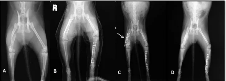
traffic accidents (n=2) or falling from high places (n=3). Other fracture causes were unknown trauma (n=5). Two cats had concomitant orthopedic injuries. Cat 5 had a contralateral distal diaphyseal comminuted tibial fracture. The tibial fracture was stabilized with UniLOCK implants (Figure 1). Cat 8 had a contralateral caput femoris fracture that was treated with excision arthroplasty. The UniLOCK implants were applied to 11 fractures in open surgery.
The fracture configurations were as: comminuted (n=3), short oblique (n=1) and transverse (n=3) and involved the proximal to mid-diaphysis in 6 cats, and the distal epiphyseal SH type I (n=3) and type II (n=1) in 4 cats.
A single 2.0 mm plate was applied to 11 fractures. One ancillary implant was used in 2 cases, including a lag screw for interfragmentary stabilization. Plate screw density was measured between 0.66 and 0.85 (mean, 0.77).
Most animals (n=8) started to use their limbs immediately after anesthesia was awake and after the effect of epidural anesthesia with bupivacaine whereas initial limb use was 2–3 days after repair in the other cats (n=2) (mean 0.5 day). SH type I and type II fractures in four cases (case 1, 2, 4 and 8) were recorded, operated upon and contralateral normal stifle joint was measured with manuel goniometer at the maximum flexion and extension positions. The flexion of contralateral normal stifle joints ranged from 20˚ to 40˚ (mean, 29˚), and extension ranged from 160˚ to 175˚ (mean, 168˚). Flexion of affected stifle joints ranged from 35˚ to 55˚ (mean, 45˚), and extension ranged from 125˚ to 165˚ (mean, 142˚). Restoration of normal joint stability was achieved in these
Figure 1. Cat 5. Preoperative craniocaudal (A), immediate postoperative craniocaudal with soft padded bandage (B) radiographic projections. Day 30 craniocaudal radiographic projection (C), the stabilization of the implant was affected and exuberant callus formation was encountered (white arrow). Therefore implants were removed from the patient immediately. Day 90 craniocaudal radiographic projection (D).
Şekil 1. Kedi 5. Preoperatif kraniokaudal (A), operasyonu takiben yumuşak destek bandajlı kraniokaudal (B) radyografik görünümler. 30. gün kraniokaudal radyografik görünüm (C), implant stabilizasyonunun etkilendiği ve eksuberant kallus formasyonu gözlenmekte (beyaz ok). Hastanın implantları hemen uzaklaştırıldı. 90. gün kraniokaudal radyografik görünüm (D).
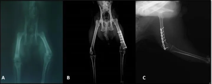
Figure 2. Cat 2. Preoperative craniocaudal (A), month 12 craniocaudal (B) and mediolateral (C) radiographic projections. Şekil 2. Kedi 2. Preoperatif kraniokaudal (A), 12. ay kraniokaudal (B) ve mediolateral (C) radyografik görünümler.
Table 1. Summary of clinical data for 10 cats with 11 long-bone fracture repaired with an UniLOCK implants. Tablo 1. UniLOCK implantlarıyla sağaltılan 11 uzun kemik kırığı bulunan 10 kediye ait klinik veri özeti.
Cases Race Age (month)
Sex Weight (kg)
History Fracture Plate-Screw density First limb use (day) Lameness Scoring 10th/30th days Functional outcome
1 tabby 11 F 3 UT Salter-Harris type I
femur 0.85 ♦ 2/1 Excellent 2 angora-persian mix 7 F 3 FHP Salter-Harris type II femur 0.85 ♦ 2/1 Excellent 3 mix 24 F 1.5 UT Proximal diaphyseal, transverse radius 0.83 ♦ 2/1 Excellent
4 tabby 7 F 3 UT Salter-Harris type I
femur
0.71 ♦ 2/1 Excellent
5 tabby 24 M 3.5 TA Right
mid-diaphyseal comminuted femur+ left distal diaphyseal comminuted tibia R: 0.66 L:0.80 ♦ 3/2 Excellent
6 mix 18 F 4 TA Distal diaphyseal,
short oblique tibia
0.80 ♦ 2/1 Excellent
7 tabby 12 M 5 UT Mid-diaphyseal,
comminuted femur
0.66 2 2/2 Good
8 mix 7.5 M 3.5 UT Left Salter-Harris
type I femur+ Right caput femoris fracture 0.80 3 3/2 Excellent 9 tabby 12 M 4.5 FHP Mid-diaphyseal, transverse femur 0.66 ♦ 2/1 Excellent 10 Turkish van 24 M 5 FHP Mid-diaphyseal, transverse tibia 0.62 ♦ 2/1 Excellent
F: female, M: male, FHP: falling from high places, UT: unknown trauma, TA: traffic accident, ♦: immediately after waking up from anesthesia.
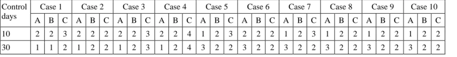
Table 2. Pain scoring on postoperatively 10th and 30th days in all cases.
Tablo 2. Tüm olgularda postoperatif 10.ve 30. gün ağrı skorları. Control
days
Case 1 Case 2 Case 3 Case 4 Case 5 Case 6 Case 7 Case 8 Case 9 Case 10 A B C A B C A B C A B C A B C A B C A B C A B C A B C A B C 10 2 2 3 2 2 2 2 2 3 2 2 4 1 2 3 2 2 2 1 2 3 1 2 2 1 2 2 1 2 2 30 1 1 2 1 2 2 1 2 3 1 2 4 3 2 2 3 2 2 3 2 2 3 2 2 3 2 2 3 2 2
Table 3. Pain scores: [Median (25-75%)] and statistical significance control according to patients' control days. Tablo 3. Hastaların kontrol günlerine göre ağrı skorları: [Median ( %25-%75)] ve istatistik önem kontrolü.
Pain Scors Control days P values
(Wilcoxon Signed Rank Test) Day 10 Median ( %25-%75) Day 30 Median ( %25-%75) A 1.5 (1- 2) 3 (1- 3) 0.105 B 2 (2- 2) 2 (2- 2) 0.317 C 2.5 (2- 3) 2 (2- 2.25) 0.083 Total 6 (5- 7) 7 (5.75- 7) 0.464
* The difference between the pain scores according to the control days was not statistically significant. The pain scores did not change. * Kontrol günlerine göre ağrı skorları arasında fark istatistiksel olarak önemli bulunmamıştır. Ağrı skorları değişiklik göstermemiştir.
patients, and all fractures healed. There were no major effects determined on the growth potential according to the functional and radiological outcomes. Osteomyelitis was not radiographically apparent in any animal. As a result of the postoperative trauma, the stabilization of the implant was affected and exuberant callus formation was encountered in cat 5 on the 30th day of control. Therefore
implants were removed from the patient immediately. There were no functional loss in this patient.
Postoperatively, the 10th day lameness score was 2 in
eight cases and 3 in two cases. The lameness score on the 30th day was 1 in eight cases and 2 in two cases (Table 1).
Lameness scoring was not done after the 30th day. The
detailed postoperative signs of pain of all cases are presented in table 2.
The implants were removed after fracture healing in nine cases. Unfortunately, the owner of case 2 refused a second surgery (Figure 2). Consequently, the functional outcome was excellent in 10 fractures and good in 1.
Discussion and Conclusion
This retrospective clinical study describes use of the UniLOCK system for stabilization of long bone fractures in cats. The UniLOCK system is originally designed for maxillofacial human surgery. These implants are commonly used in the treatment of mandibular fractures and mandibular reconstruction after tumor resections (4). In veterinary surgery, the possible applications for this system are much wider given the different small bones and joints in cats and dogs (7, 17, 18). UniLOCK instruments and implants were found to be easy and practical to use. The adaptation of the plate is easier.
In a locking system the plate does not need to be indirect contact with the underlying bone. As the screws are tightened, they interlock with the plate, this stabilizing the segments without the need to compress the plate to the bone (5, 12, 13, 15). Plate-screw density is used to determine the most appropriate length of internal fixator. This is calculated by dividing the number of plate holes by the number of screws. In humans 0.5 to 0.4 are below the recommended values (5). However, there is no literatüre data available for cats. In this study, an average value of 0.77 was obtained. This result, which will then be taken into account when determining the length of UniLOCK plate studies conclude that it will be useful.
A diminished contact between plate and bone may also preserve periosteal blood supply. Hereby, the biological advantage of internal fixators is that the screws can be inserted monocortically (5, 12, 20). The monocortical insertion of the screws was not used more often in the study of Voss et al. (17) because of the habit of surgeons to insert bicortical screws. The insertion of the screws monocortically were used more often than bicortical screws in the present study. Additionally, the distal fragment of intra-articular fractures was stabilized with monocortical screws and it thought to minimize the potential damage to the growth plate. In the present study, the short distal fragment in four cases allowed for placement of plenty of screws. Actually, the fact that the fixation remained stable supports the theory that interlocked screws offer superior stability and that fewer screws must be inserted in shorter fragments.
As reported by Voss et al. (17), using the UniLOCK implants, errors, those of particularly a technical nature, are encountered which are closely related to surgeons’
habits. Screw pull-out may occur if the screws have not been fully seated in the bone or if they only engage the caudal or cranial edge of a cortex. This problem may occur unnoticed during surgery because the surgeon’s feel for the screw holding in the bone during tightening of the screw is not as good as when conventional screws are used (18). There is an overall complication rate of 19.7% and a fixation failure rate of 10.6% in Voss’s study (17). The surgeons gained experience of the system throughout the study period, and produced seven complications in the first half and only two in the second half of the cases in the study (17). Consequently, gaining LCP experience is very important for surgeons. In the present study, the most important reason for lack of technical errors and complications, we believe, lies in our previous LCP experiences (16).
The median osseous healing time was five weeks. The fractures in all cases except for case 5 completely healed within five weeks. As a result of the postoperative trauma, exuberant callus formation was encountered in the cat 5. long term radiographic assessment of the affected limb was performed in the present study. Radiological evaluations revealed a very good framing of fracture healing for 9 cats, and good for 1 cat.
Fractures of the distal femoral physis are injuries frequently encountered in cats and usually seen in the form of SH type I and II fractures. In particular, cats have a high incidence of SH type I fractures. These fractures are often seen at 6.5 and 9.5 months of age, when a significant shortening in the femur is usually encountered. Internal fixation of SH type I and type II fractures of the distal femur may involve single or multiple intramedullary pins, static or dynamic cross-pinning, and lag screw fixation. Young animals with growth potential in particular require pin fixation to prevent premature closure of the growth plate (14). Perfect reduction was achieved in four growing cats UniLOCK implants, and it was determined that the application did not cause premature closure of the growth plate.
Succesful treatment of several musculoskeletal disorders is associated with maintaining an adequate range of joint motion. Goniometry provides a rapid and reliable method to quantify the ROM of joints (6). Joint angles were directly measured with a universal goniometer constructed of plastic. In this study, the motion of the operated and contralateral normal stifle joint was measured in four cats. The differences between the ROM values of the operated and contralateral stifle joint had no effect clinically. These values may be useful in future studies because there are no records of cat's stifle joint ROM values after plate-screw fixations in skeletally immature cats.
Titanium implants compared to other implants (vitalium, steel) were superior on bone tissue healing, do
not require removal of bone tissue, and have a better texture alignment (10, 11). Titanium UniLOCK implants are expensive orthopaedic materials. Although there are many advantages of UniLOCK plate-screws, these implants widely prevent the use of high cost of implants. These implants will be used in clinical routines and result in a fall in prices.
Gait examinations and lameness grades can be difficult for cats. They often require time and patience because cats can not to be led on a leash, and often tend to refuse to move around freely in a new enviroment. If a cat completely refuses to walk, lameness can not be assessed at the clinic, the observations of the owners have to be used gather information. Another valuable method for gait evaluation in cats is to ask the owners to film their cat at home (18). In this study, we used also “a simple lameness classification score” reported by Voss et al. It is very simple and practical soring lameness in cats. We also prepared another lameness scale (excellent, good, moderate, poor) for patient-owners. They have told that this lameness scale was very practical and simply for their pets at home. According to our opinion, this two scales are very acceptable and useful in cats for gait examinations.
The use of pain asssessment scales have been well established in humans and are becoming more useful in veterinary patients. There is, a fundamental difference; human patients score their own pain on the scales, otherwise pain assessment in animals requires an observer (2). We used to determine simple descriptive scores for assessment of pain in our patients. This scoring system is very practical in cat patients.
In this present study, the application of UniLOCK implant effectively sustained joint motion and allowed early weight bearing activity in cats with especially epiphyseal fractures. The functional outcome of epiphyseal fracture repaired with UniLOCK implant appears promising, but additional studies are necessary.
Acknowledgements
The authors wish to acknowledge the support of
Mehmet Akif Ersoy University Scientific Research Projects Commission (Project Number: 0097-NAP-10) in enabling the completion of this study.
References
1. Amato NS, Richard A, Knight TA, et al. (2008): Ex vivo
biomechanical comparison of the 2.4 mm UNILOCK Reconstruction plate using 2.4 mm locking versus Standard screws for fixation of acetabular osteotomy in dogs. Vet
Surg, 37, 741-748.
2. Cambridge AJ, Tobias KM, Newberry RC, et al. (2000):
Subjective and objective measurements of postoperative pain in cats. J Am Vet Med Assoc, 217, 685-690.
3. Franch J, Lopez C (2007): Atlas der Chirurgishen
Zugange Hund und Katze. 1st ed, 16-72, Verlag-Elsevier,
Germany.
4. Frigg R (2003): Development of the locking compression
plate. Injury, 34, 6-10.
5. Gauiter E, Sommer C (2003): Guidelines for the clinical
application of the LCP. Injury, 34, 63-76.
6. Jaeger GH, Marcellin-Little DJ, Depuy V, et al. (2007):
Validity of goniometric joint measurements in cats. Am J
Vet Res, 68, 822-26.
7. Keller M, Voss K, Montavon PM (2004): The Compact
Unilock 2.0/2.4 System and its application in small animal
orthopaedics. 5th Continuing Education Course for Japan
Small Animal Surgeons at the Small Animal Surgery Clinic, Vetsuisse Faculty, University Zurich, University, August 23-27, pg. 14-28.
8. Keller M, Voss K (2002): Unilock: Applications in small
animals. AO Dialogue, 15, 20-21.
9. Koch D (2005): Screws and Plates. In, Johnson AL, Houlton EF, Vannini R (Eds): AO Principles of Fracture Management in the Dog and Cat. 1st ed, 47, AO Publishing, Switzerland.
10. Linder L, Lundskog L (1975): Incorporation of stainless
steel, titanium and vitallium in bone. Injury, 6, 277-285.
11. Millar BG, Frame JW, Browne RM (1990): A histological
study of stainless steel and titanium screws in bone. Br J
Oral Maxillofac Surg, 28, 92-95.
12. Perren SM (2001): Evolution and rational of locked
internal fixator technology. Introductory Remarks. Injury,
32, 3-9.
13. Plecko M, Lagerpusch N, Pegel B, et al. (2012): The
influence of different osteosynthesis configurations with locking compression plates (LCP) on stability and fracture healing after an oblique 45° angle osteotomy.
Injury, 43, 1041-51.
14. Simpson DJ, Lewis DD (2003): Fractures of the femur. In, Slatter D (ed), Textbook of Small Animal Surgery Saunders, pg. 2080-2086.
15. Şengöz Ö, Olcay B (2008): A new implant system called
locked compression plate in veterinary practice. Vet
Cerrahi Derg, 14, 44-47.
16. Şengöz Ö, Kaya Ü, Olcay B (2013): Clinical and
radiological outcomes of the locking compression plate system in dogs with diaphyseal fractures: 32 cases. Kafkas
Univ Vet Fak Derg, 19 (Suppl A), 13-18.
17. Voss K, Kull MA, Haessig M, et al. (2009): Repair of
long-bone fractures in cats and small dogs with the Unilock mandible locking plate system. Vet Comp Orthop
Traumatol, 22, 398-405.
18. Voss K, Langley-Hobbs SJ, Montavon PM (2009):
Femur. In, Montavon PM, Voss K, Langley-Hobbs SJ
(Eds): Feline Orthopaedic Surgery and Musculoskeletal Disease, 1st ed, 460-473, Saunders, Switzerland.
19. Voss K, Steffen F (2009): Patient assessment. In, Montavon PM, Voss K, Langley-Hobbs SJ (Eds). Feline Orthopaedic Surgery and Musculoskeletal Disease. 1st ed, 8, Saunders, Switzerland.
20. Wagner M (2003): General principles for the clinical use
of the LCP. Injury, 34, 31-4
Geliş tarihi: 10.11.2016 / Kabul tarihi: 06.03.2017
Address for correspondence:
Asst. Prof. Dr. Özlem ŞENGÖZ ŞİRİN Mehmet Akif Ersoy University, Faculty of Veterinary Medicine,
Department of Surgery, TR-15030, Burdur,Turkey. E-mail: [email protected]