Ankara Üniv Vet Fak Derg, 57, 139-142, 2010
Short Communication / Kısa Bilimsel Çalışma
First record of Synhimantus (S.)laticeps (Rudolphi, 1819) Railliet,
Henry et Sisoff, 1912 (Nematoda, Acuariidae) in Accipiter nisus (Aves,
Accipitridae) in Turkey
Şinasi UMUR, Yunus Emre BEYHAN, Gökmen Zafer PEKMEZCİ, Mustafa AÇICI, Ali Tümay GÜRLER
Department of Parasitology, Faculty of Veterinary Medicine, Ondokuz Mayis University, Kurupelit, Samsun, Turkey
Summary: One female and two male nematodes were identified as Synhimantus (Synhimantus) laticeps (Rudolphi, 1819) Railliet, Henry et Sisoff, 1912 in the postmortem examination of a female goshawk’s gizzard (Accipiter nisus) which was brought to our Parasitology Laboratory by the Directorship of Environmental and Forestry authorities of Samsun, Turkey in September, 2008. Some morphological features, measures and photographs of the nematodes were given within this report. To our knowledge, this is the first report of Synhimantus (S.) laticeps and also the genus from Turkey.
Key words: Accipiter nisus, goshawk, Synhimantus (S.) laticeps, Turkey
Türkiye’de atmacada (Aves, Accipitridae) ilk Synhimantus (S.)laticeps (Rudolphi, 1819) Railliet, Henry et Sisoff, 1912 (Nematoda, Acuariidae) olgusu
Özet: Bu çalışmada, Samsun Çevre ve Orman İl Müdürlüğü tarafından Eylül 2008’de Fakültemiz Parazitoloji laboratuarına getirilen bir dişi atmacanın postmortem muayenesi sırasında midesinden bir dişi ve iki erkek Synhimantus laticeps (Rudolphi, 1819) Railliet, Henry et Sisoff, 1912 teşhis edilmiştir. Türkiye’de bu cins ve Synhimantus laticeps’in ilk kayıt olması nedeniyle parazitin önemli morfolojik özellikleri, ölçümleri ve fotoğrafları verilmiştir.
Anahtar sözcükler: Accipiter nisus, atmaca, Synhimantus(S.)laticeps, Türkiye.
Acuariid nematodes which belong to Synhimantus
(Synhimantus)laticeps (Rudolphi, 1819) Raillet, Henry
and Sisoff, 1912 are characterized by their anastomosed cordons, a long buccal capsule and their tricuspid deirids located posterior to the cordons (7). This species was widely reported from all around the world, especially Turkistan, Russia, Bulgaria, Spain, Algeria, USA and India in some raptor birds (Accipiter, Aegiolus, Aquila,
Asio, Bubo, Buteo, Circus, Cerchenis, Falco, Gyps, Otus,
and Strix) (1,9). Despite its cosmopolite distribution, information about this nematode morphological feature is restricted. This parasite is located in oesophagus, crop, gizzard, stomach and intestines (6,9).
Acuariidae is a very wide family which contains 25 genera and some of them are Acuaria, Dispharynx,
Echinuria, Seuratia, Streptocara, Synhimantus, etc. The Synhimantus genus resembles to Dispharynx that is seen
in Turkey. However some authors considered that
Dispharynx is a subgenus of Synhimantus (10), it can be
differentiated by anastomosed and recurrent cordons (2).
Synhimantus contains of 25 species, but just nine of
them were seen in Europe; S. affinis, S. elliptica, S. falconis,
S. hamatus, S. laticeps, S.niloticus, S. robertodollfusi, S. sirry and S. spiralis. (7,8), Synhimantus (S.) laticeps can
be defined by recurrent and anastomosed cordons, long and narrow buccal capsule, posteriorly located tricuspid cervical papilla behind the cordons, and the unequal and dissimilar male spicules, distal end of left spicule has complexity (1,3,9)
Some morphological features, measures and photographs of the nematodes were given within this report. To our knowledge, this is the first report of
Synhimantus (S.) laticeps and also the genus from
Turkey.
A wounded female goshawk had been died during the transport to our faculty clinics for treatment by the Directorship of Environmental and Forestry authorities of Samsun, Turkey in September, 2008. Initially, the gastrointestinal system was opened in the Parasitology Laboratory. Each organ contents were separately sieved
Şinasi Umur - Yunus Emre Beyhan - Gökmen Zafer Pekmezci - Mustafa Açıcı - Ali Tümay Gürler 140
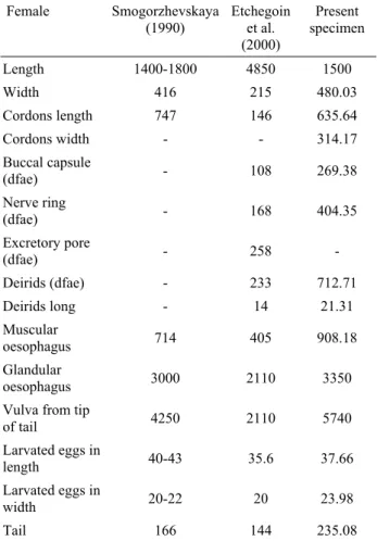
Figure 2. D. Posterior end of male and post anal papillae, E. Tricuspid cervical papilla (Scale bar 50 µm) Şekil 2. D. Erkeğin arka kısmı ve post anal papilla, E. Trikuspid servikal papilla (Bar 50 µm)
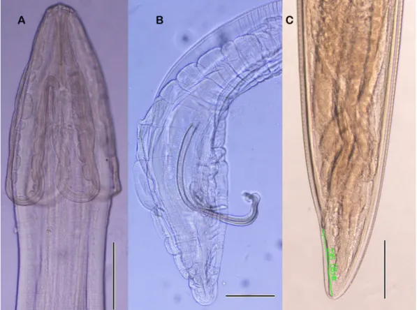
Figure 1. A. Anterior part of male, B. Posterior extremity and spicules of male, C. Posterior end of female. (Scale bars in three figures: 200 µm)
Şekil 1. A. Erkeğin ön kısmı, B. Erkeğin arka ekstremitesi ve spikülü, C. Dişinin arka kısmı. (Her üç şekildede bar 200 µm)
Ankara Üniv Vet Fak Derg, 57, 2010 141
through 100 µm pore diameter sieve and examined under the stereomicroscope (Nikon SMZ 1500). Collected parasites were washed in physiological saline, than fixed in 70% hot alcohol, and cleared in lactophenol for examination. After examination, specimens were preserved in; 92 part 70% ethanol, 5 part glycerine, and 3 part commercially available formalin solution. They were then measured and identified using a (Nikon Eclipse 80i) microscope.
Parasite identify card
Host: Accipiter nisus, goshawk (Aves, Accipitridae) Site of infection: Gizzard
Locality: Çarşamba, Samsun, (40° 50’ - 41° 51’ North latitude, 37° 08’ and 34° 25’ East longitude), Black Sea Region, Turkey.
Intensity of infection: 3 (1 female and 2 male) nematodes from Accipiter nisus.
Material deposited: Voucher specimens were deposited in the Helminth Coll. No. 2008/3 Department of Parasitology, Faculty of Veterinary Medicine, Samsun, Turkey.
Description
Cuticle with clear transverse striations. Anterior end with two lateral pseudolabia. Cordons recurrent, anastomosing on each lateral surface. Cervical papilla (deirids) tricuspid and located posteriorly to the cordons. Buccal capsule narrow, long and expanded anteriorly. Four large cephalic papillae located at the base of pseudolabia. Oral aperture oval-elongated. Spicules are clearly unequal. Five pairs of post-cloacal are pedunculated papillae and one pair of sessile papillae (1,3,7,9).
Male (n=2): Length 8.04 (8.00 – 8.08) mm. Width
235.78 µm (220.04-251.51). Cordons long 371.74 (325.01-418.47) µm. Cordon width 166.11 (139.00-193.22) µm. Buccal capsule 191.17 (176.18-206.16) µm long, nerve ring 253.29 (205.14-301.43) µm, deirids 432.35 (400.87-463.82) µm, excretory pore 386.94 (344.66- 429.21) µm from anterior extremity. Deirids 19.91 (18.46-21.35) µm long (Fig.1-A, 2-E). Spicules are dissimilar and unequal. Right spicule 251.82 (204.83-298.81) µm and left spicule 708.69 (696.30-721.08) µm in length and distal end of it is complex shape (Fig.1-B). Tail 149.06 (132.70-165.42) µm long (Fig.2-D, Table 1).
Female (n=1): Length 15 mm. Width at the level of
the vulva 480.03 µm. Cordons 635.64 µm long, 314.17 µm width, recurrent, approximately 1/3 part of the length anastomosing. Buccal capsule 269.38 µm long, nerve ring 404.35 µm, deirids 712.71 µm distance from anterior extremity. Deirids 21.31 µm long (Fig.2-E) Oesophagus 4.26 mm in length. Muscular oesophagus 908.18 µm in length and glanduler oesophagus 3.35 mm in length. Vulva 5.74 mm from tip of tail. Larvated eggs 37.66 in length and 23.98 in width. Tail 235.08 µm in length (Fig.1-C, Table 2).
Table 1: Comparative mean values measurements (µm) of male specimens of S. (S.) laticeps.
Tablo 1. Erkek S. (S.) laticeps’in karşılaştırmalı ölçüleri (µm). Male Smogorzhevskaya (1990) Etchegoin et al. (2000) Acosta et al. (2008) Present specimens Length 8000 4470 8890 8040 Width 200 145 237 235.78 Cordons length 416 149 360 371.74 Cordons width - - - 166.11 Buccal capsule (dfae) 220 107 215 191.17 Nerve ring (dfae) 260 139 - 253.29 Excretory pore (dfae) - 164 408 386.94 Deirids (dfae) 315-465 165 477 432.35 Deirids long - 11.6 19.91 Right spicule 156-184 194 202 251.82 Left spicule 660 554 796 708.69 Tail - 90 347 149.06
dfae: distance from anterior extremity
Table 2: Comparative mean values measurement (µm) of female specimens of S. (S.) laticeps
Tablo 2. Dişi S. (S.) laticeps’in karşılaştırmalı ölçüleri (µm).
Female Smogorzhevskaya (1990) Etchegoin et al. (2000) Present specimen Length 1400-1800 4850 1500 Width 416 215 480.03 Cordons length 747 146 635.64 Cordons width - - 314.17 Buccal capsule (dfae) - 108 269.38 Nerve ring (dfae) - 168 404.35 Excretory pore (dfae) - 258 - Deirids (dfae) - 233 712.71 Deirids long - 14 21.31 Muscular oesophagus 714 405 908.18 Glandular oesophagus 3000 2110 3350
Vulva from tip
of tail 4250 2110 5740 Larvated eggs in length 40-43 35.6 37.66 Larvated eggs in width 20-22 20 23.98 Tail 166 144 235.08
Şinasi Umur - Yunus Emre Beyhan - Gökmen Zafer Pekmezci - Mustafa Açıcı - Ali Tümay Gürler 142
Some authors accepted that Dispharynx genus which occurs in Turkey (4) is a subgenus of the
Synhimantus genus. Dispharynx nasuta is rarely seen as Synhimantus (Dispharynx) nasuta in the literatures (10).
However, cordons with anastomosing were accepted as
Synhimantus (Synhimantus) in contrast cordons without
anastomosing were accepted as Synhimantus (Dispharynx) genus (2).
Synhimantus (S.) laticeps were reported in different
birds in some European countries (Germany, Netherlands, Spain, France and Greece) (1,5,6). Sanmartin et al. (2004) and Acosta et al. (2008) reported that S. (S.)
laticeps is a common species which found 60% and
36.5% in Falcaniform birds in Spain, respectively. Recently Papazahariadou et al. (2008) reported
Synhimantus(S.)laticeps in Greece from free-ranging
birds (Buteo buteo).
Our morphological measurements were similar to literatures (1,3,7) except small differences (Table 1, 2).
Turkey is located at the junction of three continents, with natural important route for seasonal migration for the wild birds in the world. Also in Turkey there are some important wetlands (Kızılırmak “redriver”delta in Bafra, Yeşilırmak ”greenriver” delta in Çarşamba) which provide a habitat for a large number of migratory birds and resident species. So the new species or/and new diseases can be seen every year.
The reason of these differences is may be arisen from different hosts, so host size is effected by the parasite size in classical knowledge. Therefore, some morphological measurements of the species were similar except little differences in the literature (2,6).
A detailed description and some morphological and general structures for the definition about an adult male and a female of S. laticeps is presented with this report to guide for other Synhimantus species while S. (S.) laticeps is reported for the first time by the present study in Turkey.
References
1. Acosta I, Hernández S, Gutiérrez PN, Martínez-Cruz MS, Hernández E, Buffoni L, Martínez-Moreno FJ (2008). Acuaroid nematodes in the common kestrel (Falco
tinnunculus) in the south of Spain. Vet J. (In Press).
2. Chabaud AG (1975). Keys to the genera of the order
Spirurida. Part 2. Spiruroidea, Habronematoidea and Acuarioidea. 29-58. In: Anderson RC, Chabaud AG,
Willmott S (Eds), CIH Keys to the Nematode Parasites of Vertebrates. No.3. Commonwealth Agricultural Bureaux, UK. 3. Etchegoin JA, Cremonte F, Navone GT (2000).
Synhimantus (Synhimantus) laticeps (Rudolphi, 1819) Railliet, Henry et Sisoff, 1912 (Nematoda, Acuariidae) parasitic in Tyto alba (Gmelin) (Aves, Tytonidae) in Argentina. Acta Parasitol, 45, 99-106.
4. Gıcık Y (1997). Dispharynx nasuta (Rudolphi, 1819) in a
pigeon (Columbia livia). Vet J Ankara Univ, 44, 1-3.
5. Papazahariadou M, Diakou A, Papadopoulos E, Georgopoulou I, Komnenou A, Antoniadou-Sotiriadou K (2008). Parasites of the digestive tract in free-ranging
birds in Greece. J Nat Hist, 42, 381-398.
6. Sanmartin ML, Alvarez F, Barreiro G, Leiro J (2004).
Helminths fauna of Falcaniform and Strigiform birds of prey in Galicia, Northwest Spain. Parasitol Res, 92, 255-263.
7. Smogorzhevskaya LA (1990). Nematodes. Part 3. Acuarioidea. In: VP Sharpilo (Ed), Fauna Ukrainy. Vol.
32, Kiev, (In Russian).
8. Vicente JJ, Pinto RM, Noronha D (1996). Synhimantus
(Synhimantus) magnipapillatus n.sp. (Nematoda, Acuarioidea) from the Yellow-crowned Night-heron, Nyctanassa violacea cayennensis (Gmelin) (Aves, Ardeidae). Mem Inst Oswaldo Cruz, 91, 51-53.
9. Yamaguti S (1961). Systema Helminthum. Vol. III. Interscience Publishers Inc, London, UK.
10. Zhang L, Brooks DR, Causey D (2004). Two species of
Synhimantus (Dispharynx) Railliet, Henry et Sisoff, 1912 (Nematoda: Acuarioidea, Acuaridae), in Passerinae birds from the area de Conservacion Guanacaste, Costa Rica. J
Parasitol, 90, 1133-1138.
Geliş tarihi: 06.02.2009 / Kabul tarihi: 08.05.2009
Yazışma Adresi:
Prof.Dr. Şinasi Umur
Ondokuz Mayıs Üniveristesi, Veteriner Fakültesi Parazitoloji Anabilim Dalı, 55139 Kurupelit /Samsun E posta: [email protected]
Tel: + 90(362) 4576921 Belgeç:+90(362) 4576922