• Sonuç bulunamadı

Başlık: The Role of FDG PET/CT in a Patient with Larynx Cancer Who Developed Necrotizing Fasciitis Secondary to Tracheostomy Which is a Rare ComplicationYazar(lar):BAYAR, Ebru; BURAL, GoncaCilt: 70 Sayı: 1 Sayfa: 067-070 DOI: 10.1501/Tipfak_0000000966 Yay

N/A
N/A
Protected

Academic year: 2021

Share "Başlık: The Role of FDG PET/CT in a Patient with Larynx Cancer Who Developed Necrotizing Fasciitis Secondary to Tracheostomy Which is a Rare ComplicationYazar(lar):BAYAR, Ebru; BURAL, GoncaCilt: 70 Sayı: 1 Sayfa: 067-070 DOI: 10.1501/Tipfak_0000000966 Yay"

Copied!
4
0
0

Yükleniyor.... (view fulltext now)

Tam metin

(1)

The Role of FDG PET/CT in a Patient with Larynx Cancer Who

Developed Necrotizing Fasciitis Secondary to Tracheostomy

Which is a Rare Complication

Trakeostomiye sekonder Çok Nadir Bir Komplikasyon Olan Nekrotizan Fasiit Gelișen Larinks Kanserli Hastada FDG PET/BT’nin Rolü

Ebru Bayar

1

,Gonca Bural

1

1 Department of Nuclear Medicine, Izmir Katip Çelebi University, Atatürk

Research and Training Hospital, Izmir, Turkey Necrotizing fasciitis (NF) is characterized with rapid involvement of soft tissues and fascias and could be fatal

if not treated properly. Our patient had history of larynx cancer. He had a tracheostomy and developed NF of the neck and upper mediastinum which is a very rare complication of tracheostomy.

The patient was referred for a Positron Emission Tomography/Computed Tomography (PET/CT) scan after suspicion of residual lesion on follow up computed tomography (CT). On fluorodeoxyglucose (FDG) PET/CT images there was moderate increased uptake at the anterior glottic region, in line with residual disease. PET/CT scan showed progression of the NF involving the neck and upper mediastinum compared to the CT scan performed 10 days before. On PET/CT a new pulmonary abscess was also noted in the lower lobe of the left lung.

In our case FDG PET/CT played an effective role in the follow up of oncologic disorders. It was also useful in the follow up and treatment response of NF, which is a rare but potentially fatal infectious and inflammatory disorder. Positron Emission Tomography/Computed Tomography (PET/CT) could be preferred in the follow up of serious infectious conditions accompanying primary malignancy.

Key Words: Necrotizing Fasciitis,18F-FDG, PET / CT , Inflammation

Nekrotizan fasiit (NF) fasya ve cilt altı yumușak doku nekrozu șeklinde seyreden, hızla yayılan ve uygun tedavi edilmezse hayatı tehdit eden bir hastalıktır. Larinks kanseri olan vakamızda trakeostomi sonrasında boyun ve üst mediastinal bölgede çok nadir bir komplikasyon olan nekrotizan fasiit gelișmiș olup, takip bilgisayarlı tomografi (BT) kontrolünde larinkste rezidü șüphesi ile Pozitron Emisyon Tomografi/Bilgisayarlı Tomografi (PET/BT) çekilmesi için bölümümüze bașvurmuștur. Yapılan fluorodeoksiglukoz (FDG) PET/BT de sağ anterior glottik bölgede rezidü lezyon ile uyumlu aktivite artıșı izlenirken, 10 gün önce çekilen BT ile karșılaștırıldığında boyun ve üst mediasten bölgesinde izlenen nekrotizan fasiit ile uyumlu enfeksiyöz değișikliklerde progresyon olduğu da tespit edilmiștir. Ayrıca önceki BT de olmayan sol akciğer alt lobda pulmoner apse ile uyumlu görünüm izlenmiștir.

Vakamızda FDG PET/BT primer malign hastalığın takibinde faydalı olmuștur. Ayrıca trakeostomi sonrası gelișen, çok nadir bir komplikasyon olan, fatal seyredebilen NF’nin takibinde ve tedaviye yanıtının değerlendirilmesinde etkin bir rol almıștır. Pozitron Emisyon Tomografi/Bilgisayarlı Tomografi (PET/BT) maligniteye eșlik eden enfeksiyonun takibinde etkin bir görüntüleme yöntemidir.

Anahtar Sözcükler: Nekrotizan Fasiit,18F-FDG, PET/BT, İnflamasyon

Nekrotizan fasiit (NF) is an infectious disorder characterized with rapid involvement of soft tissues and fascia, usually involves trunk, perineum, and lower extremities and rarely can occur in the head and neck region. It characterized with high morbidity and mortality (1-3). There is infiltration of polimorphonuclear cells secondary to vascular thrombosis and obliteration (4). Necrosis of the subcutaneous fat causes air formation in the deep facial plans. Misdiagnosis or improper treatment may cause erosion of carotid artery, mediastinitis, thrombophlebitis of the

jugular vein, sepsis and aspiration pneumonia (5). Most common organisms causing NF are E.coli, Proteus, Clostridium, etc. and aerobics (6, 7). Early diagnosis is lifesaving in NF.

Computed tomography (CT) is the most sensitive method for the detection of gas due to soft tissue infections. It is also useful for the detection of extend of inflammation and follow up soft tissue involvement.

Ankara Üniversitesi Tıp Fakültesi Mecmuası 2017, 70 (1)

DOI: 10.1501/Tıpfak_000000966

DAHİLİ TIP BİLİMLERİ/ MEDICAL SCIENCES

Olgu Sunumu / Case Report

Received: March 03, 2017  Accepted: June 22, 2017 Corresponding Author:

Ebru Bayar, M.D.

E-mail: ebruyucesoy1@yahoo.com Phone: +90 532 645 66 25 Fax: ?????

Department of Nuclear Medicine, Izmir Katip Çelebi University, Atatürk Research and Training Hospital, Izmir, Turkey

(2)

Ankara Üniversitesi Tıp Fakültesi Mecmuası 2017, 70 (1)

The Role of FDG PET/CT in a Patient with Larynx Cancer Who Developed Necrotizing Fasciitis Secondary to Tracheostomy Which is a Rare Complica

68

Fluorodeoxyglucose Positron Emission Tomography/Computed Tomography (FDG PET/CT) has a valuable imaging modality on the evaluation of both newly diagnosed and previously treated patients with head and neck cancer (8). Fluorodeoxyglucose Positron Emission Tomography/ Computed Tomography (FDG PET/ CT) also has clinical value in suspected infection and inflammation. It can demonstrate the extent and severity of disease too. Fluorodeoxyglucose Positron Emission Tomography/ Computed Tomography (FDG PET/CT) is a valuable diagnostic tool, therefore could be preferred in serious infectious conditions accompanying primary malignancy (9).

Our aim was to demonstrate and discuss the role of PET/CT in the clinical work up of a patient with larynx cancer who developed NF secondary to tracheostomy.

Case

A 66 year old male patient with history of larynx cancer had total laryngectomy surgery. The patient had an urgent tracheostomy prior the surgical excision of the primary tumor. Fifteen days after the removal of the primary tumor the patient had subcutaneous oedema starting from right neck, extending to the right anterior chest wall and had intermittent fever. There was pain, oedema, rubor and purulent flow around the tracheostomy site. The culture growed Pseudomonas and coagulase-negative Staphylococci.

Systemic examination and laboratory findings were supported infection. Patient had anaemia (Hb 8gm/dl), with leucocytosis (19 mg/dl), raised CRP (32 mg/dL), and ESR (36 mm Hg/ hr). On CT scan there was air in the upper

mediastinum, posterior to the sternum and the patient was diagnosed with mediastinitis and osteomyelitis of the right sternoclavicular joint. The patient could not have surgery as he was not stable. Patient had a PET/CT scan for staging of larynx cancer.

Positron Emission Tomography/ Computed Tomography (PET/CT) images before and after antibiotherapy are given in Figure 1. In the upper anterior mediastinum immediately below the tracheostomy site there was increased metabolic activity in the soft tissue surrounding the proximal edges of both clavicles, bilateral first costasternal joints, bilateral sternoclavicular joints and the entire manubrium sterna. There were new air densities in the left sternoclavicular joint showing the progression of NF in comparison to CT 10 days ago. On the bone window there was evidence of cortical disruption and increased metabolic activity in the manubrium sterna indicating bone involvement secondary to the soft tissue infection. There was also disruption of the overlying upper mediastinal skin on the CT images to PET/CT with mild increased metabolic activity in the subcutaneous s oft tissue (Figure 1 a,b initial scan before antibiotherapy).

Positron Emission Tomography/ Computed Tomography (PET/CT) images of lung abscess before and after treatment are given in Figure 2. On the lung images there was a new, 36x24 mm soft tissue lesion in the periphery of the superior segment on the left lower lobe with central absence of metabolic activity and with high rim of surrounding FDG uptake (SUV max:6.9) in the entire periphery. This lesion was interpreted as an abscess with central necrosis (Figure 2 a,b initial scan before antibiotherapy). Patient also had suspicion of residual/ metastatic disease and referred for a PET/CT scan. PET/CT images before and after radiotherapy are given in Figure 3. On PET/CT scan there was no evidence of metastatic disease. But there was moderate activity (SUV max:5) at the anterior of the glottic region suspicious for residual disease (Figure 3 a,b initial scan before radiotherapy).

Figure 1. Axial CT and fusion slices of PET/CT at the level of uppe rmediastinum before treatment. There was high metabolic activity and air density related to NF in the upper mediastinum (arrow) and increased FDG uptake on fusion sternum and sternoclaviculary joints suggestive of osteomyelitis (a,b).

Axial CT and fusion slices of PET/CT at the level of upper mediastinum after treatment. There has been complete resolution of air densities (arrow) and interval decrease in the metabolic activity of sternum and sternoclaviculary joint, suggestive of response to treatment (c,d).

(3)

Journal of Ankara University Faculty of Medicine 2017, 70 (1)

Ebru Bayar,Gonca Bural 69

A combination of large spectrum antibiotic was started for the treatment of NF and pulmonary abscess. Laryngeal biopsy showed residual larynx tumor , therefore patient had radiation treatment to head and neck region.

There was complete remission of necrotizing fasciitis after treatment (Figure 1c,d,) and pulmonary abscess (Figure 2c,d) Laboratory finding and physical examinations were also normal simultaneously supporting our imaging findings. However residual disease persisted and showed progression (SUV max: 14) in the laryngeal area (Figure 3c,d) on PET/ CT.

Literature Review and Discussion Nekrotizan fasiit (NF) is a life treating infection

of the superficial fascia, usually spreads rapidly. The infection starts at a site of trauma which may be extensive or at the site of minor trauma (1,2).

Over 70 per cent of cases are recorded in people with at least one of the following predisposing clinical situations: immune suppression, diabetes, alcoholism/drug abuse/ smoking, malignancy, and chronic systemic diseases. People initially have signs of inflammation and fever. With progression of the disease, often within hours, tissue becomes progressively swollen, the skin becomes discoloured. Mortality rate may increase up to 73 per cent if there is delay in surgery or left untreated (10). Our patient had history of larynx cancer, as a predisposing factor. He had oedema, rubor and purulent flow around the tracheostomy and intermittent fever.

Computed Tomography (CT) criteria of soft tissue gas and collections of the fluid are valuable in evaluating suspected necrotizing fasciitis (11). On the CT images of our patient there was gas collection typical for NF in the soft tissue in the upper anterior mediastinum. Magnetic resonance imaging (MRI) is the

modality of choice for detailed evaluation of soft tissue infection but is often not performed for necrotizing fasciitis evaluation because its acquisition is time consuming and may delay treatment (12). In our case CT was preferred for diagnosis and follow up.

Figure 2. Axial CT and fusion slices of PET/CT on lung window setting before treatment. New lung abscess (arrow) in the superior segment of the left lower lobe is noted (a, b).

Axial fusion and CT slices on lung window setting after treatment. Interval complete resolution (arrow) of pulmonary abscess after treatment (c, d).

Figure 3. Axial CT and fusion slices of PET/CT at the level of glottic region on previous PET CT. Moderately increased activity (SUV max: 5 ) (arrow) was noted on the anterior glottic region suspicious for residual disease (a, b).

There has been interval increase in metabolic activity (arrow) in the same region (SUV max: 13.8) suggestive of residual disease (c, d).

(4)

Ankara Üniversitesi Tıp Fakültesi Mecmuası 2017, 70 (1)

The Role of FDG PET/CT in a Patient with Larynx Cancer Who Developed Necrotizing Fasciitis Secondary to Tracheostomy Which is a Rare Complica

70

A main superiority of PET/CT is its comprehensive imaging of the whole body in a single study and detects truly relapse and metastatic disease for malign lesions. Positron Emission Tomography/ Computed Tomography (PET/CT) also provides metabolic information and anatomic localization together. So it is known a valuable imaging modality for staging, and assessment of response to therapy for malignancies. Since tumor cells have a higher affinity for glucose than normal cells and use glycolysis as their primary mechanism of metabolism, fluorodeoxyglucose (FDG), a glucose analogue accumulates in tumor cells and can be detected by the scanner (9). In this subject who had a surgery for larynx cancer, a hyper metabolic region was noted in the region of larynx suspicious for residual disease on staging FDG PET/CT. There was no evidence of metastatic disease elsewhere. On PET/CT images after radiotherapy progression of residual disease was noted. Studies have demonstrated that PET/CT with the glucose analogue (FDG) holds great potential in the evaluation of infection and aseptic inflammation disease and thereby, can be of significant value in managing these benign but concomitant disorders to malignancies (9, 13). Under the basis for FDG uptake in the inflammatory cells,

overexpression of GLUT-1 subtype in the stimulated macrophages, neutrophils, and lymphocytes (9).

Fluorodeoxyglucose Positron Emission Tomography/Computed Tomography (FDG PET/CT) can detect the extend of infection or inflammation simultaneously with the changes noted on conventional anatomical imaging modalities. Also FDG PET/CT can determine severity of disease, confirm sites for tissue sampling and evaluate therapy response.

Our subject with larynx cancer developed NF secondary to tracheostomy. Fluorodeoxyglucose Positron Emission Tomography/Computed Tomography (FDG PET/CT) was able to determine the extent of this benign but potentially fatal disease. A new region of pulmonary abscess was also detected as an incidental finding, which did not exist on the CT images performed 10 days ago.

There is limited publish reports of NF association in patient with malignancy (14, 15). One was found about necrotizing fasciitis defined by FDG PET/CT imaging in a patient with acute myeloid leukaemia (16). There is no published data on FDG PET/CT and NF with tracheostomy.

Tracheostomy performed on the patient for larynx cancer which later was complicated with NF of the neck and upper mediastinum. He was referred for a PET/CT scan for suspicion of recurrent disease and metastases. Positron Emission Tomography/ Computed Tomography (PET/CT) not only detected relapsed tumor at laryngeal area but also showed the progression of NF and detected a new pulmonary abscess in the left lung.

Conclusion

In this case; FDG PET/ CT was helpful in the follow up malignant disorder and was useful in the follow up of superficial soft tissue inflammation / infection. Fluorodeoxyglucose Positron Emission Tomography/Computed Tomography (FDG PET/CT) determined the new pulmonary abscess.

While FDG PET CT allows whole body scan it is possible to detect different pathologies other than initial area to be studied. Fluorodeoxyglucose Positron Emission Tomography/ Computed Tomography (FDG PET/ CT) is a useful imaging modality therefore could be preferred in the follow up of serious infectious conditions accompanying primary malignancy.

REFERENCES

1- Vayvada H, Demirdöver C, Menderes A, et al. Necrotizing fasciitis: diagnosis, treatment and review of the literature. Turkish Journal and Emergency Surgery 2012;18(6): 507-513. 2- Fichev G, Kostov V, Marina M, Tzankova M.

Fornier’s gangrene: a clinical and bacteriological study. Anaerobe 1997;(3): 195-197.

3- Libanore M, Merlo R, Borin M, at al. Listeria Monocytogenes: an uncommon pathogen of cervical necrotizing fasciitis. B-ENT 2016;12(3): 245-247.

4- Stamenkovic I, Lew PD. Early recognition of potentially fatal necrotizing fasciitis. Theuse of frozen-section biopsy. N Engl J Med 1984;(310): 1689–1693.

5- Lee JW, Immerman SB, Morris LG. Techniques for early diagnosis and management of cervicofacial necrotising fasciitis. J Laryngol Otol 2010;(124): 759–764.

6- Edlich RF, Cross CL, Dahlstrom JJ, Long WB3rd. Modern concepts of the diagnosis

and treatment of necrotizing fasciitis. J Emerg Med 2010;(39): 261–265.

7- Fayad LM, Carrino JA, Fishmann EK. Musculoskeletal infection: role of CT in the emergency department. Radio Graphics 2007;(27): 1721–1735.

8- Fleming AJ, Jr, Johansen ME. The clinician’s expectations from the use of positron emission tomography/computed tomography scanning in untreated and treated head and neck cancer patients. Curr Opin Otolaryngol Head Neck Surg 2008;(16): 127–34.

9- Jamar, F.,Buscombe, J., Chiti, A et al. EANM/SNMMI guideline for 18F-FDG use in inflammation and infection. J NuclMed. 2013;(54): 647–658.

10 - Trent JT, Kirsner RS -"Necrotizing Fasciitis". Medscape 2002;11-21.

11- Turecki MB, Taljanovic MS, Stubbs AY, et al. Imaging of musculoskeletal soft tissue infections. Skeletal Radiol 2010;(39): 957–971.

12- Kim KT, Kim YJ, Lee JW, et al. Can necrotizing infectious fasciitis be differentiated from nonnecrotizing infectious fasciitis with MR imaging? Radiology 2011 Jun; 259 (3): 816–824.

13- Basu S, Torigian D, Alavi A. The role of modern molecular imaging techniques in gastroenterology. Gastroenterology 2008;(135): 1055-61.

14- Duncan BW, Adzick NS, deLorimier AA, et al. Necrotizing fasciitis in two children with acute lymphoblastic leukemia. J Pediatr Surg. 1992May; 27(5): 668-71.

15- Lou J, Low CH. Necrotising fasciitis in leukaemic children. Ann Acad Med Singapore. 1990 Mar;19(2): 290-4.

16- Padma S, Sundaram PS, Shagos GS, Kumar S Unsuspected early necrotizing fasciitis identified by 18F-FDG PET imaging in a patient with acute myeloid leukemia. Iran J Nucl Med 2015;23(1): 53-55.

Şekil

Figure 1. Axial CT and fusion slices of PET/CT at the level of uppe rmediastinum before  treatment
Figure 2. Axial CT and fusion slices of PET/CT on lung window setting before treatment

Referanslar

Benzer Belgeler

In conclusion, although the definitive diagnosis of lung cancer is established by histopathological analysis, positron emission tomography/computed

As being a functional imaging technique PET is a very sensitive and whole body imaging technique when compared to anatomical imaging modalities like CT, MRI, and

Operasyon bulgularına ve patolojik incelemeye bağlı olarak tedavisi appendektomiden sağ hemikolektomiye kadar uzanır.Bu yazıda; karın ağrısı ile tarafımıza başvuran,

Sarcoid-Like Reaction Mimicking Nodal Spread of Lung Cancer in a Silica-Exposed Patient on Positron Emission Tomography/Computed Tomography: A Case Report | Şen et al.. 15

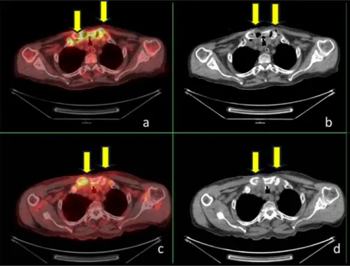
Axial slices of computed tomography (CT) (A), F-18 fluorodeoxyglucose (FDG) positron emission tomography (PET)/CT fusion (B) and F-18 FDG PET (C) images of a 64-year-old

The results of the present study (97.93%) corroborates with these three studies in terms of vocabulary coverage, and it means that the model predicting the equation

Araştırmamızda bebeklerin gestasyon yaşlarına ve doğum kilolarına göre ebeveynlerin YYBÜ Anne Baba Stres Ölçeğinden aldıkları puan ortalamaları arasında

Our results reveal that plasma-cleaned titanium surfaces provide a clean and reproducible starting condition for further plasma treatments to form well-controlled surface