Ankara Üniv Vet Fak Derg, 60, 185-187, 2013
Leishmania sp. in Visceral Leishmaniasis suspected patients in
Kayseri
Suleyman YAZAR, Salih KUK, Ulfet CETINKAYA, Yunus UYAR, Izzet SAHIN
Erciyes University, Medical Faculty, Department of Parasitology, Kayseri, Turkey.
Summary: Visceral leishmaniasis (VL), also known as dumdum fever or kala-azar, is characterized by intermittent fever,
substantial weight loss, hepatosplenomegaly and pancytopenia. If left untreated, the disease can have a fatality rate as high as 100% within two years. This study was aimed to investigate the presence of Leishmania sp. in VL suspected patients who applied to Erciyes University Medical Faculty Department of Parasitology between January 1995 and August 2012. Bone marrow samples obtained from 37 (54.4%) males and 31 (45.6%) females making a total of 68 VL suspected patients between January 1995 and January 2004 were examined by Giemsa stain technique. Microscopic examination showed Leishmania amastigotes in 4 (5.9%) patients. Bone marrow and sera samples obtained from 42 (55.3%) males and 34 (44.7%) females making a total of 76 VL suspected patients between January 2004 and August 2012 were examined by Giemsa stain technique and Indirect Fluorescent Antibody Test (IFAT). Three (3.9%) patients were found to be positive with both IFAT and Giemsa stain technique. Four (5.3%) patients were found to be positive with only IFAT. As a result, VL is a health problem in Kayseri and the fight against VL must be supported by special programs. Therefore, we believe that it should be safer to use both Giemsa stain technique and IFAT together for diagnosis of VL.
Key words: Giemsa, IFAT, Kayseri, Visceral leishmaniasis.
Kayseri'de Visseral Leishmaniasis şüpheli hastalarda Leishmania sp.
Özet: Dumdum ateşi veya kala-azar olarak bilinen, aralıklı ateş, kilo kaybı, hepatosplenomegali ve pansitopeni ile karakterize
visseral leishmaniasis (VL), tedavi edilmezse iki yılda %100 ölüm oranına sahip olabilmektedir. Bu çalışmada, Ocak 1995 ve Ağustos 2012 arasında Erciyes Üniversitesi Tıp Fakültesi Parazitoloji Anabilim Dalı'na VL şüphesi ile başvuran hastalarda
Leishmania sp. araştırılması amaçlanmıştır. Ocak 1995 ile Ocak 2004 tarihleri arasında 37 (%54.4)’si erkek ve 31 (%45.6)’i kadın
olmak üzere toplam 68 VL şüpheli hastadan alınan kemik iliği örneği Giemsa boyama tekniği ile incelenmiştir. Mikroskobik inceleme sonucunda 4 (%5.9) hastada Leishmania amastigotları tespit edilmiştir. Ocak 2004 ve Ağustos 2012 tarihleri arasında 42 (%55.3)'si erkek ve 34 (%44.7)'ü kadın olmak üzere toplam 76 VL şüpheli hastadan alınan kemik iliği ve serum örnekleri Giemsa boyama ve İndirekt Fluoresan Antikor (IFA) teknikleri ile incelenmiştir. İncelenen hastalardan 3 (%3.9)'ü hem IFA hem de Giemsa boyama tekniği ile pozitif bulunmuştur. Dört (%5.3) hasta ise yalnızca IFAT ile pozitif bulunmuştur. Sonuç olarak VL Kayseri’de bir sağlık sorunudur ve VL'ye karşı mücadele özel programlar ile desteklenmelidir. Bununla birlikte, VL tanısında IFA ve Giemsa boyamanın birlikte kullanılması daha güvenli olacaktır.
Anahtar sözcükler: Giemsa, IFAT, Kayseri, Visseral leishmaniasis.
Introduction
Leishmaniasis is a parasitic disease that is widely seen in tropical and subtropical regions of the World. It is caused by obliged intracellular protozoa of the genus
Leishmania and human become infected by the bite of
phlebotomine sandflies. These are present in three different forms: visceral leishmaniasis (VL), cutaneous leishmaniasis (CL), and mucocutaneous leishmaniasis (ML), but two forms are observed in Turkey; VL and CL. VL, also known as dumdum fever or kala-azar is a systemic infectious disease characterized by intermittent fever, substantial weight loss, hepatosplenomegaly anemia and leucopenia. If left untreated, the disease can
have a fatality rate as high as 100% within two years. Therefore, in this disease, early diagnosis is of great importance (3-5.12). The most common method of diagnosing of VL is microscopically direct examination of biopsy material of bone marrow. For definitive diagnosis of parasite, identification of the pathogen infected tissue samples is required. Additionally, serological and molecular methods are used for the diagnosis of this disease (4, 12). In this study, it was aimed to investigate the presence of Leishmania sp. in VL suspected patients who applied to Erciyes University Medical Faculty Department of Parasitology between January 1995 and August 2012.
Suleyman Yazar - Salih Kuk - Ulfet Cetinkaya - Yunus Uyar - Izzet Şahin 186
Materials and Methods
Bone marrow samples obtained from 37 (54.4%) males and 31 (45.6%) females making a total of 68 VL suspected patients between January 1995 and January 2004 were examined by Giemsa stain technique. Bone marrow and sera samples obtained from 42 (55.3%) males and 34 (44.7%) females making a total of 76 VL suspected patients January 2004 and August 2012 were examined by Giemsa stain technique and Indirect Fluorescent Antibody Test (IFAT). For IFAT, the Euroimmun (Germany) Kit was used following the manufacturer’s instructions.
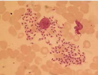
Figure 1: Bone marrow aspirate smear; Leishmania sp. within reticulo-endothelial macrophage. (Giemsa x1000).
Şekil 1: Kemik iliği yayma preparatı; Retiküloendotelial makrofajlar içerisinde Leishmania sp. (Giemsa x1000).
Figure 2: Total number and percentage of patients with visceral leishmaniasis according to region.
Şekil 2: Bölgelere gore visseral leishmaniasisli hasta sayı ve yüzdeleri.
Results
Microscopic examination showed Leishmania amastigotes in 4 (5.9%) patients between January 1995 and January 2004 were examined by Giemsa stain technique (Figure 1). Bone marrow and sera samples
obtained from 76 patients between January 2004 and August 2012 were examined by Giemsa stain technique and IFAT. Three (3.9%) patients were found to be positive with both IFAT and Giemsa stain technique. Four (5.3%) patients were found to be positive with only IFAT. Seven (63.6%) out of 11 infected patients were men while 4 (36.4%) were females. 9 (81.8%) out of 11 infected patients were in pediatric patients.
Nine patients from Kayseri, one patient from Mersin and one patient from Niğde, a total 11 patients with VL, were admitted to our hospital. Seven out of nine patients from Kayseri that were admitted to our hospital reside in Yahyalı, one of them resides in Felahiye and another one of them resides in Melikgazi (Figure 2).
Discussion and Conclusion
Leishmaniasis caused by Leishmania species occurs in five continents and is endemic in tropical and subtropical regions of 88 countries. The geographic distribution of leishmaniasis is limited by the distribution of the sandfly, its susceptibility to cold climates, and its capacity to support the internal development of specific
Leishmania spp. There are an estimated 12 million cases
of leishmaniasis worldwide, 2 million new cases occur each year, and 350 million people are at risk (6). VL is still significant public health problems in Turkey, but a decline is observed in the number of cases with both infections in recent years (10). In endemic areas, the disease tends to be relatively chronic, and children are especially affected. 81.8% in our infected patients were younger than ≤8 years. These findings were also similar to those reported in the other Mediterranean countries and Turkey (1, 7-9, 13). Examination of bone marrow smears is an easy method for diagnosing VL and was positive in 55- 80% of cases. Culturing of blood or organ aspirates increases the sensitivity of diagnosis. Liver biopsy and spleen puncture are valuable diagnostic methods, but both carry huge risks including life threatening risks. ELISA and IFAT in serological diagnostic tests are reliable diagnostic methods. IFAT is a method frequently used for the diagnosis of VL. In this test promastigot or amastigote forms as the antigen are used (12). Asymptomatic and IFAT (+) cases have been reported (11). In a study from Turkey, Yilmaz and his coworkers reported that all patients were determined to be positive by IFAT, only 69.2% of patients were determined to be positive by Giemsa stain technique (1). In another study, Balcı and his coworkers (2) reported that all patients were determined to be positive by IFAT, but 2 patients were not found to be positive by Giemsa stain technique. In this study, we determined that 4 (5.3%) patients were found to be positive with only IFAT.
Ankara Üniv Vet Fak Derg, 60, 2013 187
As a result, VL is a health problem in Kayseri and the fight against VL must be supported by special programs. Therefore, we believe that it should be safer to use both together Giemsa stain technique and IFAT for diagnosis of VL.
Acknowledgement
This article was presented at 1st National Symposium
on Vectors and Vector-Borne with International Participation, 9-10 September, 2012, Avanos, Cappadocia, Nevsehir, Turkey.
References
1. Arik Yilmaz E, Tanir G, Tuygun N, Taylan Ozkan A (2009): Visceral leishmaniasis in 13 pediatric patients in
Turkey: treatment experience. Turkiye Parazitol Derg, 33,
259-262.
2. Balcı YI, Türk M, Özgür A, Küçüktaşçı K (2011): Dört
Çocuk Hastada İndirekt Floresan Antikor Test Kullanımı ile Visceral Leishmaniasis Tanısının Değerlendirilmesi.
35(2), 114-116.
3. Belazzoug S (1992): Leishmaniasis in Mediterranean
countries. Vet Parasitol, 44, 15-19.
4. Berman JD (1997): Human Leishmaniasis: clinical,
diagnostic, and chemotherapeutic developments in the last 10 years. Clin Infect Dis, 24, 684-703.
5. Blackwell JM, Fakiola M, Ibrahim ME, Jamieson SE, Jeronimo SB, Miller EN, Mishra A, Mohamed HS, Peacock CS, Raju M, Sundar S, Wilson ME (2009):
Genetics and visceral leishmaniasis: of mice and man.
Parasite Immunol, 31, 254-266.
6. Desjeux P, (1998): UNAIDS. Leishmania and HIV in Gridlock. In PF Beales, World Health Organization and United Nations Programme on HIV/AIDS, WHO/CTD/ LEISH/98.9 Add 1.
7. Dursun O, Erişir S, Yeşilipek A (2009): Visceral
childhood leishmaniasis in southern Turkey: experience of twenty years. Turk J Pediatr, 51, 1-5.
8. Maltezou HC, Siafas C, Mavrikou M, Spyridis P, Stavrinadis C, Karpathios T, Kafetzis DA (2000):
Visceral leishmaniasis during childhood in southern Greece. Clin Infect Dis, 31, 1139-1143.
9. Ok UZ, Balcioglu IC, Taylan Ozkan A, Ozensoy S, Ozbel Y (2002): Leishmaniasis in Turkey. Acta Trop, 84, 43-48.
10. Ozensoy S, Ozbel Y, Turgay N, Alkan MZ, Gul K, Gilman-Sachs A, Chang KP, Reed SG, Ozcel MA (1998): Serodiagnosis and epidemiology of visceral
leishmaniasis in Turkey. Am J Trop Med Hyg, 59,
363-369.
11. Ozensoy Töz S, Sakru N, Ertabaklar H, Demir S, Sengul M, Ozbel Y (2009): Serological and entomological
survey of zoonotic visceral leishmaniasis in Denizli Province, Aegean Region, Turkey. New Microbiol, 32,
93-100.
12. Srivastava P, Dayama A, Mehrotra S, Sundar S (2011):
Diagnosis of visceral leishmaniasis. Trans R Soc Trop
Med Hyg, 105(1), 1-6.
13. Tanir G, Taylan Ozkan A, Dağlar E (2006). Pediatric
visceral Leishmaniasis in Turkey. Pediatr Int, 48, 66-69. Geliş tarihi: 12.02.2013 / Kabul tarihi: 28.03.2013
Address for correspondence:
Prof. Dr. Süleyman Yazar
Erciyes University, Medical Faculty Department of Parasitology, 38039 Kayseri-Turkey