R E S E A R C H A R T I C L E
Open Access
High sensitivity C-reactive protein and cerebral
white matter hyperintensities on magnetic
resonance imaging in migraine patients
Aynur Yilmaz Avci
1*, Hatice Lakadamyali
2, Serap Arikan
3, Ulku Sibel Benli
1and Munire Kilinc
1Abstract
Background: Migraine is a common headache disorder that may be associated with vascular disease and cerebral white matter hyperintensities (WMHs) on magnetic resonance imaging (MRI) scan. High sensitivity C-reactive protein (hs-CRP) is a marker of inflammation that may predict subclinical atherosclerosis. However, the relation between migraine, vascular risks, and WMHs is unknown. We evaluated hs-CRP levels and the relation between hs-CRP level and WMHs in adult migraine patients.
Methods: This case–control study included 432 subjects (216 migraine patients [without aura, 143 patients; with aura, 73 patients]; 216 healthy control subjects without migraine; age range 18–50 y). Migraine diagnosis was determined according to the International Classification of Headache Disorders II diagnostic criteria. The migraine patients and control subjects had no known vascular risk factors, inflammatory disease, or comorbid disease. The presence and number of WMHs on MRI scans were determined, and serum hs-CRP levels were measured by latex-enhanced immunoturbidimetry.
Results: Mean hs-CRP level was significantly greater in migraine patients (1.94 ± 2.03 mg/L) than control subjects (0.82 ± 0.58 mg/L; P ≤ .0001). The mean number of WMHs per subject and the presence of WMHs was significantly greater in migraine patients (69 patients [31.9%]; 1.68 ± 3.12 mg/dL) than control subjects (21 subjects [9.7%]; 0.3 ± 1.3; P ≤ .001). However, there was no correlation between hs-CRP level and WMHs in migraine patients (r = 0.024; not significant). The presence of WMHs was increased 4.35-fold in migraine patients (odds ratio 4.35, P ≤ .001). Conclusions: High hs-CRP level may be a marker of the proinflammatory state in migraine patients. However, the absence of correlation between hs-CRP level and WMHs suggests that hs-CRP is not causally involved in the
pathogenesis of WMHs in migraine patients. The WMHs were located mostly in the frontal lobe and subcortical area. Keywords: Headache; Inflammation; Vascular disease; Pathophysiology
Background
Migraine is a common neurologic disorder, typically cha-racterized by recurrent attacks of debilitating headache and symptoms of autonomic nervous system dysfunc-tion. In one third patients, migraine attacks are accom-panied by transient focal neurologic aura symptoms. The frequency of migraine is 3-fold greater in women than men [1]. Migraine prevalence is 8.6% in males, 17.5% in females, and 13.2% overall in the United States
[2]. In Turkey, migraine prevalence is 8.5% in males and 24.6% in females, and the 1-year prevalence of migraine (16.4%) is similar or higher than the prevalence world-wide [3]. The risk of having migraine is greater in women aged < 45 years, and hormonal effects may be a causal fac-tor for this female predominance [4]. Migraine is associ-ated with an increased risk of developing cardiovascular disease and a 2-fold increased risk of developing ischemic stroke [4-7]. The relation between migraine and the risk of vascular disease may be explained, in part, by the higher prevalence of multiple risk factors in migraine patients [4-6]. Additionally, the association between migraine with aura and ischemic stroke is more apparent for individuals
* Correspondence:[email protected]
1
Department of Neurology, Baskent University, Saray Mah, Yunusemre cad, No. 1, Alanya-Antalya 07400 Ankara, Turkey
Full list of author information is available at the end of the article
© 2015 Avci et al.; licensee Springer. This is an Open Access article distributed under the terms of the Creative Commons Attribution License (http://creativecommons.org/licenses/by/4.0), which permits unrestricted use, distribution, and reproduction in any medium, provided the original work is properly credited.
without vascular risk factors [8-10]. Migraine also has also been associated with hemorrhagic stroke [11].
Migraine is a neurovascular disorder associated with cortical spreading depression, neurogenic inflammation, and cranial vascular contractile dysfunction. Activation of brain tissue causes release of peptides from the peri-vascular trigeminal regions that cause inflammation and dilation of extraparenchymal vessels. Repeated migraine attacks are associated with inflammatory arteriopathy of the cranial vessels [12,13]. In migraine, specific abnor-malities of inflammatory marker levels in the systemic circulation have been observed, including increased levels of C-reactive protein (CRP), interleukins, and adhesion molecules [13-15].
Migraine is a risk factor for white matter hyperinten-sities (WMHs), which are infarct-like lesions associated with volume changes in the brain (grey matter and white matter regions) [16]. Incidental findings on brain mag-netic resonance imaging (MRI) scans are common, and incidental findings detection is more likely using high resolution MRI sequences than standard resolution se-quences [17-20]. The pathophysiology of WMHs is un-known. Increased age and atherosclerosis may be the main risk factors for the development of WMHs [17,20,21]. In migraine, the duration of disease and attack frequency are important in the development of WMHs, and comorbid disease also may contribute to the development of WMHs [18,20,21]. In migraine, cumulative effects of repeated in-tracerebral hemodynamic changes may contribute to the development of WMHs, including oligemia, focal hypo-perfusion with ischemia, and hypoxia below the ischemic threshold [22].
High sensitivity C-reactive protein (hs-CRP) is a mar-ker of inflammation, and hs-CRP levels may increase in vascular diseases such as myocardial infarction and is-chemic stroke or in healthy individuals who have no car-diovascular disease [23]. The hs-CRP level has prognostic use in the Framingham coronary risk score, severity of metabolic syndrome, severity of hypertension, and pa-tients who have or do not have subclinical atherosclerosis [23,24]. The association of CRP level with migraine has been shown in small case–control studies of migraine with vascular risk factors and a large prospective cohort study of women aged > 45 years [15,25-27]. Higher CRP levels were associated with the presence and progression of peri-ventricular and subcortical WMHs, independent of car-diovascular risk factors and carotid atherosclerosis, in old nondemented patients [28-30].
The complex mechanisms involved in migraine and mechanisms linking migraine and vascular risks are in-completely understood [4,5]. The distribution of brain WMHs in patients who have migraine is unclear, and limited information is available about the complex rela-tion between migraine, ischemia, and WMHs [18-20].
We hypothesized that hs-CRP levels may be associated with migraine disease, and that high hs-CRP levels also may be associated with the presence and distribution of cerebral WMHs on brain MRI scans in migraine pa-tients. The purpose of the present study was to analyze the relation between hs-CRP levels and WMHs in mi-graine patients.
Methods
Subjects
This case–control clinical study was performed with pa-tients who had migraine newly diagnosed at the neur-ology outpatient clinics of Baskent Medical Faculty from October 2011 to December 2013 and who had brain MRI scans as a part of their evaluation. Inclusion criteria were duration of migraine symptoms≥ 1 year, headache frequency≥ 2 attacks/month, and absence of any known vascular risk factors, inflammatory disease, chronic illness, metabolic disease, or infections. Patients were excluded for (1) migraine duration < 1 year; (2) history of cerebro-vascular or cardiocerebro-vascular disease, arterial hypertension (blood pressure > 130/80 mm Hg), diabetes mellitus, or hyperlipidemia (low-density lipoprotein cholesterol≥ 160 mg/dL); (3) body mass index < 18 kg/m2or > 35 kg/m2; (4) abnormal plasma hs-CRP level (>10 mg/L); (5) smoking cigarettes > 1 pack/day; (6) current pregnancy, lactation, or hormonal contraceptive use; (7) alcohol or substance abuse; (8) drug use such as antiplatelet agents, anticoagulants, sta-tins, or hormonal drugs; (9) renal, metabolic, psychiatric, inflammatory, infectious, or immune disease; (10) musculo-skeletal disorders or fibromyalgia; or (11) possible “symp-tomatic migraine” in which the MRI showed arteriovenous malformations, ischemic infarcts, brain tumors, or other conditions that may be associated with migraine. In ad-dition, patients who had high levels of hs-CRP (≥10 mg/L) were excluded from the study because high levels of hs-CRP (>10 mg/L) may represent nonspecific inflam-mation and lack positive predictive value [23,24]. There were 300 consecutive migraine patients considered for the study, and 84 patients were excluded (infection, 17 pa-tients; thyroid disease, 12 patients, thyroid stimulating hormone, range 3.5 - 4.9 μIU/mL; no laboratory tests available, 11 patients; declined MRI scan, 10 patients; low-density lipoprotein cholesterol > 160 mg/dL, 9 pa-tients; positive antinuclear antibody, 9 papa-tients; hs-CRP > 10 mg/L, 8 patients; antidouble-stranded DNA autoanti-body, 5 patients; silent lacunar infarct, 3 patients). The other 216 consecutive, newly diagnosed migraine patients were included in the study (165 women [76%] and 51 men [24%]; age range, 18–50 y [mean age, 31 ± 7 y]).
Healthy control subjects (216 subjects: 150 women [69.4%] and 66 men [30.6%]) aged between 18 and 50 years (mean age, 32.46 ± 7.54) were recruited consecutively from hospital staff, laboratory staff, relatives of patients,
and the general population. Inclusion criteria for the con-trol subjects were (1) absence of headaches such as mi-graine, tension-type headache, or cluster headache; (2) absence of other neurologic or systemic disease; and (3) presence of a match with migraine patients by age (±2 y), sex, body mass index, education level, and smoking habits. Exclusion criteria for control subjects were the same as for the migraine group. The study was approved by the local ethics committee of the Medical Faculty of Baskent University Hospital, and all migraine and control subjects gave informed consent to participate in the study and have MRI scans and laboratory tests.
Evaluation
Patients were diagnosed as having migraine according to the criteria of the International Classification of Headache Disorders II [1]. A detailed history of migraine was ob-tained including disease duration (y), age at onset, aver-age duration of current headache (h), presence of aura, trigger factors, accompanying symptoms, frequency per month, and location and severity of pain. Severity of headache was evaluated with visual analog score (range, 1 [minimum pain] to 10 [maximum pain]). Migraine headache attack frequency was defined as the number of attacks per month. All patients and control subjects received a complete physical and neurologic examination. Comorbidities (coronary artery disease, stroke, diabetes mellitus, or thyroid disease) and intercurrent illnesses such as respiratory or urinary infections were determined from the patient history, physical examination, and labora-tory tests (biochemical and hematologic tests). Blood pres-sure, body weight, height, smoking habits, and education level were recorded for all migraine patients and control subjects. No migraine patients took any medication within 3 days before blood sampling. Patients previously had used medications for acute pain such as acetaminophen, nonsteroidal anti-inflammatory drugs, triptans, or caffeine for headache, but patients who used analgesics daily were excluded from the study. Patients who were treated for migraine prophylaxis with drugs such as propranolol, topiramate, or valproic acid were excluded.
White matter hyperintensities
All 432 participants had cerebral MRI brain scanning (1.5 Tesla, Siemens Magnetom Vision Plus, Siemens, Munich, Germany) with the orbitomeatal line as reference. The scans included≥ 3 sequences: sagittal T1-weighted, axial T2-weighted, and axial fluid attenuated inversion re-covery (FLAIR) images. The slice thickness was 5 mm, the gap was 1 mm, and no intravenous contrast was used.
All MRI scans were reviewed and scored by a radiolo-gist who was blinded to the clinical details. The scans were visually assessed for the presence and features of WMHs including appearance, number, size, distribution
(infratentorial or supratentorial), and anatomic location. The number and size of the WMHs were determined on the FLAIR images and grouped according to location and distribution. Subgroups were delineated according to the distribution of the WMH following the method-ology previously described in multiple sclerosis patients [31]. These subgroups were juxtacortical, subcortical, and periventricular. The locations of WMHs were defined as frontal, parietal, temporal, occipital, or infratentorial. Peri-ventricular WMHs were defined as being anterior, poster-ior, or located at the lateral band. We included focal and punctate hyperintensities (size < 9 mm). Confluent and large hyperintensity lesions (>9 mm) were excluded. There were no WMHs at the corpus callosum. The McDonald and Barkhof MRI diagnostic criteria for dissemination in space in multiple sclerosis were applied to each patient to determine whether the criteria were satisfied [31,32].
Migraine and control subjects who had WMHs that were detected on brain MRI were evaluated with labora-tory tests for vasculitis (anticardiolipin antibodies, antinu-clear antibody, lupus anticoagulant, antidouble-stranded DNA autoantibody, and C3 and C4 levels). Migraine and control subjects who had WMHs underwent cardiac exa-mination and transthoracic echocardiography to exclude patent foramen ovale and atrial septal defect; only 1 pa-tient was excluded because of atrial septal defect on echocardiography.
High sensitivity C-reactive protein
Blood samples were obtained from the antecubital vein from control subjects and during a headache-free period from migraine patients. To exclude the potential effects of a recent attack, migraine patients had been free of mi-graine attack for≥ 3 days before blood sampling. No subjects had taken anti-inflammatory drugs for≥ 3 days before the study because these drugs may be associa-ted with improved endothelial function and might have affected the results. Phlebotomy tubes contained no anticoagulant. Blood was centrifuged at 3000 × g for 10 minutes and stored at−20°C until analysis. Serum hs-CRP was measured by latex-enhanced immunoturbidimetry using monoclonal anti-CRP antibodies (Architect C 800, Abbott Diagnostic Systems, Abbott Park, IL, USA) (hs-CRP reference level, ≤ 5 mg/L).
Statistical analysis
Data analysis was performed with statistical software (IBM SPSS Statistics for Windows, Version 21.0, IBM Corp., Armonk, NY, USA). Statistical analysis of the nu-meric parameters that were normally distributed was performed with independent t test. Data that did not satisfy normal distribution approximation were analyzed with nonparametric Mann–Whitney test. Average values were reported as mean ± standard deviation (SD), and
statistical analysis was performed with median values. Categorical and ordinal data were analyzed with Pearson chi-square and Fisher exact chi-square tests. Correlation analysis was performed with Spearman rank correlation. Factors affecting hs-CRP were investigated with regression analysis. However, to prove hypotheses of the regression analysis, transformation to the hs-CRP variable was ap-plied to satisfy the normal distribution condition of the parameter. Factors affecting the presence of WMHs were analyzed with multiple logistic regression. In addition, de-scriptive statistics for categorical variables were specified as number (%), and median statistics for numeric variables were reported with range (minimum to maximum) and mean ± SD. In all analyses, statistical significance was defined by P≤ .05 and decisions were at the 95% confi-dence level.
Results
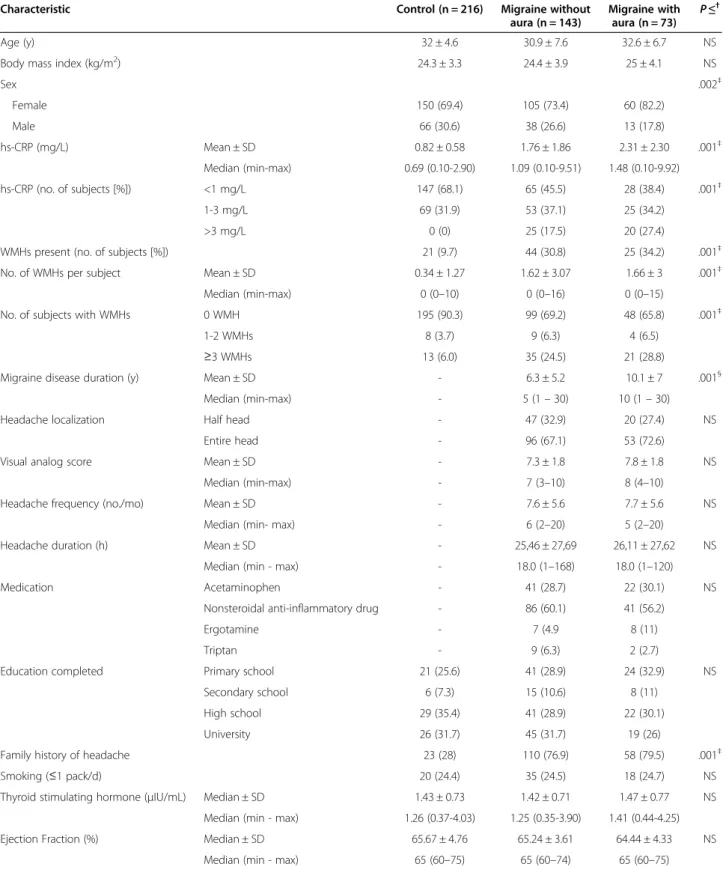
In the 216 consecutive, newly diagnosed migraine pa-tients who were included in the study, migraine without aura was diagnosed in 143 patients (66%) and migraine with aura was diagnosed in 73 patients (34%) (Table 1). The migraine and control groups were similar in mean age, body mass index, education, and frequency of smokers (Table 1). Frequency of family history of graine and mean hs-CRP levels were similar between mi-graine patients with or without aura and were greater in migraine patients than control subjects (Table 1). Mean duration of migraine disease was statistically significantly greater in patients who had migraine with than with-out aura (Table 1). There was no statistically signifi-cant difference between the migraine groups in headache localization, headache duration, visual analog score, head-ache frequency, medication, education level, or smoking habits (Table 1).
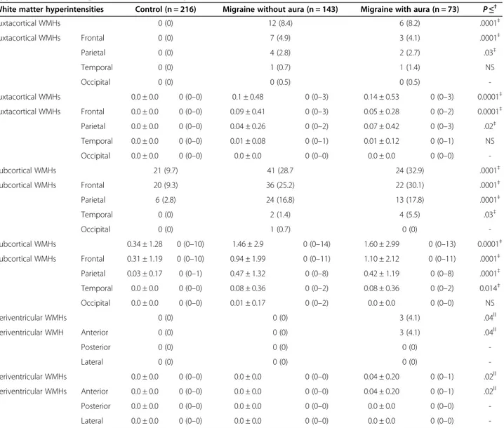
The presence of WMHs and mean number of WMHs per subject were significantly higher in migraine patients (69 patients [31.9%]; 1.68 ± 3.12 mg/dL) than control subjects (21 subjects [9.7%]; 0.3 ± 1.3; P≤ .0001) (Table 1). In the 69 migraine patients who had WMHs, 67 patients (97%) had supratentorial WMHs and 2 patients (3%) had infratentorial WMHs. In 15 of 69 (10.4%) migraine patients who had WMHs, the WMHs were present in > 1 anatomic location. In migraine patients, WMH diam-eter was≤ 3 mm in 63 patients (91%) and 4 to 9 mm in 6 patients (9%). In all control subjects who had WMHs (21 patients [9.7%]), WMH diameter was≤ 3 mm, and no infratentorial lesions were present.
The distribution of WMHs was significantly different between migraine and control subjects (Table 2). The presence and the number of WMHs per subject (juxta-cortical, sub(juxta-cortical, and periventricular) were signifi-cantly higher in migraine patients than control subjects (P≤ .001) (Table 2). The WMHs in the migraine and
control groups were detected most frequently in the frontal lobe and least frequently in the occipital lobe (Table 2) (P≤ .05). In the control group, the WMHs were detected only in the subcortical region (Table 2). Frequencies of juxtacortical and subcortical WMHs were similar between migraine patients with and without aura (not significant). Only migraine patients with aura had periventricular WMHs (Table 2), which were located in the anterior horn of the lateral ventricles; no patients had WMHs in the lateral ventricular bands or posterior horn of lateral ventricles (Table 2). In the 69 migraine patients, the WMHs were contiguous with the cortex in 18 patients (26%) and with the periventricular structure in 3 patients (4%) (Table 2). In 69 migraine patients who had WMHs, only 1 patient satisfied the 2010 McDonald criteria [32]. No patients satisfied the Barkhof criteria or radiologically isolated syndrome criteria [31,33].
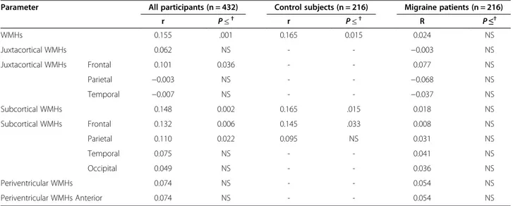
There was no statistically significant correlation bet-ween hs-CRP level and WMHs in migraine and control subjects (r = 0.155; P > .001) (Table 3). In migraine pa-tients, the hs-CRP levels were not significantly different between juxtacortical, subcortical, and periventricular WMHs (Table 4). There was no correlation between hs-CRP and headache characteristics (r < 0.042; not signifi-cant) (Table 4). The hs-CRP level was similar for females and males, and the presence and number of WMHs/ subject were similar between females and males (Table 4). The WMHs (juxtacortical, subcortical, and periventri-cular) were not correlated with headache characteris-tics (migraine duration, visual analog score, or headache duration) (not significant).
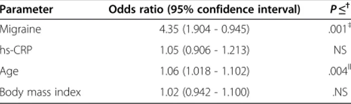
Migraine disease was associated with a 4.35-fold in-creased risk of presence of WMHs (Table 5). In addition, age 1 year was associated with a 1.06-fold increased risk of the presence of the WMHs (Table 5).
Discussion
In the present study, serum hs-CRP levels were signifi-cantly higher in migraine patients than control subjects. Based on published Framingham coronary risk criteria, 57.9% migraine patients had moderate to high vascular risk [23,24]. The present data did not show associations between hs-CRP level and distribution of WMHs in mi-graine patients (Table 3). The prevalence of WMHs was 4.35-fold higher in migraine patients than control jects (Table 5). The WMHs primarily involved the sub-cortical region and frontal lobe (Table 2). Only 1 migraine patient satisfied the revised 2010 McDonald criteria for multiple sclerosis [32].
A unique feature of the present study design was the simultaneous assessment of the associations of hs-CRP and WMHs in migraine patients. The participants had no known vascular risk factors or inflammatory disease. We included patients aged < 50 years to minimize the
Table 1 Characteristics of participants in study of migraine*
Characteristic Control (n = 216) Migraine without
aura (n = 143)
Migraine with aura (n = 73) P ≤
†
Age (y) 32 ± 4.6 30.9 ± 7.6 32.6 ± 6.7 NS
Body mass index (kg/m2) 24.3 ± 3.3 24.4 ± 3.9 25 ± 4.1 NS
Sex .002‡ Female 150 (69.4) 105 (73.4) 60 (82.2) Male 66 (30.6) 38 (26.6) 13 (17.8) hs-CRP (mg/L) Mean ± SD 0.82 ± 0.58 1.76 ± 1.86 2.31 ± 2.30 .001‡ Median (min-max) 0.69 (0.10-2.90) 1.09 (0.10-9.51) 1.48 (0.10-9.92) hs-CRP (no. of subjects [%]) <1 mg/L 147 (68.1) 65 (45.5) 28 (38.4) .001‡ 1-3 mg/L 69 (31.9) 53 (37.1) 25 (34.2) >3 mg/L 0 (0) 25 (17.5) 20 (27.4)
WMHs present (no. of subjects [%]) 21 (9.7) 44 (30.8) 25 (34.2) .001‡
No. of WMHs per subject Mean ± SD 0.34 ± 1.27 1.62 ± 3.07 1.66 ± 3 .001‡
Median (min-max) 0 (0–10) 0 (0–16) 0 (0–15)
No. of subjects with WMHs 0 WMH 195 (90.3) 99 (69.2) 48 (65.8) .001‡
1-2 WMHs 8 (3.7) 9 (6.3) 4 (6.5)
≥3 WMHs 13 (6.0) 35 (24.5) 21 (28.8)
Migraine disease duration (y) Mean ± SD - 6.3 ± 5.2 10.1 ± 7 .001§
Median (min-max) - 5 (1– 30) 10 (1– 30)
Headache localization Half head - 47 (32.9) 20 (27.4) NS
Entire head - 96 (67.1) 53 (72.6)
Visual analog score Mean ± SD - 7.3 ± 1.8 7.8 ± 1.8 NS
Median (min-max) - 7 (3–10) 8 (4–10)
Headache frequency (no./mo) Mean ± SD - 7.6 ± 5.6 7.7 ± 5.6 NS
Median (min- max) - 6 (2–20) 5 (2–20)
Headache duration (h) Mean ± SD - 25,46 ± 27,69 26,11 ± 27,62 NS
Median (min - max) - 18.0 (1–168) 18.0 (1–120)
Medication Acetaminophen - 41 (28.7) 22 (30.1) NS
Nonsteroidal anti-inflammatory drug - 86 (60.1) 41 (56.2)
Ergotamine - 7 (4.9 8 (11)
Triptan - 9 (6.3) 2 (2.7)
Education completed Primary school 21 (25.6) 41 (28.9) 24 (32.9) NS
Secondary school 6 (7.3) 15 (10.6) 8 (11)
High school 29 (35.4) 41 (28.9) 22 (30.1)
University 26 (31.7) 45 (31.7) 19 (26)
Family history of headache 23 (28) 110 (76.9) 58 (79.5) .001‡
Smoking (≤1 pack/d) 20 (24.4) 35 (24.5) 18 (24.7) NS
Thyroid stimulating hormone (μIU/mL) Median ± SD 1.43 ± 0.73 1.42 ± 0.71 1.47 ± 0.77 NS
Median (min - max) 1.26 (0.37-4.03) 1.25 (0.35-3.90) 1.41 (0.44-4.25)
Ejection Fraction (%) Median ± SD 65.67 ± 4.76 65.24 ± 3.61 64.44 ± 4.33 NS
Median (min - max) 65 (60–75) 65 (60–74) 65 (60–75)
*Data reported as mean ± SD, number of subjects (%), or median (range, minimum to maximum). Abbreviations: hs-CRP high sensitivity C-reactive protein, WMH white matter hyperintensity.
†NS, not significant (P > .05).
‡Significant difference between migraine patients and control subjects. §
effects of age-dependent WMHs. Limitations of the pre-sent study included the limited number of participants. In addition, an MRI scanner with a higher magnetic field than the scanner used may be more sensitive in detect-ing WMHs on FLAIR images. A more sensitive imagdetect-ing protocol with thin slices (thickness, 3 mm) might have improved the detection of smaller lesions. Furthermore, some risk factors such as smoking and obesity were not strictly excluded; although we excluded heavy smokers (>1 pack/d), we did not exclude all cigarette smokers. There were no statistically significant differences between the migraine and control groups in smoking habits. Al-though all participants had body mass index≤ 35 kg/m2, some participants had body mass index > 30 kg/m2, and
CRP level may increase with obesity [34]. Nevertheless, we observed no significant differences between the migraine and control groups in body mass index. In addition, it may be important to study other markers of endothelial dysfunction or inflammation such as tissue plasminogen activator antigen, von Willebrand factor activity, homo-cysteine level, or inflammatory cytokine levels to further evaluate the association of hs-CRP in the development of WMHs in migraine patients. We did not perform carotid Doppler studies to evaluate the possibility of microemboli. The observed high hs-CRP levels in migraine patients were consistent with previous reports (Table 1) [15,25-27]. In contrast with the present study, the Reykjavik study re-ported that CRP levels were similar in migraine patients
Table 2 Anatomic location and distribution of juxtacortical, subcortical, and periventricular white matter hyperintensities in migraine and control subjects*
White matter hyperintensities Control (n = 216) Migraine without aura (n = 143) Migraine with aura (n = 73) P ≤†
Juxtacortical WMHs 0 (0) 12 (8.4) 6 (8.2) .0001‡ Juxtacortical WMHs Frontal 0 (0) 7 (4.9) 3 (4.1) .0001‡ Parietal 0 (0) 4 (2.8) 2 (2.7) .03‡ Temporal 0 (0) 1 (0.7) 1 (1.4) NS Occipital 0 (0) 0 (0.5) 0 (0.5) -Juxtacortical WMHs 0.0 ± 0.0 0 (0–0) 0.1 ± 0.48 0 (0–3) 0.14 ± 0.53 0 (0–3) 0.0001‡ Juxtacortical WMHs Frontal 0.0 ± 0.0 0 (0–0) 0.09 ± 0.41 0 (0–3) 0.05 ± 0.28 0 (0–2) 0.0001‡ Parietal 0.0 ± 0.0 0 (0–0) 0.04 ± 0.26 0 (0–2) 0.07 ± 0.42 0 (0–3) .02‡ Temporal 0.0 ± 0.0 0 (0–0) 0.01 ± 0.08 0 (0–1) 0.01 ± 0.12 0 (0–1) NS Occipital 0.0 ± 0.0 0 (0–0) 0.0 ± 0.0 0 (0–0) 0.0 ± 0.0 0 (0–0) -Subcortical WMHs 21 (9.7) 41 (28.7 24 (32.9) .0001‡ Subcortical WMHs Frontal 20 (9.3) 36 (25.2) 22 (30.1) .0001‡ Parietal 6 (2.8) 24 (16.8) 13 (17.8) .0001‡ Temporal 0 (0) 2 (1.4) 4 (5.5) .03‡ Occipital 0 (0) 1 (0.7) 0 (0) -Subcortical WMHs 0.34 ± 1.28 0 (0–10) 1.46 ± 2.9 0 (0–14) 1.60 ± 2.99 0 (0–13) 0.0001‡ Subcortical WMHs Frontal 0.31 ± 1.19 0 (0–10) 0.94 ± 1.99 0 (0–11) 1.10 ± 2.12 0 (0–11) .0001‡ Parietal 0.03 ± 0.17 0 (0–1) 0.47 ± 1.32 0 (0–8) 0.42 ± 1.19 0 (0–8) .0001‡ Temporal 0.0 ± 0.0 0 (0–0) 0.08 ± 0.36 0 (0–2) 0.08 ± 0.36 0 (0–2) 0.014‡ Occipital 0.0 ± 0.0 0 (0–0) 0.01 ± 0.17 0 (0–2) 0.0 ± 0.0 0 (0–0) NS Periventricular WMHs 0 (0) 0 (0) 3 (4.1) .04ǁ Periventricular WMH Anterior 0 (0) 0 (0) 3 (4.1) .04ǁ Posterior 0 (0) 0 (0) 0 (0) -Lateral 0 (0) 0 (0) 0 (0) -Periventricular WMHs 0.0 ± 0.0 0 (0–0) 0.0 ± 0.0 0 (0–0) 0.04 ± 0.20 0 (0–1) .02ǁ Periventricular WMHs Anterior 0.0 ± 0.0 0 (0–0) 0.0 ± 0.0 0 (0–0) 0.04 ± 0.20 0 (0–1) .02ǁ Posterior 0.0 ± 0.0 0 (0–0) 0.0 ± 0.0 0 (0–0) 0.0 ± 0.0 0 (0–0) -Lateral 0.0 ± 0.0 0 (0–0) 0.0 ± 0.0 0 (0–0) 0.0 ± 0.0 0 (0–0)
-*Data reported as number of patients (%), mean ± SD, or median (range, minimum to maximum). Abbreviation: WMHs deep white matter hyperintensities.
†NS, not significant (P > .05).
‡Comparison between control subjects and migraine patients (with and without aura). ǁComparison between migraine patients with and without aura.
and control subjects; vascular risk factors and concomi-tant disease were not eliminated, and the mean age of participants was 55 years [35]. Furthermore, another study reported no association between hs-CRP levels and migraine, and patients had no vascular risk factors; however, the sample was small, mean age of partici-pants was older than in the present study, and head-ache frequency and migraine disease duration were not reported [36]. In the present study, inclusion criteria ensured that headache frequency was≥ 2 headaches/mo
and migraine disease duration was > 1 year. Elevated hs-CRP level is a sensitive marker of inflammation and arteriosclerosis [23]. The higher mean level of hs-CRP in migraine patients than control subjects is evidence that inflammation may contribute to migraine (Table 1). In migraine, elevation of CRP may be caused by oxida-tive stress, leukocyte activation, and inflammatory dila-tion of blood vessels, and inflammatory cytokines are increased during acute attacks of migraine and between attacks [13,14].
Table 3 Relation between high sensitivity C-reactive protein and cerebral white matter hyperintensities on magnetic resonance imaging in migraine patients and control subjects*
Parameter All participants (n = 432) Control subjects (n = 216) Migraine patients (n = 216)
r P ≤† r P ≤† R P ≤† WMHs 0.155 .001 0.165 0.015 0.024 NS Juxtacortical WMHs 0.062 NS - - −0.003 NS Juxtacortical WMHs Frontal 0.101 0.036 - - 0.077 NS Parietal −0.003 NS - - −0.068 NS Temporal −0.007 NS - - −0.037 NS Subcortical WMHs 0.148 0.002 0.165 .015 0.018 NS Subcortical WMHs Frontal 0.132 0.006 0.145 .033 0.008 NS Parietal 0.110 0.022 0.095 NS 0.031 NS Temporal 0.075 NS - - 0.041 NS Occipital 0.049 NS - - 0.036 NS Periventricular WMHs 0.074 NS - - 0.054 NS Periventricular WMHs Anterior 0.074 NS - - 0.054 NS
*Data reported as correlation coefficient r between hs-CRP and parameter. Abbreviation: WMHs white matter hyperintensities.
†NS, not significant (P > .05).
Table 4 Relation between high sensitivity C-reactive protein and location of cerebral white matter hyperintensities, headache characteristics, and sex differences in migraine patients*
Parameters hs-CRP (mg/L) P ≤† Juxtacortical WMHs Present 1.95 ± 2,22 1.13 (0.21– 9.40) NS Absent 1.94 ± 2,01 1.25 (0.10– 9.92) Subcortical WMHs Present 2.01 ± 2.20 1.35 (0.10– 9.92) NS Absent 1.91 ± 1,95 1.18 (0.19– 9.80) Periventricular WMHs Present 1.91 ± 0.83 2.04 (1.02– 2.67) NS Absent 1.94 ± 2.04 1.24 (0.10– 9.92) Sex Female 1.99 ± 2.14 1.21 (0.01– 9.92) NS Male 1.80 ± 1.62 1.35 (0.10– 8.30)
Migraine disease duration (y) ≤1 1.46 ± 2.04 0.75 (0.15– 9.51) NS
2-6 1.92 ± 1.87 1.35 (0.10– 9.40)
≥7 2.10 ± 2.18 1.31 (0.10– 9.92)
Headache duration (h) r =−0.002 NS
Visual analog score r =−0.028 NS
Headache frequency (no./mo) r = 0.042 NS
*Data reported as mean ± SD, number of subjects (%), median (range, minimum to maximum), or correlation coefficient r between hs-CRP and parameters. Abbreviations: hs-CRP high sensitivity C-reactive protein, WMH white matter hyperintensity.
Although migraine is a risk factor for WMHs, inciden-tal findings on brain MRI scans are common, prevalence increases with age, and detection is more likely using high resolution MRI sequences than standard resolution sequences [16,17]. The prevalence of WMHs in adult migraine patients varies from 14% to 59% [16,20,21]. In the present study, WMHs were detected with 4.35-fold greater frequency on brain MRI scans in migraine pa-tients than control subjects (Table 5). The reason for the association between headache and WMHs is unknown. The WMHs may be an indirect marker of focal cerebral hypoperfusion induced by migraine attacks. Repeated and prolonged oligemia during and after migraine attacks may affect the vulnerable small deep penetrating arteries, and the WMHs may represent minor brain injury caused by reduced local perfusion [22,37].
In the present study, hs-CRP levels were not asso-ciated with the presence and severity of WMHs on brain MRI scans in migraine patients (Tables 2 and 5), consist-ent with previous studies [30,38,39]. In contrast with our study, several previous studies reported a relation be-tween hs-CRP levels and WMHs in elderly nondemen-ted patients with unknown migraine history [28,29]. The Cardiovascular Health Study reported that CRP levels were associated with the presence of WMHs; partici-pants were aged > 65 years, vascular risk factors were not eliminated, and patients had cerebral ischemic infarcts [29]. The Rotterdam Scan Study described a relation be-tween CRP and WMHs (severity and 3-year progression of WMHs); the participants were aged 60 to 90 years and had cardiovascular risk factors [28]. Increased age and vas-cular risk factors have been associated with high levels of hs-CRP and the presence of WMHs [21,23,24]. Differ-ences in the composition of study participants may have been important. In the present study, the participants were younger and apparently healthier than those of pre-vious studies. The present participants had only migraine headache and no other health conditions, and possible confounding factors that could have affected hs-CRP levels and WMHs were avoided such as vascular risk factors, inflammatory disease, or chronic illness. We did
not include patients who had cerebral ischemic infarcts. Therefore, it is likely that we studied earlier stages of WMHs in migraine patients. The absence of an associ-ation between hs-CRP levels and WMHs may support the hypothesis that the hs-CRP level does not affect the brain pattern or severity of WMHs that are associated with mi-graine disease. The WMHs on MRI in mimi-graine patients may be associated with migraine disease and may be de-termined genetically [18,40].
In the present study, the anatomic location of WMHs was different between patients who had different mi-graine subtypes and control subjects. Only mimi-graine pa-tients with aura had periventricular WMHs. The WMHs were located mostly in the subcortical region and frontal lobe (Table 2) The WMHs of the control subjects were located only in the subcortical region. The distribution of subcortical and juxtacortical WMHs was not different between migraine patients with or without aura (Table 2). A previous cross-sectional population study also showed a higher association between deep WMHs and migraine in patients who had migraine with than without aura [20]. In contrast with our study, there were no differ-ences in the location of periventricular WMHs between migraine patients and control subjects. It was proposed that different mechanisms may affect the development of deep and periventricular WMHs in patients who have migraine with or without aura [18]. In addition, we did not find any differences between female and male mi-graine patients in the presence, number, or distribution of WMHs (Table 4). In contrast with the present study, the CAMERA 1 and 2 studies showed that women who had migraine, especially migraine without aura, had a higher incidence of deep WMHs and deep WMH pro-gression than control subjects [18,19]. In the previous studies, migraine patients were older and had vascular risk factors, and the WMH classification was different than in the present study.
In the present study, we observed no associations bet-ween WMHs and migraine headache characteristics, simi-lar to previous studies [19]. In contrast with the present findings, previous studies reported a positive correlation between WMHs and migraine headache characteristics, but in the previous studies, the patients were older and had more comorbid conditions than the present study participants [14,18,20].
Although revisions in 2010 simplified the McDonald criteria, allowing an earlier diagnosis of multiple sclerosis (enabling establishment of dissemination in space (DIS) and dissemination in time (DIT) even with a single scan), they imposed an importance to the issue of excluding al-ternative diagnoses. With the increasing use of brain MRI, the finding of asymptomatic intracranial abnormalities has increased, and there is increased awareness of MRI find-ings suggestive of multiple sclerosis in patients without
Table 5 Factors affecting the presence of white matter hyperintensity*
Parameter Odds ratio (95% confidence interval) P ≤†
Migraine 4.35 (1.904 - 0.945) .001‡
hs-CRP 1.05 (0.906 - 1.213) NS
Age 1.06 (1.018 - 1.102) .004ǁ
Body mass index 1.02 (0.942 - 1.100) .NS
*Data reported as odds ration and 95% confidence interval. Abbreviation: hs-CRP high sensitivity C-reactive protein.
†NS, not significant (P > .05).
‡Migraine increased the risk of the presence of WMH by 4.35-fold. ǁAge 1 y increased the risk of the presence of WMH by 1.06-fold.
typical multiple sclerosis symptoms [41]. The importance of this problem was highlighted by the definition of the entity termed radiologically-isolated syndrome (RIS) [33]. Half of the patients who have RIS had their initial MRI be-cause of headache, and characteristic imaging features and clinical associations of WMHs in migraine should be de-termined [21]. Therefore, many studies have suggested new and different definitions of periventricular and juxta-cortical lesions because they were not precisely defined previously [42]. However, newer definitions may cause confusion instead of enabling an accurate classification of WMHs that are observed incidently on brain MRI scans. In our study, we also tried to differentiate inci-dental MRI lesions from imaging characteritics and fac-tors associated with migraine; we observed that the prevelance of WMHs was 4.35-times higher in migraine patients than control subjects. Although our cohort in-cluded few patients, only 1 of 69 patients with migraine (1.4%) satisfied the revised 2010 McDonald criteria for multiple sclerosis [32], consistent with another study that reported that the 2010 McDonald criteria were satisfied in 4 of 44 patients with migraine (9%) [21,32]. In another headache study, 2.4%-7.1% patients satisfied the Barkhof criteria and 24.4%-34.5% patients satisfied the McDonald criteria; in that study, the Barkhof and McDonald criteria were modified and included migraine patients with un-known medical history [42]. Therefore, these findings may have represented a false positive finding. Yet, it is impor-tant to be aware of WMHs because of the potential for diagnostic confusion. Aging, vascular risk factors, and in-flammatory disease are associated with WMHs [21,20]. Nevertheless, none of our patients with migraine satisfied the Barkhof criteria. Therefore we suggest caution in interpreting asymptomatic MRI findings, especially in migraine patients, to avoid overdiagnosis. The Barkhof cri-teria may be more sensitive than McDonald 2010 multiple sclerosis criteria. The size of WMHs may be useful in dif-ferentiating migraine from demyelinating disease.
Conclusions
The present study showed that high levels of hs-CRP may be a marker of the proinflammatory state in migraine patients. However, the absence of correlation between hs-CRP and WMHs suggests that hs-CRP is not caus-ally involved in the pathogenesis of WMHs in migraine patients. The WMHs were located mostly in the frontal lobe and subcortical area. The size and location of the WMHs may be important to differentiate WMHs from demyelination. Further prospective studies are justified to evaluate the relation between migraine and WMHs. Abbreviations
CRP:C-reactive protein; WMHs: white matter hyperintensities; FLAIR: fluid attenuated inversion recovery; hs-CRP: high sensitivity C-reactive protein; MRI: magnetic resonance imaging.
Competing interests
The authors declare that they have no competing interests.
Authors’ contributions
AYA, HL, and SA, conceived and designed the study and acquired and interpreted the data; AYA, USB, and MK revised the manuscript critically for important intellectual content. All authors read and approved the final manuscript.
Authors’ information
A. Yilmaz Avci, assistant professor; H. Lakadamyali, associate professor; S. Arikan, assistant professor, U.S. Benli, professor; M. Kilinc, associate professor.
Acknowledgements
The authors thank Nagehan Kacar for statistical analysis.
Author details
1Department of Neurology, Baskent University, Saray Mah, Yunusemre cad,
No. 1, Alanya-Antalya 07400 Ankara, Turkey.2Department of Radiology,
Baskent University, Ankara, Turkey.3Department of Biochemistry, Baskent
University, Ankara, Turkey.
Received: 21 August 2014 Accepted: 22 December 2014 Published: 16 January 2015
References
1. Headache Classification Subcommittee of the International Headache Society (2004) The international classification of headache disorders: 2nd edition. Cephalalgia 24(suppl 1):9–160
2. Victor TW, Hu X, Campbell JC, Buse DC, Lipton RB (2010) Migraine prevalence by age and sex in the United States: a life-span study. Cephalalgia 30:1065–1072
3. Ertas M, Baykan B, Orhan EK, Zarifoglu M, Karli N, Saip S, Onal AE, Siva A (2012) One-year prevalance and the impact of migraine and tension-type headache in Turkey: a nationwide home-based study in adults. J Headache Pain 13:147–157
4. Schürks M, Rist PM, Bigal ME, Buring JE, Lipton RB, Kurth T (2009) Migraine and cardiovascular disease: systemic review and meta-analysis. BMJ 339:b3914, doi:10.1136/bmj.b3914
5. Bigal ME, Kurth T, Hu H, Santanello N, Lipton RB (2009) Migraine and cardiovascular disease: possible mechanisms of interaction. Neurology 72:1864–1871
6. Spector JT, Kahn SR, Jones MR, Jayakumar M, Dalal D, Nazarian S (2010) Migraine headache and ischemic stroke risk: an updated meta-analysis. Am J Med 123:612–624
7. Goldstein LB, Bushnell CD, Adams RJ, Appel LJ, Braun LT, Chaturvedi S, Creager MA, Culebras A, Eckel RH, Hart RG, Hinchey JA, Howard VJ, Jauch EC, Levine SR, Meschia JF, Moore WS, Nixon JV, Pearson TA, American Heart Association Stroke Council; Council on Cardiovascular Nursing; Council on Epidemiology and Prevention; Council for High Blood Pressure Research, Council on Peripheral Vascular Disease, and Interdisciplinary Council on Quality of Care and Outcomes Research (2011) Guidelines for the primary prevention of stroke: a guideline for healthcare professionals from the American Heart Association/American Stroke Association. Stroke 42:517–584 8. Kurth T, Schürks M, Logroscino G, Gaziano JM, Buring JE (2008) Migraine,
vascular risk, and cardiovascular events in women: prospective cohort study. BMJ 337:a636, doi:10.1136/bmj.a636
9. MacClellan LR, Giles W, Cole J, Wozniak M, Stern B, Mitchell BD, Kittner SJ (2007) Probable migraine with visual aura and risk of ischemic stroke: the stroke prevention in young women study. Stroke 38:2438–2445 10. Henrich JB, Horwitz RI (1989) A controlled study of ischemic stroke risk in
migraine patients. J Clin Epidemiol 42:773–780
11. Sacco S, Ornello R, Ripa P, Pistoia F, Carolei A (2013) Migraine and hemorrhagic stroke: a meta-analysis. Stroke 44:3032–3038
12. Bolay H, Reuter U, Dunn AK, Huang Z, Boas DA, Moskowitz MA (2002) Intrinsic brain activity triggers trigeminal meningeal afferents in migraine model. Nat Med 8:136–142
13. Waeber C, Moskowitz MA (2005) Migraine as an inflammatory disorder. Neurology 64(10 suppl 2):S9–S15
14. Sarchielli P, Alberti A, Baldi A, Coppola F, Rossi C, Pierguidi L, Floridi A, Calabresi P (2006) Proinflammatory cytokines, adhesion molecules, and
lymphocyte integrin expression in the internal jugular blood of migraine patients without aura assessed ictally. Headache 46:200–207
15. Tietjen GE, Herial NA, White L, Utley C, Kosmyna JM, Khuder SA (2009) Migraine and biomarkers of endothelial activation in young women. Stroke 40:2977–2982
16. Bashir A, Lipton RB, Ashina S, Ashina M (2013) Migraine and structural changes in the brain: a systematic review and meta-analysis. Neurology 81:1260–1268
17. Morris Z, Whiteley WN, Longstreth WT Jr, Weber F, Lee YC, Tsushima Y, Alphs H, Ladd SC, Warlow C, Wardlaw JM, Al-Shahi Salman R (2009) Incidental findings on brain magnetic resonance imaging: systemiic review and meta-analysis. BMJ 339:b3016, doi:10.1136/bmj.b3016
18. Kruit MC, van Buchem MA, Hofman PA, Bakkers JT, Terwindt GM, Ferrari MD, Launer LJ (2004) Migraine as a risk factor for subclinical brain lesions. JAMA 291:427–434
19. Palm-Meinders IH, Koppen H, Terwindt GM, Launer LJ, Konishi J, Moonen JM, Bakkers JT, Hofman PA, van Lew B, Middelkoop HA, van Buchem MA, Ferrari MD, Kruit MC (2012) Structural brain changes in migraine. JAMA 308:1889–1897
20. Kurth T, Mohamed S, Maillard P, Zhu YC, Chabriat H, Mazoyer B, Bousser MG, Dufouil C, Tzourio C (2011) Headache, migraine, and structural brain lesions and function: population based Epidemiology of Vascular Ageing-MRI study. BMJ 342:c7357, doi:10.1136/bmj.c7357 21. Seneviratne U, Chong W, Billimoria PH (2013) Brain white matter
hyperintensities in migraine: clinical and radiological correlates. Clin Neurol Neurosurg 115:1040–1043
22. Bednarczyk EM, Remler B, Weikart C, Nelson AD, Reed RC (1998) Global cerebral blood flow, blood volume, and oxygen metabolism in patients with migraine headache. Neurology 50:1736–1740
23. Bassuk SS, Rifai N, Ridker PM (2004) High-sensitivity C-reactive protein: clinical importance. Curr Probl Cardiol 29:439–493
24. Ridker PM, Cook N (2004) Clinical usefulness of very high and very low levels of C-reactive protein across the full range of Framingham risk scores. Circulation 109:1955–1959
25. Kurth T, Ridker PM, Buring JE (2007) Mgraine and biomarkers of cardiovascular disease in women. Cephalalgia 28:49–56
26. Welch KM, Brandes AW, Salerno L, Brandes JL (2006) C-reactive protein may be increased in migraine patients who present with complex clinical features. Headache 46:197–199
27. Vanmolkot FH, de Hoon JN (2007) Increased C-reactive protein in young adult patients with migraine. Cephalalgia 27:843–846
28. van Dijk EJ, Prins ND, Vermeer SE, Vrooman HA, Hofman A, Koudstaal PJ, Breteler MM (2005) C-reactive protein and cerebral small-vessel disease: the Rotterdam Scan Study. Circulation 112:900–905
29. Fornage M, Chiang YA, O’Meara ES, Psaty BM, Reiner AP, Siscovick DS, Tracy RP, Longstreth WT Jr (2008) Biomarkers of inflammation and MRI-defined small vessel disease of the brain: the Cardiovascular Health Study. Stroke 39:1952–1959
30. Schmidt R, Schmidt H, Pichler M, Enzinger C, Petrovic K, Niederkorn K, Homer S, Ropele S, Watzinger N, Schumacher M, Berghold A, Kostner GM, Fazekas F (2006) C-reactive protein, carotid atherosclerosis, and cerebral small-vessel disease: results of the Austrian Stroke Prevention Study. Stroke 37:2910–2916
31. Barkhof F, Filippi M, Miller DH, Scheltens P, Campi A, Polman CH, Comi G, Adèr HJ, Losseff N, Valk J (1997) Comparison of MRI criteria at first presentation to predict conversion to clinically definite multiple sclerosis. Brain 120(pt 11):2059–2069
32. Polman CH, Reingold SC, Banwell B, Clanet M, Cohen JA, Filippi M, Fujihara K, Havrdova E, Hutchinson M, Kappos L, Lublin FD, Montalban X, O'Connor P, Sandberg-Wollheim M, Thompson AJ, Waubant E, Weinschenker B, Wolinsky JS (2011) Diagnostic criteria for multiple sclerosis: 2010 revisions to the McDonald criteria. Ann Neurol 69:292–302
33. Okuda DT, Mowry EM, Beheshtian A, Waubant E, Branzini SE, Goodin DS, Hauser SL, Pelletier D (2009) Incidental MRI anomalies suggestive of multiple sclerosis: the radiologically isolated syndrome. Neurology 72:800–805 34. Rexrode KM, Pradhan A, Manson JE, Buring JE, Ridker PM (2003) Relationship of
total and abdominal adiposity with CRP and IL-6 in women. Ann Epidemiol 13:674–682
35. Gudmundsson LS, Aspelund T, Scher AI, Thorgeirsson G, Johannsson M, Launer LJ, Gudnason V (2009) C-reactive protein in migraine sufferers similar to that of non-migraineurs: the Reykjavik study. Cephalalgia 29:1301–1310
36. Guldiken B, Guldiken S, Demir M, Turgut N, Kabayel L, Ozkan H, Ozcelik E, Tugrul A (2008) Insulin resistance and high sensitivity C-reactive protein in migraine. Can J Neurol Sci 35:448–451
37. Cutrer FM, Sorensen AG, Weisskoff RM, Ostergaard L, Sanchez del Rio M, Lee EJ, Rosen BR, Moskowitz MA (1998) Perfusion-weighted imaging defects during spontaneous migrainous aura. Ann Neurol 43:25–31
38. Wada M, Nagasawa H, Kurita K, Koyama S, Arawaka S, Kawanami T, Tajima K, Daimon M, Kato T (2008) Cerebral small vessel disease and C-reactive protein: results of a cross-sectional study in community-based Japanese elderly. J Neurol Sci 264:43–49
39. Reitz C, Berger K, de Maat MP, Stoll M, Friedrichs F, Kardys I, Witteman JC, Breteler MM (2007) CRP gene haplotypes, serum CRP, and cerebral small-vessel disease: the Rotterdam Scan Study and the MEMO Study. Stroke 38:2356–2359
40. Atwood LD, Wolf PA, Heard-Costa NL, Massaro JM, Beiser A, D'Agostino RB, DeCarli C (2004) Genetic variation in white matter hyperintensity volume in the Framingham Study. Stroke 35:1609–1613
41. Granberg T, Martola J, Kristofferson-Wiberg M, Aspelin P, Fredrikson S (2013) Radiologically isolated syndrome - incidental magnetic resonance imaging findings suggestive of multiple sclerosis, a systematic review. Mult Scler 19:271–280
42. Liu S, Kullnat J, Bourdette D, Simon J, Kraemer DF, Murchison C, Hamilton BE (2013) Prevalence of brain magnetic resonance imaging meeting Barkhof and McDonald criteria for dissemination in space among headache patients. Mult Scler 19:1101–1105
doi:10.1186/1129-2377-16-9
Cite this article as: Avci et al.: High sensitivity C-reactive protein and cerebral white matter hyperintensities on magnetic resonance imaging in migraine patients. The Journal of Headache and Pain 2015 16:9.
Submit your manuscript to a
journal and benefi t from:
7 Convenient online submission 7 Rigorous peer review7 Immediate publication on acceptance 7 Open access: articles freely available online 7 High visibility within the fi eld
7 Retaining the copyright to your article