Original Article
Investigation of the Relationship between Asthma and
Visceral Obesity by Epicardial Fat Thickness Measurement
INTRODUCTION
Asthma is a chronic inflammatory, heterogeneous disease that is considered as a public health problem [1]. Almost 300 million people worldwide were affected by asthma in 2005 according to the World Health Organization. Asthma causes approximately 255,000 deaths worldwide annually [2]. Two types of risk factors have been identified for asthma: genetic and environmental [1]. Obesity is a risk factor defined in recent years for asthma. As the severity of obesity increases, the risk of developing asthma increases as well [3]. It was shown in the National Health and Nutrition Examination Survey that nearly one in three patients with asthma is obese, and the prevalence of obesity in patients with asthma ranges from 21% to 32.8% [4]. Obesity, which has been increasing in prevalence, is a health problem worldwide and is associated not only with asthma but also with many atherosclerotic cardiovascular diseases (ASCVDs) [5]. A number of recent studies have shown that visceral obesity is a more significant risk factor than general obesity in cardiovascular diseases [6,7]. Although the association of visceral obesity with cardiovascular diseases is well known, the relationship in patients with asthma is not fully understood. Epicardial fat thickness (EFT) is defined as the fat tissue located between the visceral layer of the pericardium and the myocardium and is the visceral fat tissue of the heart [6]. EFT reflects visceral fat rather than general fat and has been shown to be a significant risk factor for cardiovascular disease in many studies [8,9].
In a previous study, it was shown that pre-atherosclerosis might be higher in patients with asthma. Moreover, EFT mea-surements were similar between patients with asthma and controls. Visceral adiposity by itself in patients with asthma may not be responsible for the elevated risk of pre-atherosclerosis [10], but we are not sure whether the study population is sufficient. For this reason, we planned the present study. The aim of our study was to investigate whether there is a relationship between asthma and visceral fat by using EFT measurement. We also examined the relationship between EFT and the severity of asthma.
Hatice Eylül Bozkurt Yılmaz1 , Mustafa Yılmaz2 , Nazan Şen1 , Zuhal Ekici Ünsal1 , Füsun Öner Eyüboğlu3 , Şule Akçay3
1Department of Pulmonary Medicine, Başkent University School of Medicine, Adana, Turkey 2Department of Cardiology, Başkent University School of Medicine, Adana, Turkey
3Department of Pulmonary Medicine, Başkent University School of Medicine, Ankara, Turkey
Our preliminary results were presented in Turkish Thoracic Society 20th annual congress, 5-9 April 2017 Antalya, Turkey.
Address for Correspondence: Hatice Eylül Bozkurt Yılmaz, Department of Pulmonary Medicine, Başkent University School of
Medicine, Adana, Turkey E-mail: [email protected]
©Copyright 2019 by Turkish Thoracic Society - Available online at www.turkthoracj.org
1
Cite this article as: Bozkurt Yılmaz HE, Yılmaz M, Şen N, et al. Investigation of the Relationship between Asthma and VisceralObesity by Epicardial Fat Thickness Measurement. Turk Thorac J 2019; 20(1): 1-5.
OBJECTIVES: Obesity is a risk factor defined in recent years for asthma. It is associated not only with asthma but also with many
car-diovascular diseases. Visceral obesity is a more significant risk factor than general obesity in carcar-diovascular diseases. Although the as-sociation of visceral obesity with cardiovascular diseases is well known, the relationship in patients with asthma is not fully understood. The aim of the present study was to investigate whether there is a relationship between asthma and visceral fat by using epicardial fat thickness (EFT) measurement.
MATERIALS AND METHODS: A total of 401 subjects (229 patients with persistent asthma and 172 controls) were enrolled in the study.
In our study, EFT was measured, recorded by echocardiography, and was evaluated whether there was a statistical significant difference between the two groups.
RESULTS: The mean EFT was 5.84±0.79 mm in the patient group and 5.71±0.93 mm in the control group. There was no statistically
significant difference between the groups (p=0.145). Similarly, when we compared control and asthma severity subgroups, we did not find statistically significant differences (control group mean 5.71±0.93 mm, mild group mean 5.86±0.81 mm, moderate group mean 5.8±0.84 mm, and severe group mean 5.83±0.67 mm, p=0.505).
CONCLUSION: In the present study, we observed that the EFT did not increase in patients with asthma compared with the normal
popu-lation. Based on our results, we suggest that visceral obesity may not be a significant risk factor for asthma.
KEYWORDS: Asthma, allergy, clinical problems, diagnostic methods
Abstract
MATERIALS AND METHODS
The study was performed as a cross-sectional study. We includ-ed 401 subjects (229 patients with asthma and 172 healthy controls) who had been admitted to Başkent University Adana Hospital outpatient clinic between January 1, 2015 and April 30, 2017 into the study. The diagnosis of persistent asthma was described according to clinical findings (history and physical examination), pulmonary function tests, and the criteria in the Global Initiative for Asthma (GINA) guidelines [1].
The following patients were excluded from the study: ASCVD (coronary, cerebral, or peripheral artery disease), heart failure, high degree heart valve disease, asset of active infection, preg-nancy, maligpreg-nancy, end-stage kidney disease, diabetes mellitus, advanced liver disease, autoimmune diseases, use of systemic steroids, chronic lung disease other than asthma, age >65 years or <18 years, and patients with asthma other than persistent. We categorized patients into three groups based on asthma severity in accordance with the GINA 2016 guidelines as mild, moderate, and severe [1]. The baseline clinical characteristics of the subjects were recorded. Pulmonary function tests were performed according to the American Thoracic Society guide-lines [11] by a single physician using a spirometer (SensorMed-ics Vmax spectra 229; Bilthoven, The Netherlands) to deter-mine forced expiratory volume in 1 second (FEV1) % of pre-dicted, forced vital capacity (FVC) % of prepre-dicted, and FEV1/
FVC ratio. The same operator made height and weight mea-surements, and the same weighing and measuring instruments were used for each individual. The date of onset of symptoms and the date of first diagnosis of the disease were recorded for each patient. EFT was measured and recorded during echocar-diographic evaluation in both groups. The statistical difference between the two groups was examined.
We used a Philips EPIQ 7 (Philips, Seattle, USA) ultrasound system and a Philips X5-1 (Philips, Seattle, USA) probe (broad-band transducer) for echocardiographic assessment. All sub-jects were evaluated by standard two-dimensional and Doppler evaluation according to the recommendations of the American Society of Echocardiography [12] and the European Association of Cardiovascular Imaging. The modified Simpson method was used to the calculate ejection fraction.
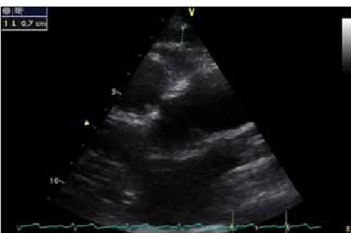
Epicardial fat thickness is described as the echo-free distance between the outer surface of the myocardium and the vis-ceral stratum of the pericardium [6]. We measured EFT from the parasternal long-axis imaging at the end of the diastole and at three cardiac cycles, vertical to the right ventricular free wall (Figure 1). Two different operators who were blind-ed to subjects performblind-ed echocardiographic evaluations twice at different times, and the average of the measurements of both operators was obtained.
The study was performed in accordance with the criteria of the Declaration of Helsinki. The study was approved by Başkent University Research Ethics Committee and support-ed by Başkent University Research Fund (project no.: KA16/335). All participants signed a written consent form. Statistical Analysis
A statistician performed a power analysis before starting the study. According to the power analysis result, we had to have at least a total of 90 patients (power analysis of two-sample T-test for testing equivalence using differences, numeric
results for testing equivalence using a parallel-group design, reference group sample size n1=45, treatment group sample size n2=45, alpha=0.0500, beta=0.1948). Continuous vari-ables are expressed as mean±standard deviation or median (range, interquartile range). Categorical data are presented as proportion. Chi-square test was used to analyze categorical parameters. One-way analyses of variance (ANOVA) or unpaired t-test was used to analyze continuous variables with normal distribution. Mann-Whitney U test was used to ana-lyze continuous variables with non-normal distribution. Continuous variables were checked by Kolmogorov-Smirnov test to assess whether they were normally distributed. A p value of <0.05 was considered as statistically significant. The SPSS statistical software was used for statistical analysis Statistical Package for the Social Sciences (SPSS version 21.0 for Windows; SPSS IBM Corp.; Armonk, NY, USA).
RESULTS
A total of 401 patients were enrolled in the study, including 229 patients with a diagnosis of asthma and 172 healthy volunteers. There was no significant difference between the groups in terms of baseline clinical characteristics (p>0.05). Table 1 shows the baseline clinical characteristics of the
2
Figure 1. Measurements of epicardial fat thickness on the free wall of
the right ventricle from the parasternal long-axis views
Table 1. Baseline characteristics of the study population Asthma Control (n=229) (172) p
Age, years 43.86±13.18 45.11±11.36 0.318
Sex, female gender, 135 (59) 100 (58.7) 0.870 n (%) Hypertension, n (%) 25 (10.9) 21 (12.2) 0.688 Smoking, n (%) 65 (28.4) 40 (23.3) 0.248 BMI (kg/m2) 29.72±3.98 29.18±3.62 0.167 Creatinine (mg/dL) 0.78±0.14 0.76±0.12 0.096 Hemoglobin (g/dL) 13.72±1.41 13.8±1.38 0.560 White blood cell 8234±1717 8085±1409 0.353 count (/mm3)
Platelets (100/mm3)* 280 280 0.439
(245–317.5) (246–324)
*Data are expressed as median with interquartile range BMI: body mass index
groups. Table 2 shows the asthma control grade, grade of severity, and duration of asthma of the study subjects. Baseline echocardiographic measurements and cardiac void measurements were similar in both groups. Table 3 shows the echocardiographic measurements.
The mean EFT was 5.84±0.79 mm in the patient group and 5.71±0.93 mm in the control group. There was no statisti-cally significant difference between these two groups (p=0.145). Similarly, when we compared control and asthma severe subgroups (mild, moderate, and severe asthma groups), there were no statistically significant differences in the control and asthma subgroups (control group 5.71±0.93 mm, mild group 5.86±0.81 mm, moderate group 5.8±0.84 mm, and severe group 5.83±0.67 mm, p=0.505).
DISCUSSION
In the present study, we observed that the EFT did not increase in patients with asthma when compared with the normal population. The second finding of the study was that there was no association of asthma severity subgroups with visceral adiposity assessed by EFT. Based on our results, we suggest that visceral obesity may not be a significant risk fac-tor for asthma.
Obesity and asthma are two health problems that are increasing in frequency worldwide. For the first time, Seidell et al. [13] reported a relationship between obesity and asth-ma, especially in female patients. A lot of study has been performed since the present study, and the relationship between asthma and obesity has been shown clearly in chil-dren, adolescents, and adults [3, 14]. Although the relation-ship between asthma and obesity is well known, it is not fully understood whether general obesity or visceral obesity is the more important risk factor and what the mechanism explains the relationship between obesity and asthma. This point should be discussed and clarified.
Some suggestions for possible mechanisms by which general obesity leads to asthma have been explained in previous studies. One of these potential mechanisms is the mechani-cal effect of obesity on the lungs. General obesity may cause dyspnea by preventing diaphragm movements. It has also been shown that functional residual capacity (FRC) decreas-es in obdecreas-ese patients, and there is an inverse relationship between body mass index and FRC [15].
Apart from the mechanical effect of obesity on the dia-phragm, immunological and inflammatory effects may cause asthma or its worsening. For example, levels of C-reactive protein, interleukin 6, tumor necrosis factor-α, plasminogen activator inhibitor 1, eotaxin, vascular endothelial growth factor, and monocyte chemotactic protein 1 were found to be high in obese individuals and obese asthmatics. These mark-ers have been shown to cause chronic systemic inflammation and are one of the causes of low-grade chronic airway inflammation in patients with asthma [16,17].
A number of possible mechanisms discussed above may explain the relationship between asthma and general obesity. The results of our study suggest that visceral obesity may not be an additional risk factor for asthma. There are also other studies that contradict the results of our work. Capelo et al. [18] showed that visceral obesity is negatively correlated with asthma control and lung function in female patients in a study of 83 women with asthma. Reproducibility is very important in operator-dependent techniques. In the present study, a single operator made measurements of visceral fat and subcutaneous fat using an ultrasound device. The intrao-bserver and interointrao-bserver variability are the biggest problems in operator-dependent measurements. Several different mea-surements with multiple operators followed by calculation of the averages can be a solution in order to obtain more accu-rate results. Performing the study only in women and the measurements once by a single operator might affect the results. In another study, Brumpton et al. [19] found that general obesity is a risk factor for asthma in men and women; additionally, they reported that waist circumference is high in women with asthma, and abdominal obesity increases the incidence of asthma in addition to general obesity in women. The measurement of the waist circumference is frequently used as a parameter to assess abdominal obesity; however, its predictor value for visceral obesity is weak [20], and its sen-sitivity, specificity, and reproducibility are low. It gives inac-curate results, especially in the aged and morbidly obese patients [21,22]. Fenger et al. [23] speculated that the effect of adiposity on asthma is mainly seen in non-atopics and did not seem to be attached on the distribution of adiposity.
3
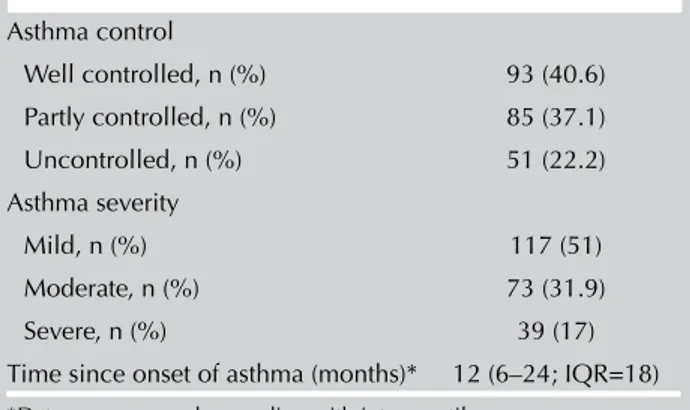
Table 2. Asthma control, severity, and time since onset ofasthma Asthma control Well controlled, n (%) 93 (40.6) Partly controlled, n (%) 85 (37.1) Uncontrolled, n (%) 51 (22.2) Asthma severity Mild, n (%) 117 (51) Moderate, n (%) 73 (31.9) Severe, n (%) 39 (17)
Time since onset of asthma (months)* 12 (6–24; IQR=18)
*Data are expressed as median with interquartile range IQR: interquartile range
Table 3. Measurements of two-dimensional echocardiographic parameters Asthma Control (n=229) (172) p EF (%) 59.07±2.96 58.77±2.97 0.316 LVEDD (mm) 43.06±3.28 42.90±3.32 0.644 LVESD (mm) 26.59±1.56 26.54±1.57 0.737 Septum (mm) 0.91±0.15 0.92±0.15 0.868 Posterior wall (mm) 0.91±0.15 0.91±0.15 1 Left atrium (mm) 37.50±2.52 37.39±2.55 0.664 Right atrium (mm) 37.41±2.57 37.72±2.58 0.225 Right ventricle (mm) 28.15±2.06 28.48±2.12 0.117
EF: ejection fraction; LVEDD: left ventricle end diastolic diameter; LVESD: left ventricle end systolic diameter
Increasing adiposity was associated with lower pulmonary function independent of atopic condition. In another study, Song et al. showed that the relationships between obesity and asthma in the elderly population may be mediated by factors including abdominal subcutaneous adiposity [24]. Our study has some limitations. First, the study was not a prospective study. Our study population consists of patients who have been already diagnosed with asthma and have been on appropriate treatment. We do not know whether the EFT has changed with treatment. The number of patients is relatively limited, and the study was not a multicenter study and so may not reflect the general population. EFT in the study was measured by echocardiography from the right ventricular free wall. The site where the measurements were obtained may not reflect the volume of all subepicardial fat tissue; however, it has been shown to be highly correlated in previous studies [6-8]. EFT is not a gold standard method to measure visceral fat, but it is cheap and easy to perform and was found to be reflecting correctly to the visceral obesity measured by magnetic resonance imaging and/or computed tomography.
CONCLUSION
Although it has been shown in many studies that obesity is a risk factor for asthma, it is not clear whether general obesity or visceral obesity is a more important risk factor. We did not find EFT, a good indicator of visceral obesity, to be high in patients with asthma. We conclude that visceral obesity may not be a risk factor for asthma.
Ethics Committee Approval: Ethics committee approval was received
for this study from the ethics committee of Başkent University.
Informed Consent: Written informed consent was obtained from
patients who participated in this study.
Peer-review: Externally peer-reviewed.
Author Contributions: Concept - H.E.B.; Design - H.E.B., M.Y.;
Supervision - H.E.B., Z.E.Ü.; Resources - H.E.B., Z.E.Ü.; Materials - N.Ş., M.Y.; Data Collection and/or Processing - H.E.B., N.Ş., M.Y.; Analysis and/or Interpretation - M.Y., F.Ö.E.; Literature Search - H.E.B., Ş.A.; Writing Manuscript - H.E.B.; Critical Review - Ş.A., F.Ö.E.
Acknowledgments: The authors would like to thank Mr. M. Agah
Tekindal, Mr.Tansel Erol and the cardiology fellows for their valuable contributions.
Conflict of Interest: The authors have no conflicts of interest to
declare.
Financial Disclosure: This study was approved by Başkent University
Institutional Review Board and Ethics Committee and supported by Başkent University Research Funding (KA 16/335).
REFERENCES
1. Global Initiative for Asthma (GINA) [homepage on the In-ternet]. Bethesda: National Heart, Lung and Blood Institute. National Institutes of Health, US Department of Health and Human Services; 2016 update. Available from: http://www. ginasthma.org
2. World Health Organization (WHO). Asthma fact sheet (WHO web site); 2010. Available from http://www.who.int/mediacen-tre/factsheets/fs307/en/
3. Forno E, Celedón JC. The effect of obesity, weight gain, and weight loss on asthma inception and control. Curr Opin Allergy Clin Immunol 2017;17:123-30. [CrossRef]
4. Ford ES, Mannino DM. Time trends in obesity among adults with asthma in the United States: findings from three national surveys. J Asthma 2005;42:91-5. [CrossRef]
5. Flegal KM, Kit BK, Orpana H, et al. Association of all cause mor-tality with over weight and obesity using standard body mass index categories: a systematic review and meta analysis. JAMA 2013;309:71-82. [CrossRef]
6. Sinha SK, Thakur R, Jha MJ, et al. Epicardial Adipose Tissue Thickness and Its Association With the Presence and Severity of Coronary Artery Disease in Clinical Setting: A Cross-Sectional Observational Study. J Clin Med Res 2016;8:410-9. [CrossRef] 7. Altin C, Sade LE, Gezmiş E, et al. Assessment of Subclinical
Atherosclerosis by Carotid Intima-Media Thickness and Epi-cardial Adipose Tissue Thickness in Prediabetes. Angiology 2016;67:961-9. [CrossRef]
8. Nabati M, Saffar N, Yazdani J, et al. Relationship between epi-cardial fat measured by echocardiography and coronary athero-sclerosis: a single-blind historical cohort study. Echocardiogra-phy 2013;30:505-11. [CrossRef]
9. Sengul C, Cevik C, Ozveren O, et al. Epicardial fat thickness is associ-ated with non-dipper blood pressure pattern in patients with essential hypertension. Clin Exp Hypertens 2012;34:165-70. [CrossRef] 10. Yılmaz M, Bozkurt Yılmaz HE, Sen N, et al. Investigation of the
relationship between asthma and subclinical atherosclerosis by carotid/femoral intima media and epicardial fat thickness mea-surement. J Asthma 2018;55:50-6. [CrossRef]
11. American Thoracic Society. Standardization of spirometry: 1994 update. Am J Respir Crit Care Med 1995;152:1107-36. [CrossRef] 12. Lang RM, Badano LP, Mor-Avi V, et al. Recommendations for
cardiac chamber quantification by echocardiography in adults: an update from the American Society of Echocardiography and the European Association of Cardiovascular Imaging. J Am Soc Echocardiogr 2015;28:1-39. [CrossRef]
13. Seidell JC, de Groot LC, van Sonsbeek JL, et al. Associations of mod-erate and severe overweight with self-reported illness and medical care in Dutch adults. Am J Public Health 1986;76:264-9. [CrossRef] 14. Rzehak P, Wijga AH, Keil T, et al. GALEN-WP 1.5 Birth Co-horts. Body mass index trajectory classes and incident asthma in childhood: results from 8 European Birth Cohorts--a Global Allergy and Asthma European Network initiative. J Allergy Clin Immunol 2013;131:1528-36. [CrossRef]
15. Abdeyrim A, Zhang Y, Li N, et al. Impact of obstructive sleep apnea on lung volumes and mechanical properties of the respi-ratory system in over weight and obese individuals. BMC Pulm Med 2015;15:76. [CrossRef]
16. Sutherland TJ, Cowan JO, Young S, et al. The association between obesity and asthma: interactions between systemic and airway in-flammation. Am J Respir Crit Care Med 2008;178:469-75. [CrossRef] 17. Scott HA, Gibson PG, Garg ML, et al. Airway inflammation is
augmented by obesity and fatty acids in asthma. Eur Respir J 2011;38:594-602. [CrossRef]
18. Capelo AV, da Fonseca VM, Peixoto MV, et al. Visceral adipos-ity is associated with cytokines and decrease in lung function in women with persistent asthma. Rev Port Pneumol (2006) 2016;22:255-61. [CrossRef]
19. Brumpton B, Langhammer A, Romundstad P, et al. General and abdominal obesity and incident asthma in adults: the HUNT study. Eur Respir J 2013;41:323-9. [CrossRef]
20. Roever LS, Resende ES, Diniz AL, et al. Abdominal obesity and association with atherosclerosis risk factors: the Uberlândia heart study. Medicine (Baltimore) 2016;95:e1357. [CrossRef]
21. Natale F, Tedesco MA, Mocerino R, et al. Visceral adiposity and arterial stiffness: echocardiographic epicardial fat thickness reflects, better than waist circumference, carotid arterial stiff-ness in a large population of hypertensives. EurJ Echocardiogr 2009;10:549-55. [CrossRef]
22. Altin C, Sade LE, Gezmis E, et al. Assessment of epicardial adi-pose tissue and carotid/femoral intima media thickness in insu-lin resistance. J Cardiol 2017;6:843-50. [CrossRef]
23. Fenger RV, Gonzalez-Quintela A, Vidal C, et al. Exploring the obesity-asthma link: do all types of adiposity increase the risk of asthma?. Clin Exp Allergy 2012;42:1237-45. [CrossRef] 24. Song WJ, Kim SH, Lim S, et al. Association between obesity and
asthma in the elderly population: potential roles of abdominal subcutaneous adiposity and sarcopenia. Ann Allergy Asthma Immunol 2012;109:243-8. [CrossRef]