Retinal nerve fiber layer thickness in patients with
essential tremor
1
Yakup Turkel,
2Nurgul Ornek,
1Ersel Dag,
2Kemal Ornek,
1Murat Alpua,
2Tevfik Ogurel,
2Yasar Olmez
1Department of Neurology and 2Departments of Ophthalmology, Faculty of Medicine, Kirikkale
University,Kirikkale, Turkey Abstract
Objective: To investigate the retinal nerve fiber layer (RNFL) thickness in essential tremor (ET). Methods: seven eyes of 27 patients with essential tremor were included in this study. Twenty-seven eyes of 27 healthy volunteers served as controls. All eyes were examined with spectral domain optical coherence tomography (OCT) (Retinascan Advanced RS-3000; NIDEK, Gamagori, Japan) using image filling software program (NAVIS-EX, NIDEK, Tokyo, Japan). Results: No statistically significant difference was detected between ET patients and control group for overall (RNFL) and foveal retinal thickness parameters. [RNFL thickness (Average thickness p=0.86, superior average p=0.22, inferior average p=0.24, nasal average p=0.06, temporal average p=0.88), foveal retinal thickness p=0.63] There was no relationship between OCT parameters and age, gender and duration of ET (all p>0.05).
Conclusion: We did not find altered RNFL and foveal thickness values in patients with ET compared to controls. Retinal thickness changes do not seem to be a potentially useful biomarker in ET patients.
Address correspondence to: Yakup Turkel, MD, Kirikkale University, Faculty of Medicine, Department of Neurology, Kirikkale, Turkey.
INTRODUCTION
Essential tremor (ET) is characterized with postural and/or kinetic tremor, and is the most common movement disorder on the world. Although it may be seen at any age, the incidence increases with age.1 While the prevalence of
ET for all ages is 0.9%, this value was shown as 4.6% in over 65 years. The main clinical manifestation of ET is 8-12 Hz kinetic and postural tremor emerging during voluntary movement. Other anatomical sites affected by tremor are respectively head, vocal cords, trunk, legs and face regions.1 The diagnosis still depends on
medical history and neurological examination. There are no specific biological marker for the diagnosis based on laboratory tests, or imaging methods.1,2 The genetic transmission of ET is
mostly autosomal dominant with incomplete penetration. However, polygenic inheritance has been shown in some families.3
The pathogenesis of ET has not been fully understood yet. Researchers have proposed that ET may be a result of mainly central abnormal oscillator dysfunction located in Guillain Mollaret triangle. Also, the cerebellum is thought to have an important role in the pathogenesis of the disease.4,5
Although ET has been considered a monosymptomatic disease for a long time, non-motor symptoms of ET such as cerebellar symptoms, olfactory dysfunction, hearing impairment, cognitive deficits, sleep disorders, behavioral symptoms and different personality traits have been identified in recent studies. Lewy bodies in locus coerulus of the brainstem have also been shown in ET patients. Therefore ET is currently thought to be a complex, neurodegenerative and heterogeneous disease.6,7
Optical coherence tomography (OCT) is used to assess retinal nerve fiber layer (RNFL) of the anterior visual pathway. Retinal nerve fiber layer (RNFL) thickness in Parkinson’s disease (PD) patients has been documented in several studies.8-10 It has been found that the thickness of
the RNFL was reduced in PD patients. Abnormal OCT measures have also been shown in other neurodegenerative diseases, like Alzheimer’s disease, multiple sclerosis, and spinocerebellar ataxias, most likely related to the loss of retinal ganglion cells and axons, with decreased thickness in RNFL.11-13
Previous studies have shown the value of RNFL and macular examination as a method of detecting neurodegenerative disease progression
Neurology Asia December 2015
364
and facilitating diagnosis of diseases like multiple sclerosis and PD. The aim of the present study was to analyze whether a selective combination of RNFL and macular thickness parameters are altered in ET patients.
METHODS
Simple Random Sampling was used in this study. Twenty-seven eyes of 27 patients with ET were included in this study. Twenty-seven eyes of 27 healthy volunteers served as controls. Essential tremor was diagnosed if the subject had a grade 2 postural and/or kinetic tremor of the hands or forearms without identifiable secondary cause or other exclusion criteria (e.g. prominent unilateral tremor, rigidity or bradykinesia). All patients were recruited from the Neurology Department of the Kirikkale University School of Medicine. Age-matched, healthy, subjects with normal ophthalmological and neurological examinations were included as controls. All study procedures were in accordance with the revised form of the Helsinki Declaration 2008, and all participants gave informed consent. The study protocol was approved by the local ethical committee. Exclusion criteria included those with glaucoma, pseudoexfoliation syndrome, high myopia, diabetes, anomalous optic disc, age-related macular degeneration, peripheral vasospasm, sleep-related breathing disorder, history of ocular trauma, previous intraocular surgery like cataract and corticosteroid use. Each subject underwent a complete ophthalmological examination including best-corrected visual acuity and measurement of intraocular pressure. Refraction of each eye was obtained using an autorefractor (Topcon Auto Ref-Keratometer, Tokyo, Japan). For OCT, one eye was chosen randomly for each patient and control subject. The tests were conducted by a clinician blinded to the subject’s condition, and a second blinded investigator reviewed the images and reported the results independently.
All eyes were examined with spectral domain OCT (Retinascan Advanced RS-3000; NIDEK, Gamagori, Japan) using image filling software
program (NAVIS-EX, NIDEK, Tokyo, Japan). This was performed in the peripapillary area following pupillary dilation. In the peripapillary area, a circular scan centered on the optic disc (3.45 mm diameter, “disc circle” option) was used. We quantitated the thickness of the mean peripapillary RNFL (360°), superior quadrant (46°–135°), inferior quadrant (226°–315°), nasal quadrant (136°–225°), and temporal quadrant (316°–45°). In the macula, foveal thickness was measured with OCT setting: macula map X-Y (6.0 mm · 6.0 mm [256 · 256] automatically. The results were analyzed using the computer software (SPSS version 16.0). A p<0.05 was considered statistically significant in all tests. Continuous variables were presented in titer of mean and ±SD. The normality of the distribution for all variables was assessed by the Kolmogorov–Smirnov test. Student’s t- test was used for normally distributed variables, and Mann–Whitney U test was used for nonparametric variables. Chi-square test was used for categorical parameters. Bivariate analyses were performed using Pearson’s correlation.
RESULTS
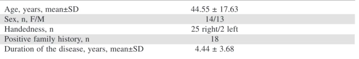
Twenty-seven patients with ET (14 women and 13 men) were included in this study and 27 eyes of 27 healthy controls (17 women and 10 men) served as controls. Mean age of the patients with ET was 44.55 ± 17.63 (range, 20–70) years and mean age of the control group was 44.25 ± 16.25 (range, 21-77) years. There were no significant differences in terms of age (p=0.9) and sex (p=0.5) between the patients and healthy controls. The mean duration of the disease in patients with ET was 4.44 ± 3.68 (range, 1–15) years. Baseline characteristics of ET patients were shown in Table 1.
Mean intraocular pressure of the patients was 16.40 ± 2.93 mm Hg and mean intraocular pressure of the control group was 16.07 ± 2.83 mm Hg. There was no statistically significant difference in the mean intraocular pressure between patients and controls (p = 0.6).
Table 1: Baseline characteristics of essential tremor patients
Age, years, mean±SD 44.55 ± 17.63
Sex, n, F/M 14/13
Handedness, n 25 right/2 left
Positive family history, n 18
No statistically significant difference between ET patients and control group was detected for overall RNFL and foveal retinal thickness parameters (all p>0.05). There was no relationship between OCT parameters and age, gender and duration of ET (all p>0.05). The OCT results are summarized in Table 2.
DISCUSSION
Ocular manifestations are rare in ET patients. Eye movement abnormalities have been shown in severely affected patients. To our knowledge, this is the first study investigating the changes in the RNFL of patients with ET using high-resolution OCT images. Our results suggested that patients with ET do not seem to have significant changes in OCT parameters.
There is only one study evaluating foveal retinal thickness using time domain (TD)-OCT in ET patients in the current literature. In this study, the Authors showed that foveal retinal thickness was asymmetric and thinner in the eye contralateral to the side more affected by tremor and parkinsonism in the ET and PD groups, respectively. However, there was no significant difference between the ET patients and controls.14 Retinal thickness
changes in PD using OCT have been reported in several studies.8-10,15-22 Many of them analyzed
peripapillary thickness and reported RNFL thinning in PD, although some did not support these findings.17-19 As local dopaminergic neuronal
groups in the retina, basal ganglia and cortical memory system are affected in PD patients, RNFL thinning seems to be a result of DA depletion in the retina of PD patients .
There is considerable evidence that ET may precede the onset of PD or is present in family members of patients with PD with higher-than-expected frequency suggesting that ET may be
a risk factor for PD. Although there is some controversy whether ET should be viewed as a monosymptomatic disorder, a variety of neurological disorders have been described in patients with ET. In a recent study by Antonini et al, low putamenal dopamine transporter values were found in a significant proportion of ET cases, more commonly in those with familial ET.23 The Authors concluded that abnormalities
are frequent in the dopaminergic system of ET patients particularly with positive family history and these subjects may be at risk for future development of parkinsonism.
OCT provides non-contact and non-invasive RNFL thickness measurements and has become an essential clinical tool. In our study, we used spectral domain OCT and through measurement of RNFL, the results revealed that RNFL was slightly thinner only in the nasal quadrant. Retinal thickness was thinner in the study by Cubo et al.14 However we measured thicker RNFL in
ET patients except for nasal RNFL, and foveal retinal thickness in patient group was similar to controls. RNFL is an anatomic part of retinal thickness, and thinner RNFL may cause thinner retinal thickness. Therefore thicker RNFL in our patients is in accordance with the normal retinal thickness. These findings suggest that there is no significant cellular damage in the retinal layers or optic nerve in ET patients.
In ET patients, we found thinner RNFL only in the nasal quadrant compared to controls. There is ISNT rule in RNFL measurements which means that RNFL is thicker in inferior and superior quadrants and thinner in temporal and nasal quadrants. In rare clinical conditions like longer eyes, high myopia and glaucoma, nasal RNFL may be much thinner than other quadrants. None of the ET patients had these ocular comorbidities. Therefore, it is hard to conclude that this localized
OCT Parameters Patients Controls p value (n=27) (n=27) RNFL thickness (mean ± SD), µm Average thickness 103.33±11.38 102.81±10.58 0.8* Superior average 133.55±23.58 126.51±18.17 0.2* Inferior average 132.66±17.90 126.70±19.59 0.2* Nasal average 79.66±14.52 88.22±18.97 0.06* Temporal average 69.18±9.38 69.62±12.24 0.8*
Foveal retinal thickness (mean ± SD), µm 262.88±22.14 262.07±15.35 0.6** OCT: Optical coherence tomography, ET: Essential tremor, RFNL: Retinal nerve fiber layer
Neurology Asia December 2015
366
difference is due to a specific tissue loss at this site. Further studies with larger sample size and longer follow up could help to clarify further this question.
This study has some limitations. First, our study group was relatively small. Second, essential tremor severity was not measured in this study. To conclude, we did not find altered RNFL and foveal thickness values in patients with ET compared to controls. Therefore, our results do not support the observation that retinal thickness changes may be a potentially useful biomarker in patients with ET. Further studies with larger sample size are needed to clarify this issue. DISCLOSURE
Conflict of interest: None REFERENCES
1. Louis ED, Ferreira JJ. How common is the most
common adult movement disorder? Update on the worldwide prevalence of essential tremor. Mov Disord 2010;25: 534-41.
2. Louis ED, Ford B, Frucht S, Barnes LF, X-Tang
M, Ottman R. Risk of tremor and impairment from tremor in relatives of patients with essential tremor: a community-based family study. Ann Neuro 2001; 49:761-9.
3. Deng H, Le W, Jankovic J. Genetics of essential tremor. Brain 2007;130:1456-64.
4. Louis ED, Faust PL, Vonsattel JP, et al.
Neuropathological changes in essential tremor: 33 cases compared with 21 controls. Brain 2007; 130:3297-307.
5. Louis ED, Faust PL, Vonsattel JP, et al. Torpedoes
in Parkinson’s disease, Alzheimer’s disease, essential tremor, and control brains. Mov Disord 2009; 24:1600-5.
6. Louis ED, Vonsattel JP, Honig LS, Ross GW, Lyons
KE, Pahwa R. Neuropathologic findings in essential tremor. Neurology 2006; 66:1756-9.
7. Chandran V, Pal PK. Essential tremor: beyond the
motor features. Parkinsonism Relat Disord 2012; 18: 407-13.
8. Altintas O, Iseri P, Ozkan B, Caglar Y. Correlation
betweenr retinal morphological and functional findings and clinical severity in Parkinson’s disease. Doc Ophthalmol 2008; 116:137-46.
9. Moschos MM, Tagaris G, Markopoulos I, et
al. Morphologic changes and functional retinal impairment in patients with Parkinson disease without visual loss. Eur J Ophthalmol 2011; 21; 24-9.
10. Kirbas S, Turkyilmaz K, Tufekci A, Durmus M.
Retinal nerve fiber layer thickness in Parkinson disease. J Neuroophthalmol 2013; 33: 62-5.
11. Berisha F, Feke GT, Trempe CL, McMeel JW,
Schepens CL. Retinal abnormalities in early Alzheimer’s disease. Invest Ophthalmol Vis Sci 2007; 48:2285-9.
12. Lamirel C, Newman N, Biousse V. The use of optical
coherence tomography in neurology. Rev Neurol Dis 2009: 6;105-20.
13. DagE, Örnek N, Örnek K, Erbahçeci-Timur IE. Optical coherence tomography and visual field findings in patients with Friedreich ataxia. J Neuroophthalmol 2014; 34: 118-21.
14. Cubo E, Tedejo RP, Rodriguez Mendez V, et al. Retina thickness in Parkinson’s disease and essential tremor. Mov Disord 2010; 25:2461-77.
15. Inzelberg R, Ramirez JA, Nisipeanu P, Ophir A.
Retinal nerve fiber layer thinning in Parkinson disease. Vis Res 2004; 44: 2793-7.
16. Hajee ME, March WF, Lazzaro DR, et al. Inner
retinal layer thinning in Parkinson’s disease. Arch Ophthalmol 2009; 127: 737-41.
17. Aaker GD, Myung JS, Ehrlich JR, Mohammed M,
Henchcliffe C, Kiss S. Detection of retinal changes in Parkinson’s disease with spectral-domain optical coherence tomography. Clin Ophthalmol 2010; 4:1427-32.
18. Archibald NK, Clarke MP, Mosimann UP, Burn
DJ. Retinal thickness in Parkinson’s disease. Parkinsonism Relat Disord 2011; 17: 431-6.
19. Albrecht P, Muller AK, Sudmeyer M, et al. Optical
coherence tomography in parkinsonian syndromes. PLoS One 2012; 7: e34891.
20. Garcia-Martin E, Satue M, Fuertes I, et al. Ability and
reproducibility of Fourier-domain optical coherence tomography to detect retinal nerve fiber layer atrophy in Parkinson’s disease. Ophthalmology 2012; 119: 2161-7.
21. Schrier EM, Adam CR, Spund B, Glazman S,
Bodis-Wollner I. Interocular asymmetry of foveal thickness in Parkinsons disease. J Ophthalmol 2012; 2012:728457.
22. Spund B, Ding Y, Liu T, Selesnick I, Glazman S,
Shrier EM, Bodis-Wollner I. Remodeling of the fovea in Parkinson disease. J Neural Transm 2013; 120:745-53.
23. Antonini A, Moresco RM, Gobbo C, et al. The status
of dopamine nerve terminals in Parkinson’s disease and essential tremor: a PET study with the tracer [11-C]FE-CIT. Neurol Sci 2001; 22:478.