THE ASSOCIATION BETWEEN VERRUCA VULGARIS AND VITAMIN D: IS THERE A CASUAL LINK?
COŞKUNÖZTEKIN1, AYNUREÖZTEKIN2, KENANTAŞTAN3, GÜLSENGÜLÖZMEN4, SUZANDEMIRPEKTAŞ5
1MD, Assistant Professor, Department of Family Medicine, Hitit University Medical School, Çorum, Turkey - 2MD, Assistant Professor,
Department of Dermatology, Hitit University Medical School, Çorum, Turkey - 3MD, Assistant Professor, Department of Family
Medicine, Atatürk University Medical School, Erzurum, Turkey - 4MD, Ali Osman Sönmez Onkology State Hospital, Department of
Biochemistry, Bursa, Turkey - 5MD, Assistant Professor, Department of Dermatology, Muğla Sıtkı Koçman University Medical School,
Muğla, Turkey
Introduction
Verruca vulgaris (warts) is a common derma-tologic disease caused by human papillomavirus (HPV). Most people experience warts at least in one form at some time in their lives. It is transmit-ted through direct contact with the infectransmit-ted people or contaminated materials and surfaces. Infection is also associated with anatomical site, the amount of infectious virus, trauma, duration of contact, and HPV specific immune status(1).
Several studies highlighted the importance of the expression of antimicrobial peptides working as a defense mechanism against verruca vulgaris(2-4).
LL-37 is a peptide belonging to the cathelicidin
family of antimicrobial peptides and is induced within the epidermis during the development of verruca vulgaris(2). Human beta-defensin-2 and 3
are also peptides which exhibits antimicrobial activity and may be responsible from the sponta-neous regression in verruca vulgaris(3). Vitamin D
may induce the expression of some antimicrobial peptides including LL-37(4,5). Therefore, vitamin D
may have a role in the treatment of verruca vul-garis.
Insufficiency of vitamin D have been demon-strated up to 50% of the population worldwide in epidemiological studies(6-8). Recently, vitamin D or
its derivatives have been recommended as potential therapeutic regimen for a variety of neoplastic,
Acta Medica Mediterranea, 2018, 34: 1047
Received December 30, 2017; Accepted February 20, 2018 ABSTRACT
Introduction: Verruca vulgaris is a well-known skin lesion caused by human papillomavirus. There are topical, local invasion
and immune therapy options for verruca vulgaris. The role of vitamin D levels in patients with verruca vulgaris is not clear. We investigated the serum vitamin D levels in patients with verruca vulgaris.
Materials and methods: Fifty-five patients with verruca vulgaris (Group I) and 60 healthy controls (Group II) included in the
study. The characteristics of verruca vulgaris (duration, clinical type, the number of lesions, family history) and demographics of the participants were recorded. Serum 25-hydroxycholecalciferol levels were measured using electrochemiluminescence binding method.
Results: After covariance analysis, we found no statistically significant difference between the groups in terms of mean age
(23.38±4.68 years in Group I versus 27.35±8.14 years in Group II). Other baseline characteristics were not significantly different between the groups (p > 0.05 for all). The mean duration of verruca vulgaris was 12 (4-18) years and the most common sites were hand (26 patients, 47.3%) and foot (23 patients, 41.8%). Serum vitamin D levels were 8.35±6.03 ng/ml in Group I and 18.08±10.01 ng/ml in Group II. There was a statistically significant difference between the groups in terms of serum vitamin D levels (p < 0.001).
Conclusion: In this study, vitamin D levels in patients with verruca vulgaris were found to be decreased compared to healthy
controls. Low vitamin D levels may play an etiological role in the development of verruca vulgaris.
Keywords: Verruca Vulgaris, Vitamin D, Human Papilloma Virus, Serum.
inflammatory, infectious and immunologic dis-eases(9). Recently, Raghukumar et al.(10) reported
favorable outcomes with intralesional vitamin D3 injections in the treatment of extragenital recalci-trant warts. However, serum levels of vitamin D in patients with verruca vulgaris has not been investi-gated so far.
The aim of the present study is to investigate the possible relationship between verruca vulgaris and serum vitamin D levels. Table
Materials and methods
This prospective, cross-sectional, randomized study complied with the tenets of the Declaration of Helsinki and was approved by the Ethical Committee of Atatürk University, Erzurum, Turkey. Written informed consent was obtained from all the participants before the study. This study was con-ducted in Dermatology Clinic of Palandöken State Hospital, Erzurum, Turkey between December 2016 and February 2017.
A total of 55 patients with verruca vulgaris (Group I) and 60 healthy controls (Group II) were included in the study. Exclusion criteria were as fol-lows: (1) age < 18 years and > 50 years, (2) liver disease, (3) kidney disease, (4) pregnancy and lac-tation (5) use of vitamin D supplemenlac-tation last six months before the study. Eligible patients with ver-ruca vulgaris who meet the inclusion criteria were chosen using “random number generator” from the medical records. Participants in the control group consisted of healthy volunteers who had no com-plaints and systemic disease.
Age, sex, marital status, level of education, place of residence (rural or urban), smoking and alcohol use, drug use, and verruca vulgaris charac-teristics (duration, clinic type, the number of lesions, family history) were recorded. Diagnosis of verruca vulgaris was based on physical examina-tions by an experienced dermatologist.
Serum collection
Blood from the forearm vein was collected into 5-ml vacutainer tubes with no anticoagulant. The blood samples were centrifuged (1000 × g, 15
min, 4 °C) to separate serum. Serum was removed and immediately stored at -80 °C until analyzed.
Biochemical measurements
Serum 25-hydroxycholecalciferol measure-ments were performed using
electrochemilumines-cence binding method (COBAS reagent kit; COBAS e601 analyzer series, Roche Diagnostics, Basel, Switzerland). The results were expressed in ng/dl.
Statistical analysis
All data were entered into a spreadsheet, and statistical analyses were performed using R 3.3.2v (open source). Data are shown as mean ± standard deviation for continuous variables, as median (min-imum-maximum) for ordinal variables, and as fre-quency with percent for categorical variables. To evaluate the level of data normality for continuous variables, the Shapiro-Wilk test was used when n<50 and Kolmogorov Smirnov test was used when n>50. Categorical comparisons were made by chi-square test. Independent sample t test was used for comparing the means of continuous variables.
There was a mean age difference between the groups (p = 0.002). Therefore, age corrected covari-ance analysis was performed to eliminate age-relat-ed mixing effects on the results. Study groups and age were defined as “fixed factor” and “covariate” variables, respectively. For more than two indepen-dent groups, the Kruskal Wallis test was used for non-normally distributed variables. A Spearman’s rho correlation was used to analyze the association between non-normally distributed variables. Correlations between normally distributed variables were analyzed by Pearson’s correlation coefficient. A p value of <0.05 was considered statistically sig-nificant.
Results
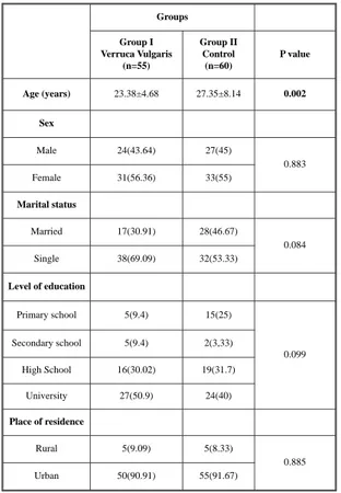
A total of 55 patients (31 females, 24 males) were included in Group I and 60 healthy individu-als in Group II (33 females and 27 males). There was a statistically significant difference between the groups in terms of mean age (23.38±4.68 years in Group I versus 27.35±8.14 years in Group II). Other baseline characteristics including sex, marital status, level of education, and place of residence were not significantly different between the groups (p > 0.05 for all). Table 1 summarizes the demo-graphics and baseline characteristics of the groups. Seventeen patients (30.91%) were smoking and 1 patient (1.82%) was using alcohol in Group I. Six patients (10.91%) had a history of drug use in Group I.
The mean duration of verruca vulgaris was 12 (4-18) years. Family history of verruca vulgaris was
evident in 16 patients (29.09). The most common sites for verruca vulgaris was hand (26 patients, 47.3%), foot (23 patients, 41.8%), face (one patient, 1.8%), and other locations (5 patients, 9.1%). The mean num-ber of verruca vulgaris per patient was 3 (1-7).
Serum vitamin D levels were 8.35±6.03 ng/ml in Group I and 18.08±10.01 ng/ml in Group II. There was a statistically significant difference between the groups in terms of serum vitamin D levels (p < 0.001). Covariance analysis showed that age did not affect the serum vitamin D levels difference between the groups (Fig.1). shows the mean serum vitamin D concentra-tions of the groups in a box plot graph.
Discussion
This is the first study evaluating the vitamin D levels in the serum of patients with verruca vul-garis. The results demonstrated decreased levels of vitamin D in the serum of patients with verruca vul-garis compared with healthy control subjects. Vitamin D regulates cell proliferation and differen-tiation via the vitamin D receptor (VDR) which is present keratinocytes and lymphocytes of the skin(11). Although the exact mechanism of action of
vitamin D activity against verruca vulgaris remains to be elucidated, it may have protective and/or pre-ventive role.
Vitamin D has an important role in bone min-eralization and calcium homeostasis. Recent evi-dence suggests that it has beneficial pleiotropic effects on both the innate and adaptive immune
sys-tem(12,13). Even though numerous studies suggest that
vitamin D has beneficial effects on the immune defense and on allergic diseases, the exact mecha-nisms are not well understood yet.
Vitamin D increases the expression of several antimicrobial peptides including cathelicidin (LL-37). LL-37 is not only an antimicrobial peptide against bacteria, virus and fungi, but also an immune modulator(14,15). Elenius et al.(15) found that
higher Vitamin D and E levels were associated with less allergic disorders in patients undergoing tonsil-lectomy. Bucak et al.(16)found that lower vitamin D
levels are associated with rotaviral diarrhea in chil-dren. Recent works highlights vitamin D’s potential role in fighting viral infections(17,18). Bhat et al.(19)
reported lower levels of vitamin D in patients with alopecia areata and found significant negative cor-relation between the levels of serum Vitamin D and severity of alopecia.
Despite there have been many studies on vita-min D in recent years, experience related with HPV and verruca vulgaris are very limited. Labendeira et al.(20) reported successful outcome with the use of
topical calcipotriol in the treatment of a giant viral wart. A more recent study concluded the role of intralesional vitamin D3 injection in sixty-four patients with recalcitrant warts of varying sizes and duration.
Complete response was seen in 54 of 60 (90%) of the patients. The authors reported the average number of injections required to achieve a complete resolution as 3.66. Aktas et al.(11) used
intralesional Vitamin D3 for plantar warts. Twenty patients were included in that study, and 7.5 mg of The association between verruca vulgaris and vitamin D: Is there a casual link? 1049
Groups Group I Verruca Vulgaris (n=55) Group II Control (n=60) P value Age (years) 23.38±4.68 27.35±8.14 0.002 Sex Male 24(43.64) 27(45) 0.883 Female 31(56.36) 33(55) Marital status Married 17(30.91) 28(46.67) 0.084 Single 38(69.09) 32(53.33) Level of education Primary school 5(9.4) 15(25) 0.099 Secondary school 5(9.4) 2(3,33) High School 16(30.02) 19(31.7) University 27(50.9) 24(40) Place of residence Rural 5(9.09) 5(8.33) 0.885 Urban 50(90.91) 55(91.67)
Table 1: The demographics and baseline characteristics
of the groups.
Fig. 1: The mean serum vitamin D concentrations of the
groups in a box plot graph.
.,
•
Groupso" ., 0 ,.
Vitamin D3 injection was given at monthly inter-vals for a maximum of 2 sessions. They reported complete clearance in 80% of patients at the end of 8 weeks. In the present study, we found decreased levels of serum vitamin D in patients with verruca vulgaris compared to healthy controls (8.35±6.03 ng/ml versus 18.08±10.01 ng/ml). In the light of these studies, the role of vitamin D in the treatment of HPV infections particularly in verruca vulgaris is an indisputable fact.
Although several studies have reported favor-able results with intralesional vitamin D analogs in skin warts, the role of vitamin D insufficiency in the pathogenesis of verruca vulgarism is unknown. Furthermore, it is not known whether elimination of vitamin D deficiency has a role in the treatment of verruca vulgaris. This study basically showed that serum vitamin D levels in verruca vulgaris patients were lower than in healthy individuals. Further studies are needed to clarify the role of vitamin D (whether protective or therapeutic) on HPV skin infections. The main limitation of this study was the mean age difference between the study and control groups. However, covariance analysis eliminated the age-related mixing effects on the results. The second limitation was the relatively small study population which might hinder extrapolation of the results.
In conclusion, this study is the first to demon-strate decreased levels of vitamin D in patients with verruca vulgaris. Decreased vitamin D levels may play an aetiopathogenic role in verruca vulgaris. Further studies with larger sample size are neces-sary to confirm our results and the potential protec-tive/therapeutic role of vitamin D from verruca vul-garis.
References
1) Kranjec C, Doorbar J. Human papillomavirus infection and induction of neoplasia: a matter of fitness. Curr Opin Virol 2016; 20: 129-36.
2) Conner K, Nern K, Rudisill J, O’Grady T, Gallo RL. The antimicrobial peptide LL-37 is expressed by ker-atinocytes in condyloma acuminatum and verruca vul-garis. J Am Acad Dermatol 2002; 47: 347-50.
3) Meyer-Hoffert U, Schwarz T, Schröder JM, Gläser R. Expression of human beta-defensin-2 and -3 in verru-cae vulgares and condylomata acuminata. J Eur Acad Dermatol Venereol 2008; 22 :1050-4.
4) Wang G. Human antimicrobial peptides and proteins. Pharmaceuticals 2014; 7: 545-94.
5) Wang TT, Nestel FP, Bourdeau V, Nagai Y, Wang Q, et al. Cutting edge: 1,25-dihydroxyvitamin D3 is a direct inducer of antimicrobial peptide gene expression. J Immunol 2004; 173: 2909-12.
6) Hoseinzadeh E, Taha P, Wei C, Godini H, Ashraf GM, Taghavi M, Miri M. The impact of air pollutants, UV exposure and geographic location on vitamin D defi-ciency. Food Chem Toxicol 2018; 113: 241-54. 7) Lucas RM, Gorman S, Black L, Neale RE. Clinical,
Research, and Public Health Implications of Poor Measurement of Vitamin D Status. J AOAC Int 2017; 100: 1225-29.
8) Schramm S, Lahner H, Jöckel KH, Erbel R, Führer D, Moebus S, Heinz Nixdorf Recall Study Group. Impact of season and different vitamin D thresholds on preva-lence of vitamin D deficiency in epidemiological cohorts-A note of caution. Endocrine 2017; 56: 658-66. 9) Caccamo D, Ricca S, Currò M, Ientile R. Health Risks of Hypovitaminosis D: A Review of New Molecular Insights. Int J Mol Sci. 2018; 19. doi: 10.3390/ijms19030892
10) Raghukumar S, Ravikumar BC, Vinay KN, Suresh MR, Aggarwal A, Yashovardhana DP. Intralesional Vitamin D(3) Injection in the Treatment of Recalcitrant Warts: A Novel Proposition. J Cutan Med Surg 2017; 21: 320-4.
11) Aktas H, Ergin C, Demir B, Ekiz Ö. Intralesional Vitamin D injection may be an effective treatment option for warts. J Cutan Med Surg 2016; 20: 118-22. 12) Bikle DD. What is new in vitamin D: 2006-2007. Curr
Opin Rheumatol 2007; 19: 383-8.
13) Beard JA, Bearden A, Striker R. Vitamin D and the anti-viral state. J Clin Virol 2011; 50: 194-200. 14) Schauber J, Gallo RL. Antimicrobial peptides and the
skin immune defense system. J Allergy Clin Immunol 2008; 122: 261-6.
15) Elenius V, Palomares O, Waris M, Turunen R, Puhakka T, Rückert B. The relationship of serum vitamins A, D, E and LL-37 levels with allergic status, tonsillar virus detection and immuneresponse. PLoS One 2017; 12 e0172350.
16) Bucak IH, Ozturk AB, Almis H, Cevik MÖ, Tekin M, et al. Is there a relationship between low vitamin D and rotaviral diarrhea? Pediatr Int 2016; 58: 270-3. 17) Hansdottir S, Monick MM, Hinde SL, Lovan N, Look
DC, Hunninghake GW. Respiratory epithelial cells con-vert inactive vitamin D to its active form: potential effects on host defense. J Immunol 2008; 181: 7090-9. 18) Hansdottir S, Monick MM, Lovan N, Powers L, Gerke
A, Hunninghake GW. Vitamin D decreases respiratory syncytial virus induction of NF-kB chemokines and cytokines in airway epithelium while maintaining the antiviral state. J Immunol 2010; 184: 965-74.
19) Bhat YJ, Latif I, Malik R, Hassan I, Sheikh G, et al. Vitamin D Level in Alopecia Areata. Indian J Dermatol 2017; 62: 407-10.
20) Labandeira J, Vázquez-Blanco M, Paredes C, Suárez-Penaranda JM, Toribio J. Efficacy of topical calcipotri-ol in the treatment of a giant viral wart. Pediatr Dermatol 2005; 22: 375-6.
_________
Corresponding author COŞKUNÖZTEKIN, MD Department of Family Medicine Hitit University Medical School Çorum
(Turkey)