SELF-ASSEMBLED PEPTIDE NANOSTRUCTURE DELIVERY SYSTEMS FOR OLIGONUCLEOTIDE THERAPY
A THESIS
SUBMITTED TO THE MATERIALS SCIENCE AND NANOTECHNOLOGY PROGRAM OF GRADUATE SCHOOL OF ENGINEERING AND SCIENCE OF BILKENT
UNIVERSITY
IN PARTIAL FULFILLMENT OF THE REQUIREMENTS FOR THE DEGREE OF
MASTER OF SCIENCE
By
Zahide Didem Mumcuoğlu June, 2014
2
I certify that I have read this thesis and that in my opinion it is fully adequate, in scope and in quality, as a thesis of the degree of Master of Science.
...
Assoc. Prof. Dr. Ayşe Begüm Tekinay (Advisor)
I certify that I have read this thesis and that in my opinion it is fully adequate, in scope and in quality, as a thesis of the degree of Master of Science.
... Assoc. Prof. Dr. Mustafa Özgür Güler
I certify that I have read this thesis and that in my opinion it is fully adequate, in scope and in quality, as a thesis of the degree of Master of Science.
... Assist. Prof. Dr. Deniz Atasoy
Approved for the Graduate School of Engineering and Science: ...
Prof. Dr. Levent Onural
3 ABSTRACT
SELF-ASSEMBLED PEPTIDE NANOSTRUCTURE DELIVERY SYSTEMS FOR OLIGONUCLEOTIDE THERAPY
Zahide Didem Mumcuoğlu
M.S. in Materials Science and Nanotechnology Supervisor: Assoc. Prof. Ayşe Begüm Tekinay
June, 2014
Oligonucleotides are potent therapeutic agents in the treatment of cancer, metabolic, cardiovascular and various hereditary diseases. Despite their great potential, oligonucleotide-based drugs have failed in clinical and pre-clinical studies due to their low cell penetration capacities, short plasma half-lives and rapid clearances. As such, development of delivery systems for oligonucleotide drugs is necessary to protect oligonucleotide based-drugs from renal and reticulo-endothelial system (RES) clearance and as well as to facilitate their delivery within target sites. In this thesis, a peptide nanostructure delivery system was developed to improve these deficiencies and to create an effective carrier for oligonucleotide therapy. Cell penetration capacity and silencing efficiency of a model oligonucleotide drug, Bcl-2 antisense oligonucleotide, was shown to be increased following encapsulation within cell penetrating peptides. In addition, the importance of the geometry of the delivery system in cellular internalization was investigated. The geometry of the nanostructure was shown to be critical in cellular internalization, where nanofibers were observed to be internalized to a greater extent compared to nanospheres. Their cellular uptake mechanisms were also studied and internalization of nanofibers was found to depend on an endocytic pathway whereas nanospheres were internalized via a non-endocytic pathway.
Keywords: Peptide amphiphiles, oligonucleotide delivery, self-assembly, geometry of the delivery system, nanofibers, nanospheres, cellular internalization mechanism, cell penetrating peptides, transfection.
4 ÖZET
KENDİLİĞİNDEN BİR ARAYA GELEN PEPTİT NANOYAPI TAŞIMA SİSTEMİ İLE OLİGONÜKLEOTİT TEDAVİSİ
Zahide Didem Mumcuoğlu
Malzeme Bilimi ve Nanoteknoloji Programı, Yüksek Lisans Tez Yöneticisi: Doç. Dr. Ayşe Begüm Tekinay
Haziran, 2014
Oligonükleotitler, kanser, metabolik, kardiyovasküler ve birçok kalıtsal hastalığın tedavisi için potansiyel terapötik ajanlardır. Ancak yüksek potansiyele sahip olmalarına rağmen, bu ilaçların çoğu düşük hücre penetrasyon kapasitesi, kısa plazma yarı-ömrü, hızla vücuttan atılımı nedenleriyle klinik ve klinik öncesi araştırmalarda başarısız olmuştur. Dolayısıyla oligonükleotit ilaçlarının idrar ve RES (retikülo-endotel system) yoluyla atılmasının engellenmesi ve hücre penetrasyon kabiliyetinin artırılması için uygun ilaç taşıma sistemlerinin geliştirilmesi gereklidir. Bu çalışmada, oligonükleotitler için peptit nanoyapı taşıma sistemi geliştirilerek, bu etkiler engellenmiş ve oligonükleotitler için etkili bir taşıma sistemi yaratılmıştır. Hücre içine girebilen peptitlerin içine yerleştirilen model oligonükleotit ilacının (Bcl-2 antisens oligonükleotitin) hücre penetrasyon kapasitesinin ve susturma etkisinin artırıldığı gözlemlenmiştir. Ayrıca, taşıma sisteminin geometrisinin, ilacın hücre içine girmesindeki önemi araştırılmıştır. Nanoşekillerin geometrilerinin hücre içine girişi için önemli olduğu bulunurken, nanofiberlerin nanokürelere göre daha çok hücre içine alındığı gözlemlenmiştir. Hücre içine alım mekanizmaları incelendiğinde, nanofiberlerin endositoz yolağı ile hücre içine girişini gerçekleştirdiği, nanokürelerin endositoz olmayan mekanizmalarla hücre içine girdiği bulunmuştur.
Anahtar kelimeler: Peptit amfifil, oligonükleotit iletimi, kendiliğinden oluşma, taşıma sisteminin geometrisi, nanofiberler, nanoküreler, hücre içine girme mekanizması, hücre içine girebilen peptitler, transfeksiyon.
5
ACKNOWLEDGEMENTS
First of all, I would like to thank my advisor Dr. Ayşe Begüm Tekinay for her support throughout two years. She not only supported my thesis with her ideas but also she contributed immensely to my personal scientific development, and was helpful and patient with me. I would also like to thank Dr. Mustafa Özgür Güler, for his guidance and support in my studies. His ideas and invaluable discussions contributed greatly to my research.
All members of NBT and BML had been very kind and nice to me; I have never felt alone during my academic endeavors, for they have been like a family for me. I would like to thank Öncay Yaşa, Ceren Garip, Elif Arslan, Yasin Tümtaş, Berna Şentürk, Gözde Uzunallı, Gülistan Tansık, Melis Şardan, Göksu Çınar, Melike Sever, Melis Göktaş, Gülcihan Gülseren and all other members of my laboratory for their friendship and endless support.
I would like to thank Melis Şardan, Göksu Çınar, Gülcihan Gülseren, Hakan Ceylan and Murat Kılınç for their help in peptide synthesis and other experiments, and to Alper Devrim Özkan for reading my thesis.
I would like to thank UNAM (National Nanotechnology Research Center) for providing facilities and equipments for my research and to thank TUBITAK (The Scientific and Technological Research Council of Turkey) for financial support, provided in the form of a BIDEB 2210 MSc fellowship.
It is to my family that I owe my greatest thanks for they have supported me throughout my life. Without my parents, I could never follow my dreams and most certainly would have failed in my quest to become a researcher and person that I now am.
6 TABLE OF CONTENTS ABSTRACT ... 3 ÖZET ... 4 ACKNOWLEDGEMENTS ... 5 CHAPTER 1. INTRODUCTION ... 15 1.1. Therapeutic oligonucleotides ... 15
1.1.1. AONs and siRNAs ... 15
1.1.2. Mechanism of action of AON and siRNA ... 16
1.1.3. In vivo barriers for oligonucleotide delivery ... 18
1.1.4. Chemical Modifications ... 19
1.2. Drug Delivery Strategies... 20
1.2.1. Advantages of encapsulated delivery systems ... 21
1.2.2. Properties of the delivery systems ... 22
1.2.3. Biomaterials as delivery systems ... 22
1.2.3.1. Lipids ... 23
1.2.3.2. Polymers ... 23
1.2.3.3. Nanoparticles ... 24
1.2.3.4. Peptides ... 24
1.2.4. Cellular internalization strategies of nanocarriers ... 26
1.2.5. Targeted Delivery Strategies ... 28
1.2.5.1. Covalent binding strategies ... 29
1.2.5.2. Non-covalent binding strategies ... 29
1.2.6. Peptide amphiphiles (PA)... 30
CHAPTER 2. MATERIALS AND METHODS ... 32
2.1. Materials ... 32
2.2. Synthesis and purification of peptide amphiphiles ... 32
2.2.1. Synthesis of peptide amphiphiles ... 32
2.2.2. Purification of peptide amphiphiles ... 33
2.3. Characterization of PAs and PA/AON complexes ... 33
2.3.1. Circular Dichroism ... 33
7
2.3.3. Morphological characterization of peptide amphiphiles and peptide amphiphile
oligonucleotide complexes ... 34
2.4. Maintanance of cells and transfection with PA and PA/AON complexes ... 34
2.5. Cell viability assay ... 35
2.6. Cellular internalization of PA/AON complexes observed with confocal microscopy ... 35
2.7. Cellular internalization of PA/AON complexes quantified with flow cytometry ... 35
2.8. Mechanisms of cellular internalization ... 36
2.9. Gene Expression studies ... 36
2.10. Monitoring internalization of PA/AON complexes with confocal microscopy ... 37
CHAPTER 3. PEPTIDE AMPHIPHILES AS DELIVERY SYSTEM FOR OLIGONUCLEOTIDES: COMPARISON OF NANOFIBERS AND NANOSPHERES IN CELLULAR INTERNALIZATION ... 38
3.1. Background ... 38
3.2. Design and synthesis of peptide amphiphiles ... 38
3.3. Characterization of PA/AON complexes ... 46
3.4. Cytotoxicity of PAs... 51
3.5. Cellular internalization of PA/AON complexes ... 52
3.6. Cellular internalization mechanisms ... 55
3.7. Cellular internalization mechanisms of glyco-PA/AON complexes ... 57
3.8. Expression of Bcl-2 in MCF-7 cells incubated with PA/AON complexes ... 59
CHAPTER 4. OLIGONUCLEOTIDE DELIVERY WITH BIOACTIVE PEPTIDE AMPHIPHILES ... 61
4.1. Background ... 61
4.2. Design and synthesis of peptide amphiphiles ... 61
4.3. Characterization of the PA/AON complexes ... 67
4.4. Cytotoxicity of PAs... 73
4.5. Cellular internalization of PA/AON complexes ... 73
4.6. Cellular internalization mechanisms of PA/AON complexes ... 74
4.7. Expression of Bcl-2... 78
CHAPTER 5. OLIGONUCLEOTIDE DELIVERY WITH BIOACTIVE NANOFIBER FORMING PEPTIDE AMPHIPHILES ... 80
8
5.2. Design and synthesis of peptide amphiphiles ... 80
5.3. Characterization of PAs and PA/AON complexes ... 84
5.4. Cytotoxicity of peptide amphiphiles ... 85
5.5. Cellular internalization of PA/AON complexes ... 85
5.6. Expression of Bcl-2 in MCF-7 cells treated with PA/AON complexes ... 86
CHAPTER 6. CONCLUSION AND FUTURE PERSPECTIVES ... 88
9
LIST OF FIGURES
Figure 1.1. Antisense mechanism. Reproduced from Robinson R. 7 ... 16 Figure 1.2. siRNA-based RNA interference mechanism. Reproduced from Robinson R. 7 ... 17 Figure 1.3. Different antisense oligonucleotide silencing mechanisms. Reproduced from Bennett
et al6. ... 18 Figure 1.4. Chemical modifications in antisense oligonucleotides. Reproduced from Bennett et
al 6. ... 20 Figure 1.5. Mechanisms in oligonucleotide uptake. Reproduced with permission from Juliano et
al.52 ... 27 Scheme 3.1. Schematic representation of peptide amphiphiles that can self-assemble to form nanospheres or nanofibers... 40 Figure 3.1. PAs used in this study. Glc-K-PA (C12-VVAGKS(Glc)-Am), K-PA (C12 -VVAGK-Am), Glc-P-PA (C12-P3GKS(Glc)-Am), P-PA (C12-P3GK-Am) ... 41 Figure 3.2. Liquid chromatogram of C12-VVAGK-Am after subtraction of water, the change of response units with respect to time at 220 nm. Purity is calculated as 89%. ... 42 Figure 3.3. Mass spectrum of C12-VVAGK-Am. Mass data [M+H]+ (calculated): 654.48,
[M+H]+ (observed): 654.49, [2M+H]+ (calculated): 1307.96, [2M+H]+ (observed): 1307.98 .... 42 Figure 3.4. Liquid chromatogram of C12-VVAGKS(Glc)-Am after subtraction of water, the change of response units with respect to time at 220 nm. ... 43 Figure 3.5. Mass spectrum of C12-VVAGKS(Glc)-Am. Mass data [M+H]+ (calculated): 903.57, [M+H]+ (observed): 903.58, 2[M+H]+ (calculated): 1807.14, 2[M+H]+ (observed): 1807.16. Purity is calculated as 87%. ... 43 Figure 3.6. Liquid chromatogram of C12-P3GK-Am after subtraction of water, the change of response units with respect to time at 220 nm. Purity is calculated as 98%. ... 44 Figure 3.7. Mass spectrum of C12-P3GK-Am. Mass data [M+H]+ (calculated): 676.47, [M+H]+ (observed): 676.48, [2M+H]+ (calculated): 1351.94, [2M+H]+ observed): 1351.95 , [M+2H]+/2 (calculated): 338.73, [M+2H]+/2 (observed): 338.74. ... 44 Figure 3.8. Liquid chromatogram of C12-P3GKS(Glc)-Am after subtraction of water, the change of response units with respect to time at 220 nm. Purity is calculated as 99%. ... 45
10
Figure 3.9. Mass spectrum of C12-P3GKS(Glc)-Am. Mass data [M+H]+ (calculated): 925.55, [M+H]+ (observed): 925.56, 2[M+H]+ (calculated): 1851.10, 2[M+H]+ (observed): 1851.12 , [M+2H]+/2 (calculated): 463.27, [M+2H]+/2 (observed): 463.28. ... 45 Figure 3.10. CD spectra of PA/AON complexes. ... 47 Figure 3.11. TEM of PA/AON complexes. ... 49 Figure 3.12. a. TEM image of Glc-P-PA/AON nanosphere. b. Electron mapping of the
corresponding nanosphere for phosphorus. c. EDX spectra of the corresponding area. ... 50 Figure 3.13. Hydrodynamic size distribution of PA and PA/AON nanospheres measured with DLS. ... 51 Figure 3.14. Viability of MCF-7 cells treated with peptide amphiphiles in serum free media. a 24 h MCF-7 viability treated with P-PA and Glc-P-PA. b 48 h MCF-7 viability treated with K-PA and Glc-K-K-PA. (Error bars show SEM, n=4)... 52 Figure 3.15. 4 h cellular internalization of PA/AON complexes in MCF-7 cells. FAM-AON (red), phalloidin (green), TO-PRO-3 (blue). Scale bar 20 µm. ... 53 Figure 3.16. 24 h cellular internalization of PA/AON complexes in MCF-7 cells. FAM-AON (red), phalloidin (green), TO-PRO-3 (blue). Scale bar 20 µm. ... 54 Figure 3.17. Cellular internalization of PA/AON complexes after 24 h quantified with flow cytometry. (Error bars show SEM, two independent experiments were repeated with n=3 in each experiment. Student’s t-test shows statistical significance with * p<0.05, ** p<0.01, ***
p<0.001) ... 55 Figure 3.18. Uptake mechanism of PA/AON complexes. Percent uptake in MCF-7 cells after 24 h normalized to without inhibitor group (wo inh). Amiloride (Amil), dynasore (Dyna),
chlorpromazine (Chlor) and nystatin (Nys) were administered to cells to inhibit endocytosis pathways. (Error bars show SEM, two independent experiments were repeated with n=3 in each experiment. One way ANOVA shows statistical significance with * p<0.05, ** p<0.01, *** p<0.001) ... 57 Figure 3.19. Glucose transporter-mediated uptake mechanisms of Glc-PA/AON complexes. Percent uptake in MCF-7 cells after 24 h normalized to without inhibitor group (wo inh). Phloridzin dihydrate (Phlor) and cytochalasin B (Cyto B) were administered to cells to inhibit glucose transporters. (Error bars show SEM with n=3. Two way ANOVA shows statistical significance with * p<0.05, ** p<0.01, *** p<0.001) ... 59
11
Figure 3.20. Bcl-2 expression of MCF-7 cells transfected with PA/AON or PA/MM complexes for 48h. Gene expression is normalized to GAPDH. (Error bars show SEM, two independent experiments were repeated with n=3 in each experiment. Student’s t-test shows statistical
significance with * p<0.05, ** p<0.01, *** p<0.001) ... 60 Figure 4.1. Chemical structures of PAs that are used in this study: C12-P4GK (K-Pro-PA), C12 -P4GKRSR (KRSR-Pro-PA), C12-P4R4 (R4-Pro-PA)and C12-P4K2R8 (R8-Pro-PA). ... 62 Figure 4.2. Liquid chromatogram of C12-P4GK-Am after subtraction of water, the change of response units with respect to time at 220 nm. Purity is calculated as 98%. ... 63 Figure 4.3. Mass spectrum of C12-P4GK-Am. Mass result [M+H]+ (calculated): 773.52, [M+H]+ (observed): 773.53, [2M+H]+ (calculated): 1546.04, [M+H]+ (observed): 1546.05 , [M+2H]+/2 (calculated):387.26, [M+2H]+/2 (observed): 387.27. ... 63 Figure 4.4. Liquid chromatogram of C12-P4GKRSR-Am after subtraction of water, the change of response units with respect to time at 220 nm. Purity is calculated as 90%. ... 64 Figure 4.5. Mass spectrum of C12-P4GKRSR-Am. Mass result [M+H]+ (calculated): 1172.76, [M+H]+ (observed): 1172.77, [M+2H]+/2 (calculated):586.88, [M+2H]+/2 (observed): 586.89, [M+3H]+/3 calculated): 391.59, [M+3H]+/3 observed): 391.59. ... 64 Figure 4.6. Liquid chromatogram of C12-P4R4-Am after subtraction of water, the change of response units with respect to time at 220 nm. Purity is calculated as 91%. ... 65 Figure 4.7. Mass spectrum of C12-P4R4-Am. Mass result [M+H]+ (calculated): 1212.81, [M+H]+ (observed): 1212.83; [M+3H]+/3 (calculated): 404.94, [M+3H]+/3 (observed): 404.95. ... 65 Figure 4.8. Liquid chromatogram of C12-P4K2R8-Am after subtraction of water, the change of response units with respect to time at 220 nm. Purity is calculated as 94%. ... 66 Figure 4.9.Mass spectrum of C12-P4K2R8-Am. Mass result [M+3H]+/2 (calculated): 1047.70, [M+3H]+/2 (observed): 1047.71, [M+4H]+/3 (calculated):698.80, [M+4H]+/3 (observed):698.81, [M+6H]+/5 (calculated): 419.68, [M+6H]+/5 (observed): 419.69. ... 66 Figure 4.10. CD spectra of PAs and PA/AON complexes ... 67 Figure 4.11. TEM images of PAs and PA/AON complexes. (Scale bar: 50 nm) ... 70 Figure 4.12. Size distribution of PA and PA/AON nanospheres measured with DLS (dynamic light scattering) part of zetasizer. ... 72 Figure 4.13. Cytotoxicity of PAs administered to MCF-7 cells for 24 h. (Error bars show SEM, n=4) ... 73
12
Figure 4.14. Cellular internalization of PA/AON compexes in MCF-7 cells after 24 h. FAM-AON (red), phalloidin (green), TO-PRO-3 (blue). (Scale bar: 20µm.) ... 75 Figure 4.15 Uptake of PA/AON complexes by MCF-7 cells quantified with flow cytometry. (Error bars show SEM, two independent experiments were repeated with n=3 in each
experiment.) ... 76 Figure 4.16. Mechanisms of cellular internalization of PA/AON complexes in MCF-7 cells. Amiloride (Amil), Dynasore (Dyna), Chlorpromazine (Chlor), Nystatin (Nys) were administered to cells to inhibit endocytosis pathways, uptake was normalized cells without inhibitor (wo inh). (Error bars show SEM, two independent experiments were repeated with n=3 in each
experiment. One way ANOVA shows statistical significance with * p<0.05, ** p<0.01, *** p<0.001) ... 77 Figure 4.17. Bcl-2 expression in MCF-7 cells treated with PA/AON or PA/MM complexes. Gene expression is normalized to GAPDH. AON: antisense oligonucleotide MM: mismatch control. (Error bars show SEM, two independent experiments were repeated with n=3 in each experiment. Student’s t-test shows statistical significance with * p<0.05, ** p<0.01, ***
p<0.001) ... 79 Figure 5.1. Bioactive fiber forming peptide amphiphiles that are used in this study:
VVAGKKRGD (C12-VVAGKKRGD-Am) and VVAGK(H)H (C12-VVAGK(H)H-Am). ... 81 Figure 5.2. Liquid chromatogram of C12-VVAGKKKRGD-Am after subtraction of water, the change of response units with respect to time at 220 nm. Two peaks demonstrated the same product with same mass. Purity is calculated as 90%. ... 82 Figure 5.3. Mass spectrum of C12-VVAGKKKRGD-Am. Mass data [M+H]+ (calculated): 1110.73, [M+H]+ (observed): 1110.74, [M+3H]+/2 (calculated): 556.36, [M+3H]+/2 (observed): 556.37, [M+4H]+/3 (calculated): 371.24, [M+4H]+/3 (observed): 371.25. ... 82 Figure 5.4. Liquid chromatogram of C12-VVAGK(H)H-Am after subtraction of water, the change of response units with respect to time at 220 nm. Purity is calculated as 94%. ... 83 Figure 5.5. Mass spectrum of C12VVAGK(H)H-Am. Mass data [M+H]+ (calculated): 970.61, [M+H]+ (observed): 970.62, [M+2H]+/2 (calculated): 485.80, [M+2H]+/2 (observed): 485.82. . 83 Figure 5.6. Circular dichroism spectra of PA and PA/AON complexes at pH 7. ... 84 Figure 5.7. 48 h viability of MCF-7 treated with VVAGKKRGD or VVAHK(H)H in serum free media. (Error bars show SEM, n=5). ... 85
13
Figure 5.8. 4 h and 24 h uptake of PA/AON complexes in MCF-7 cells. FAM-AON (red), phalloidin (green), TO-PRO-3 (blue). (Scale bar: 20µm.) ... 86 Figure 5.9. Bcl-2 expression of MCF-7 cells transfected with fiber forming PA/AON or PA/MM complexes for 48 h. Gene expression is normalized to GAPDH. (Error bars show SEM, n=3.) . 87
14
LIST OF TABLES
Table 3.1. Charges of PAs and PA/AON complexes at different molar ratio. At 30:1 molar ratio
charges of the system is slightly negative. ... 48
Table 4.1. Charges of PAs and PA/AON complexes. (n=3) ... 68
Table 4.2. Charges of KRSR-Pro-PA/R8-Pro-PA/AON complexes. (n=3) ... 68
15
CHAPTER 1. INTRODUCTION 1.1. Therapeutic oligonucleotides
Therapeutic oligonucleotides are short nucleic acids classified under three different subclasses; antisense oligonucleotides (AON), small interfering RNAs (siRNA), aptamers and CpG oligonucleotides. In general, oligonucleotides are single stranded or double stranded ribonucleic acids (RNA) or deoxyribonucleic acids (DNA), and may have different mechanisms of action1. AONs and siRNA drugs act by modulating the expression level of a target gene and are described in the following section. Aptamers and CpG oligonucleotides are also regarded as oligonucleotide therapeutics but their function does not involve direct regulation of gene expression. Aptamers are short DNA or RNA molecules that are designed to bind specific target molecules. Unlike AON and siRNA, target molecule of an aptamer is not necessarily a nucleic acid, instead any biomolecule can be targeted using the SELEX (systemic evolution of ligands by exponential enrichment) strategy2. CpG oligonucleotides are short DNA molecules containing CpG motifs which induce specific inflammatory responses in cells. Based on this mechanism, CpG oligonucleotides are designed for the treatment of various diseases by boosting the immune system3,4.
1.1.1. AONs and siRNAs
AONs and siRNAs can be designed to bind a specific messenger RNA (mRNA) and decrease the expression of the corresponding gene. Thus, they are promising therapeutics for treatment of several diseases including cancer, AIDS, cardiovascular diseases and metabolic diseases. In addition, antisense oligonucleotides can be designed to correct mis-splicing and used for the treatment of genetic disorders5.
Differences between AONs and siRNAs are present in their respective structures and mechanisms of action. Structure-wise, AONs are single stranded DNA molecules that are designed to complement with target gene. The length of an AON can be longer than that of an
16
siRNA but is less than 40 bases. siRNAs, on the other hand, are double stranded RNA molecules that mimic the endogenous microRNA (miRNA) mediated RNA interference mechanism6.
1.1.2. Mechanisms of action of AONs and siRNAs
Expression of a gene starts with the synthesis of messenger RNA (mRNA) molecules from genomic DNA. Synthesized mRNA is processed and transported to the cytosol where it can be used as a template for protein synthesis. Generally, AONs are designed to decrease the expression of their targets by undergoing Watson-Crick base pairing with their corresponding mRNAs. The general mechanism of gene expression and the inhibition thereof by AONs is illustrated in Figure 1.1.
Figure 1.1. Antisense mechanism. Reproduced from Robinson R. 7
siRNAs, on the other hand, are designed to mimic the endogenous RNA interference mechanism. In nature, microRNAs (miRNA) are double stranded RNA molecules that can bind to target mRNAs generally in the 3’ UTR (untranslated region). After dsRNA (the miRNA progenitor) is cleaved by the protein Dicer, one strand of miRNA can bind to mRNA to initiate the interference pathway. Following binding, this strand recruits proteins to form what is called RISC (RNA-Induced Silencing Complex). The protein AGO2, a component of RISC, then cleaves the target mRNA, thus effecting the modulation of gene expression by mRNA degradation8. The same
17
RNA interference mechanism is valid for synthetic siRNA. Fig 1.2. illustrates the silencing mechanism of siRNA.
Figure 1.2. siRNA-based RNA interference mechanism. Reproduced from Robinson R. 7
Unlike siRNA silencing, AONs are fully complementary to target mRNA and bind to it by Watson-Crick base-pairing. Following binding, several mechanisms are responsible for disabling the target mRNA molecule. In the RNase H dependent pathway, the target mRNA is degraded by the enzyme RNase H, thus gene expression is decreased. In addition to RNase H dependent gene silencing, AONs can promote the dsRNase or RISC dependent degradation of mRNA. AONs can also be designed to inhibit polyadenylation, or translation to silence their target. AONs, designed to bind mRNA in exon-intron boundaries, can also alter the intrinsic RNA splicing patterns, and are used in the treatment of congenital splicing disorders. As such, genetic diseases such as muscular dystrophy can be treated with splice-correction antisenses9. (Figure 1.3.)
18
Figure 1.3. Different antisense oligonucleotide silencing mechanisms. Reproduced from Bennett
et al6.
Steric blocking is another antisense-mediated mechanism and does not require RNase H or RISC dependent pathways for silencing. Nonsense-mediated decay was proposed as a mechanism for steric blocking AONs 10. Steric blocking AONS might be advantageous due to their increased chemical stability, as chemical modifications such as 2’ MOE are not restricted to the 5’ and 3’ ends and can be utilized to confer greater stability to these molecules..
In terms of their specificity, siRNAs are more prone to binding non-target molecules compared to AONs. While they show their effects via base-pairing, siRNAs are not required to be fully complementary with target mRNAs, and may tolerate mismatches and gaps except in a critical “seed region” of 5-6 bases11
. Therefore, siRNAs can bind to different target mRNA and may have off-target effects12.
19
In order to show its activity, an oligonucleotide drug has to overcome several in vivo barriers following its administration. When administered intravenously, the oligonucleotide might be degraded to some extent by enzymes in the blood plasma and tissues. These enzymes are primarily nucleases which cleave the phosphodiester bond of the oligonucleotide13. In order to combat these effects, the stability of the drug can be enhanced with chemical modifications that are discussed in the next section.
Due to their small size, oligonucleotides are readily excreted by renal filtration and their half lives in the plasma are generally short. In addition to enzymatic and excretory barrier, a third obstacle that drug has to overcome is the reticulo-endothelial system (RES). Due to the recognition of oligonucleotides as foreign molecules by phagocytic cells, RES clearance may lead to oligonucleotide accumulation in liver and spleen14. Yet another impediment to effective oligonucleotide function is the physical barrier formed by endothelial cells. The drug or the delivery system needs to exit from the blood vessel in order to function. After reaching the target tissue, the drug must also be internalized into cells and escape from endolysosomal complex inside the cell. Only after overcoming these barriers, will the oligonucleotide be functional and able to decrease the gene expression in the cytosol. There is another barrier for a specific type of AON, which is splice-correction antisense. As this AON functions in the nucleus, it needs to pass nuclear membrane as well15.
1.1.4. Chemical Modifications
To improve the stability of the drug and decrease its cleavage by nucleases, chemical modifications are frequently introduced into the structures of the nucleic acid drugs. These modifications serve to endow oligonucleotide drugs with improved stability, longer half life, better tissue distributions and better pharmacokinetic and pharmacodynamic properties. Two common strategies are used in nucleic acid modification: Backbone modifications (such as phosphorothioate modification) and sugar modifications (such as 2’-O-methyl or 2′-O-methoxyethyl)16. (Figure 1.4.)
Backbone modifications are generally introduced throughout the oligonucleotide, whereas sugar modifications are limited to the 5’ and 3’ ends of the nucleic acid, because sugar modifications may decrease the affinity of the drug to the target transcript.
20
Figure 1.4. Chemical modifications in antisense oligonucleotides. Reproduced from Bennett et
al 6.
1.2. Drug Delivery Strategies
An efficient delivery strategy is indispensible in oligonucleotide therapy due to the limitations of naked oligonucleotide delivery and the undesired side effects often displayed by the free drug. These limitations include the low stability of the drug, poor pharmacodynamic and pharmacokinetic properties, and low cellular internalization14. As biological macromolecules, oligonucleotides are prone to nuclease attacks in biological fluids. Although the stability of the drug is improved with chemical modifications, the half life can still be below the desired level.
21
Many clinical trials have failed due to low pharmacokinetic and pharmacodynamic properties of these naked oligonucleotides. Due to the small size of oligonucleotide drugs, they can be eliminated by renal filtration or RES clearance. In many in vivo studies and clinical trials, oligonucleotides were observed to accumulate in liver and spleen17. In addition to these problems, tissue distributions of oligonucleotide drugs are often poor, and the cellular internalization of free nucleic acids is challenging due to their high negative charges. Even in in
vitro studies, greater concentrations of the oligonucleotide drug are needed for naked
oligonucleotide delivery to obtain same efficiency as carrier delivered oligonucleotide. Higher concentrations, however, may create more non-specific binding, rendering them disadvantageous 18
. Up to now, only two AONs were able to obtain Food and Drug Administration (FDA) approval while a vast majority of the clinical trials were terminated due to the ineffectiveness of the tested drugs. Biodistribution of the drug and low plasma half life appeared to be major hurdle in clinical studies, and low pharmacokinetic and pharmacodynamic properties were frequently observed in intravenous (IV) administration because of renal and RES clearance. Only two FDA approved drugs were successful in clinical trials according to their administration route and efficient localization to the target tissue. From these two, Fomivirsen was locally administered to the patients; therefore, the biodistribution of the drug was not a major concern as it is for IV administration. The target molecule of Mipoversen, the other approved molecule, was a liver protein, thus, liver accumulation of the IV-injected drug turned out to be an advantage for the effectiveness of drug. Apart from these two drugs, the IV injected AONs could be re-developed with a suitable targeted drug delivery strategy to overcome aforementioned obstacles.
1.2.1. Advantages of encapsulated delivery systems
The above-mentioned limitations in free nucleic acid delivery can be avoided with the use of a biocompatible and bioactive delivery system. There are several advantages of drug delivery systems, and these advantages are particularly effective when used to remedy the deficits of free oligonucleotide delivery systems. Carrier-based drug delivery approaches are capable of increasing drug efficiency by several means. The encapsulated drug is less prone to nucleases, so delivery system protects the oligonucleotide from degradation. Plasma half-lives are also extended due to greater protection from nucleases, less RES clearance and less renal excretion. As cellular internalization is enhanced under carried-enhanced delivery systems, lower drug
22
concentrations are required to be administrated in these approaches, which prevent non-specific interactions and unforeseen side effects. With a targeted delivery system, oligonucleotides can be delivered to a specific tissue or cell type, local concentrations of the drug in the target tissue can be increased and therefore side-effects are largely eliminated. Intracellular localization can also be adjusted by altering the design of the carrier material. Finally, the slow release of the drug can be achieved with an appropriate delivery system 19.
1.2.2. Properties of the delivery system
The choice of carrier material is an important issue, as the carrier is the major determinant of the composite drug’s biocompatibility and biodegradability. The material used must be inert, but more importantly, it must be biodegradable.
The charge of the system is another property that is important for oligonucleotide complexation. Cationic lipids and polymers have been proven to complex with DNA more efficiently. Their positive charges can better facilitate electrostatic interactions between the material and the highly-negatively charged DNA molecules. Further, cationic materials can enhance cell surface binding and are shown to be efficient in transfection.
Bioactivity can be provided to the system by appending biologically important molecules on the surface. These molecules can be introduced for targeting strategies or for cellular internalization purposes. Stimuli responsive materials are also used for targeting or endosomal escape-related purposes.
1.2.3. Biomaterials as delivery systems
Different biomaterials such as lipids, polymers, peptides and inorganic nanoparticles are used as a non-viral nucleic acid carrier. More primitive delivery system designs were based on cationic lipids and polymers20. These molecules can bind to negatively charged oligonucleotides via electrostatic interactions. Also positive charge of the molecule can increase cell surface binding and cellular internalization. Due to non-specific binding of cationic liposomes and polymers, neutral or negatively charged materials were also developed. In oligonucleotide delivery, there
23
has been an effort to balance off the transfection efficiency of the material and cytotoxicity with different delivery systems which are described below.
1.2.3.1. Lipids
Lipids can form monolayers, bilayers or multilayers in water. The formation of these molecules depends on the chemical structure of the lipid. Liposomes, various type of micelles and lipid nanoparticles have previously been used for drug delivery purposes21,22,23. In addition, nucleic acid delivery using lipid based formulations has been the subject of extensive research24-26. As a lipid based delivery system, cationic lipids have been widely used for oligonucleotide and gene delivery. Cationic molecules can form complexes with nucleic acids via electrostatic interactions. In addition, interaction of cationic complexes with the cell membrane is enhanced compared to naked nucleic acids. The use of lipids for DNA delivery was first accomplished in 1987 for DNA delivery with a N-[1-(2,3-dioleyloxy)propyl]-N,N,N-trimethylammonium chloride (DOTMA) based cationic lipid27. This was followed by several other lipid formulations such as 2,3-dioleyloxy-N-[2-spermine carboxamide] ethyl-N,N-dimethyl-1-propanammonium trifluoroacetate (DOSPA); dioleoyl-sn-glycero-3-phosphatidylethanolamine (DOPE); 1,2-dioleoyl-3-trimethylammonium-propane (DOTAP); dioctadecyl dimethylammonium bromide (DODAB); N-[1-(2,3-dimyristyloxy) propyl]-N,N-dimethyl-N-(2-hydroxyethyl) ammonium bromide (DMRIE) and dioctadecyl amidoglycyl spermine (DOGS)24-26. Although cationic lipids can provide high transfection efficiencies, their positive charge may increase their binding to serum proteins and lead to aggregation28,29.
1.2.3.2. Polymers
Cationic polymers have also been extensively studied for gene delivery. Polymers have been an area of interest due to their ease of synthesis, low costs and ability to form stable complexes with DNA (polyplexes). Cationic polymers such as poly-L-lysine (PLL), polyethyleneimine (PEI) and polyamidoamine dendrimers (e.g. PAMAM) have been efficient vectors for DNA delivery. PEI is a pH responsive polymer; it is protonated in the acidic organelles and hence facilitates the endosomal escape of its cargo20. However, the cytotoxicity of cationic polymers such as PEI is proportional to their transfection efficiency30,31. Besides their toxicity, polymers possess other
24
disadvantages as delivery vehicles, such as concerns about quality control and strong batch-to-batch variances in their molecular weight32.
1.2.3.3. Nanoparticles
Gold nanoparticles33,34, iron oxide nanoparticles35 and silica nanoparticles36 are the main inorganic nanoparticles investigated for DNA delivery applications These nanoparticles have been functionalized with different biocompatible materials; usually with polymers like chitosan, poly-L-lysine (PLL), poly-L-lactic acid(PLLA) and polylactic-co-glycolic acid (PLGA)37,36. Different strategies were used to allow the covalent binding of oligonucleotide to the particles; for example, oligonucleotides were reported to be conjugated to gold nanoparticles using dithiolphosphoroamidite (DTPA)34.
1.2.3.4. Peptides
Peptide based materials have been widely used in DNA delivery38 due to their biocompatibility and bioactivity. Among bioactive peptides, cell penetrating peptides (CPPs) in particular have received much attention as potential delivery vectors 39. CPPs, also known as protein transduction domains (PTD), facilitate transport of the cargo through the cell membrane. There are different classes of CPPs and one classification is made depending on the chemical structure of the peptide: amphipathic, hydrophobic and cationic. Cationic CPPs constitute 83% of all CPPs, amphiphatic peptides 44% and hydrophobic peptides only constitute 15% of CPPs 40. Another classification system is based on the design of the CPP: protein derived, synthetic or chimeric 41. Penetratins and TAT derived peptides are well known examples of protein-derived CPPs. Penetratins are derived from the homeodomains of homeoproteins, which are transcriptional factors that show strong DNA-binding capacity. Tat is a transcription activating factor involved in the replication of HIV virus. It contains three domains; an acidic domain for transactivation, a DNA binding region and a basic domain for nuclear transport which functions as a PTD42.
Peptide sequences derived from signal sequences (membrane translocation sequences) of proteins are another major source of protein derived CPPs. Nuclear localization sequence (NLS)
25
is a short peptide signal recognized by the nuclear pore complex. NLS is composed of arginine proline and lysine rich motifs. There are a variety of NLS derived sequences such as PKKKRKV (derived from simian virus 40), VQRKRQKLMP (derived from NF-κb), GRKRKKRT (derived from Oct-6) and SKKKKTKV (derived from TFIIE-beta) 40.
Transportan, as an example of a chimeric peptide, is designed from galanin and mastoparan with an amino acid substitution. There are also completely synthetic peptides such as KLALKLALKALKAALKLA, which is a CPP designed to model alpha-helical amphipathic peptide43. In addition, CPPs may be designed to bind specific molecules on the cell surface, or to bind nucleic acids, and the peptide sequences of such peptide vectors are derived from protein domains that are known to be responsible for these types of binding. Some cationic peptides, for example, were designed to bind to negatively charged proteoglycans (heparin proteoglycans) on the cell surface. Their sequence therefore contained positively charged amino acids; arginine and lysine.
The most well known example of synthetic peptides is oligoarginines which are designed as cationic model peptides. Oligoarginines are known to be adsorbed to the negatively charged cell surface and after accumulation on the membrane, can penetrate through it. The accumulation of oligoarginines on phospholipid membranes was studied with molecular dynamics by Vazdar et
al. Their model illustrated that oligoarginines aggregate more on the phospholipid membrane
compared to oligolysines which are also cationic peptides44. Oligoarginines can form complexes with nucleic acids via electrostatic interaction. It is also proposed that the stable complex formation between arginine and the phosphate backbone of nucleic acids is favored through hydrogen bonding45. The entry mechanism of PHEA-Arg8 was suggested to be adsorptive endocytosis by Yang et al., which outlined a potential cell entry method involving the adsorption of a cell-penetrating peptide on the cell membrane followed by internalization46.
There are also proline rich amphipathic CPPs in nature and proline, alongside arginine and lysine, is considered to be one of the most important amino acid residues for membrane penetration 40. Proline is a rigid hydrophobic amino acid that readily assembles into polyproline II secondary structure which is a left-handed helix with 3.0 amino acids per turn. This type of secondary structure (PPII) is specific to prolines and it is different from well known right handed alpha helix secondary structure. The membrane penetration capacity of proline-rich sequences is
26
also enhanced by their association with recognition sites in protein-protein interactions 47. Several proline-rich CPPs have been investigated; bactenecin 7 (Bac-7) and sweet arrow peptide (SAP) are well known examples of this group. Bac-7 sequences from different bacterial species were compared in the literature, and the general structure of this protein was shown to be composed of a (PR) region, one or more (PX) repeats, and a basic site. The proline rich sequence has been reported to be important not only for largely determining the protein secondary structure, but also for providing hydrophobic domains that are important for membrane penetration 47. The SAP sequence (VRLPPPVRLPPPVRLPPP) is also composed of proline- rich regions and features several P3 repeats. While proline provides hydrophobic domain and creates a distinct secondary structure in large assemblies, single prolines are also important for providing a hinge to the structure. For instance, the single proline residue in buforin II was shown to be important for protein folding and the determination of its final structure; when the proline residue was removed, peptide lost its cell penetrating ability48.
CPPs that are derived from natural proteins might be associated with toxicity depending on the concentration and membrane permeability49,50. Thus, optimization of the delivery system with a non-toxic but efficient CPP is of great importance in the field of oligonucleotide delivery.
1.2.4. Cellular internalization strategies of nanocarriers
After reaching the target tissue, the carrier should be internalized into cells to display its effects. While there are many different pathways for internalization, nanocarriers are typically taken up by cells via endocytosis. Clathrin mediated endocytosis, caveole mediated endocytosis and micropinocytosis are well known types of endocytosis51. (Figure 1.5.) Phagocytosis and direct transduction are other pathways that might be involved in the internalization of the drug-encapsulated molecules.
27
Figure 1.5. Mechanisms in oligonucleotide uptake. Reproduced with permission from Juliano et
al.52
Unlike other types of nanocarriers, cell penetrating peptides can enter cells via penetration or pore formation on the membrane. It was initially believed that CPPs can form pores on the membrane or directly translocate through membrane, and that endocytosis was not a common mechanism. However, it was later demonstrated that CPPs can be internalized by various endocytosis pathways as well, including clathrin-mediated endocytosis, caveolae-mediated endocytosis and micropinocytosis53.
The corpus of knowledge regarding the internalization of CPPs has been contradictory and not very well-established. The entry mechanism may differ depending on the effect of peptide concentration, serum presence, cell type and the cargo carried by the peptide vector. Regarding the effect of peptide concentration, it was previously claimed that endocytosis might be the main mechanism at high concentrations, whereas direct translocation is the dominant mechanism at
28
lower concentrations54. On the other hand, it was also shown that fluorescently labeled arginine-rich peptides can enter cells directly at high peptide concentrations53,55-57.
The internalization was also shown to depend on the type of cargo. For TAT peptide, direct translocation was claimed to be the preferred mechanism only for small molecules. Moreover, CPP-mediated uptake of large molecules such as proteins depends on the endocytic ability of the cell type58. Richard et al. studied the mechanism of unconjugated TAT peptide internalization and they proposed that the uptake is mediated by clathrin dependent endocytosis and that it involves heparan sulfate receptors59.
In addition to cell penetration, CPPs can facilitate the endosomal escape of the cargo due to their membrane penetration ability. Several modifications have been made to CPPs to enhance the endosomal escape of their cargo60. Recently one of these modifications, stearic acid was shown to be effective for oligonucleotide delivery61.
Multivalency is another strategy to induce endosomal escape in CPP-mediated delivery systems. Multivalent CPPs that are presented on the surface of liposomes or dendrimers have been reported to decrease endosomal entrapment62.
1.2.5. Targeted Delivery Strategies
The targeted delivery approach has been a major focus of nucleic acid delivery investigations due to the poor biodistribution and rapid elimination of oligonucleotides. Specific targeting of an oligonucleotide drug to its target tissue would serve to decrease drug over-accumulation in the liver and kidney, side effects associated with non-specific binding, and high dose-related toxicity 63
.
A variety of strategies have been devised to direct oligonucleotides to a specific organ or tissue. One such strategy is the conjugation of a ligand to the oligonucleotide. Small molecules, peptides and aptamers are suitable ligands for this purpose. Another method is the decoration of the delivery system (liposomes, polymers or nanoparticles) with a ligand or bulky proteins64. This strategy is based on the non-covalent interaction between the carrier and oligonucleotide, which is loaded to the delivery system instead of chemical coupling to the targeting molecule.
29 1.2.5.1. Covalent binding strategies
In this approach, the targeting ligand must be a highly selective molecule that recognizes a specific receptor or protein. The conjugation of cholesterol to the nucleic acid is one example of this method. siRNA-cholesterol conjugates have been shown to target low-density lipoproteins (LDL) and high-density lipoproteins (HDL), and are, therefore, efficient in silencing apolipoprotein B (apoB) in the liver of mice65. There are other ligands that have been used to target tumor tissue, benefiting from the fact that specific cell surface markers are overexpressed in cancer cells. One of these ligands is folic acid, which binds to folate receptors that are overexpressed in many cancer cells66. In the breast cancer model Her-2, AONs were delivered with a liposome functionalized with a folate ligand. Under in vitro conditions, this system was shown to be more effective than Lipofectin transfection and the stability of the system was shown to be increased in circulation67.
The best-known strategy of covalent binding is the peptide modification of oligonucleotides, also known as peptide nucleic acids (PNA)68. Targeting peptides can be used in this modification 69. For example, the muscle targeting peptide ASSLNIA, identified by phage display,70 has been used for delivery of a splice correcting morpholino oligonucleotide for the treatment of muscular dystrophy71. Upon the systemic administration of the peptide-oligonucleotide conjugate, dystrophin expression was reported to be restored in muscular dystrophy model (mdx) mice. Compared to traditional delivery systems, the bioelimination of PNA complexes are expected to be more rapid, owing to their small sizes. Therefore, including a targeting moiety on the surface of a conventional delivery system might be a better strategy for combining the advantages of the delivery vesicle and the targeting molecule. The major concern about the conjugation of the ligand to the oligonucleotide is that the conjugation may decrease the efficiency of the oligonucleotide by decreasing its binding to the complementary strand. Non-covalent interactions between peptide and oligonucleotide is more advantageous in terms of the ease under which the carrier complex can be generated and the nuclease protection that the carrier system affords to its oligonucleotide cargo.
30
Antibody targeting (immunotargeting) is a specific targeting mechanism for oligonucleotides64. A delivery system can be functionalized with an antibody to selectively bind to a specific tissue or cell type that possesses a specific protein on its cell membrane. In breast cancer, for instance, the Her-2 receptor is frequently overexpressed, and carrier molecules can be functionalized with the Her-2 antibody to selectively deliver oligonucleotides to the tumor site72. Although immunotargeting is very specific, it has a significant drawback in that it is difficult to attach a bulky antibody on the surface of the delivery system. Instead of proteins, the targeting peptides or cell surface binding epitopes of the proteins can be used. For example, the RGD peptide, which targets integrin alpha (V) beta (3) that is overexpressed in certain tumors, has been used for functionalization of carriers and the delivery of the cargo to the tumor tissue73.
Targeting ligands are also used for the functionalization of the carrier. For cancer targeting, glucose has been widely used as a targeting ligand to deliver cargo selectively to the tumor tissue74,75. Increased glucose uptake is one of the hallmarks of cancer76 and occurs as a result of increased metabolic rates, also known as the Warburg effect. The Warburg hypothesis describes enhanced glucose metabolism in cancer cells, in which glucose uptake is increased due to overexpression of glucose transporters. The metabolic switch from oxidative phosphorylation to glycolysis occurs due to the high energy demands of dividing cancer cells77. Based on this property, glucose analogues are used in clinics for monitoring tumor progression78. Similarly, glucose-conjugated molecules are more inclined to localize in tumor tissues74.
1.2.6. Peptide amphiphiles (PA)
Bioinspiration, the mimicry of natural designs for material engineering, has allowed the design of molecules that can self-assemble into well-defined macromolecular structures. Peptide amphiphiles, which consist of a hydrophobic alkyl tail and a hydrophilic peptide sequence, form micelles in the aqueous media due to electrostatic interactions that force their hydrophobic sections away from the water79. β-sheet forming amino acids facilitate the formation of elongated micelles and, by extension, nanofibers80. Proline rich peptide amphiphiles, on the other hand, can form spherical nanostructures instead of nanofibers81. The shape of a self-assembled peptide nanostructure can therefore be modulated by changing the amino acid compositions. Self-assembling peptide amphiphiles have been shown to be effective carriers in drug delivery82,83. In
31
addition, controlled release of AON from self-assembled hydrogel was demonstrated previously84.
The present study details the first investigation of the effect of self-assembled PA nanocarrier morphology on the cellular delivery of AONs. In addition to forming two distinct nanostructure geometries, the delivery system was decorated with targeting peptides and cell penetrating peptides to enhance AON delivery. The effect of different bioactive epitopes on the delivery of oligonucleotides was investigated, and the internalization of the vehicle, the intracellular localization, the mechanism of internalization and the effect of the drug in gene silencing were evaluated. Overall, our conclusions from these investigations have allowed the design and development of a novel biocompatible bioactive oligonucleotide delivery system using peptide amphiphiles.
32
CHAPTER 2. MATERIALS AND METHODS 2.1. Materials
9-Fluorenylmethoxycarbonyl (Fmoc) and tert-butoxycarbonyl (Boc) protected amino acids except glyco amino acid, [4-[α-(2’,4’-dimethoxyphenyl) Fmoc- aminomethyl]phenoxy]acetamidonorleucyl-MBHA resin (Rink amide MBHA resin), and 2-(1H-Benzotriazol-1-yl)-1,1,3,3 tetramethyluronium hexafluorophosphate (HBTU) were purchased from NovaBiochem. Fmoc-Ser[β-Glc(OAc)4]-OH was purchased from AAPPTec. Lauric acid and N,N- Diisopropylethylamine (DIEA) were purchased from Merck. Other chemicals were purchased from Alfa Aesar or Sigma-Aldrich and used as provided. All water used was deionized water with a resistance of 18 MΩ.cm (Millipore Milli-Q). Cell culture media, fetal bovine serum, penicillin-streptomycin and trypsin were purchased from Gibco, Life Technologies. Lipofectamine and Alamar Blue were purchased from Invitrogen, uptake inhibitors were purchased from Sigma. Bcl-2 antisense oligonucleotide (G-3129, Genasense) and Fluorescein-6-amide (FAM) labeled antisense oligonucleotide having the sequence: 5′-tct ccc agc gtg cgc cat-3′ were donated by Genta Inc. G4126, the mismatch (MM) control with a sequence 5′-tct ccc agc atg tgc cat-3′, was used in expression studies.
2.2. Synthesis and purification of peptide amphiphiles 2.2.1. Synthesis of peptide amphiphiles
Peptide amphiphiles were synthesized with standard Fmoc solid phase peptide synthesis method. As a solid phase Rink Amide MBHA (0.59 mmol g-1 loading) resin was used. The resin was swelled in DCM (Dichloromethane) and the solvent was subsequently exchanged to DMF (N,N-dimethylformamide), in which all remaining reactions were carried out. Between each step, the solid phase was washed three times with DMF, three times with DCM and three times with DMF to remove unreacted chemicals. The first step of synthesis comprised the cleavage of Fmoc groups, and was done by addition of 20% piperidine in DMF and treating for a period of 20 minutes. Then, Fmoc protected amino acids were dissolved in 8 ml DMF and amino acids were
33
activated with O-Benzotriazole-N,N,N’,N’-tetramethyl-uronium-hexafluoro-phosphate (HBTU), and N-ethyl-diisopropylamine (DIEA). Fmoc protected amino acid, HBTU and DIEA were added in an equivalency of 2:1.95:3 respectively. Dissolved and activated amino acid was coupled to resin or previous amino acid for 2 hours. Third, to determine the completeness of coupling, Kaiser test was performed after each coupling. Forth, to block the unreacted amine groups, 10% acetic anhydride was used for 30 minutes. After repeating these four steps for coupling of each amino acid, peptide chain grew on the resin and alkyl tail (lauric acid) was coupled with same method. To collect the peptide amphiphile from solid phase, it was cleaved from resin for 2 hours with trifluoroacetic acid (TFA): triisopropylsilane (TIS): water at the ratio of 95:2.5:2.5. The solvent (DCM) and TFA were evaporated with rotary evaporator. Ice cold diethylether was added on the remaining viscous solution and left overnight at -20 ˚C. After centrifugation at 8000 rpm for 15 minutes, peptide amphiphile precipitated, diethylether was decanted and remaining was evaporated in chemical hood. Peptide amphiphile was dissolved in ddH2O, after leaving -80 ˚C four hours, it was freeze dried for three days.
2.2.2. Purification of peptide amphiphiles
Peptide amphiphiles were purified on an Agilent 1200 HPLC having Agilent Zorbax 300SB-C8 (21.2 x 150 mm) column with water (0.1% TFA)/acetonitrile(0.1% TFA) gradient. The purified peptides were characterized by reverse phase HPLC on an Agilent 6530 accurate-Mass Q-TOF LC/MS equipped with an Agilent 1200 HPLC. A phenomenex Luna 3µ C8 100A (50 x 3.00 mm) column and water (0.1% formic acid)/acetonitrile (0.1% formic acid) gradient were used as stationary phase and mobile phase, respectively. After HPLC purification and lyophilization, remaining TFA was exchanged with chloride ions by addition of 1 mL, 1 mM HCl to 5 mg peptide. Then peptide was lyophilized again and stored at -20 ˚C.
2.3. Characterization of PAs and PA/AON complexes 2.3.1. Circular Dichroism
PA samples were prepared at a final concentration of 200 µM and for the PA/AON combinations samples were prepared to have 30:1 PA/AON molar ratio keeping the PA concentration same. Quartz cuvette with 1 mm pathlength was used for measurements. Circular Dichroism analysis
34
was performed with J-815 Jasco spectrophotometer in the far UV region. Parameters were selected as following: Integration time of 4 s, bandwidth of 1 nm, and data pitch of 0.1 nm. The average of three readings between 190 and 300 nm were collected. Ellipticity was converted to molar ellipticity with the unit degree cm2 mol-1 using following formula.
[θ]: Molar ellipticity, θ: Ellipticity in degrees, C: Concentration in M, l: length in cm
2.3.2. Zeta Potential and DLS
A Malvern Zetasizer nano-ZS ZEN 3600 (Malvern Instruments, USA) instrument was used for zeta potential and Dynamic Light Scattering (DLS) experiments with detector angle of 173°. Glass cuvette was used for DLS measurements and zeta potential disposable cuvette was used for zeta potential measurements. Samples were prepared to have AON concentration of 1 µM. Peptide concentration was calculated accordingly to have a 3:1, 10:1, 30:1, 100:1 and 300:1 PA/AON concentration. Only PA sample was prepared to have a 100 µM PA concentration. At least three measurements were done with automatic subreadings.
2.3.3. Morphological characterization of peptide amphiphiles and peptide amphiphile oligonucleotide complexes
TEM samples were prepared on a 200-mesh copper grid. Dilute solutions of PA to have 0.05% (w/v) and PA/AON complexes to have 0.05% (w/v) and 50 ng/µL AON concentrations were prepared. Sample was dropped onto the grid after 5 min, sample is removed and grid is stained with 2% uranyl acetate for 3 min. After washing, samples were air dried and images were obtained with a FEI Tecnai G2 F30 TEM.
2.4. Maintenance of cells and transfection with PA and PA/AON complexes
Cells were maintained in 75 cm2 and 162 cm2 flasks with standard medium, containing Dulbecco's modified eagle medium (DMEM) with 10% fetal bovine serum (FBS), and 1%
35
penicillin–streptomycin and passaged at a cell confluency between 80 and 90% using trypsin– EDTA. In transfection experiments PA/AON complexes were prepared in ddH2O to have molar ratio of 30:1 and these complexes were administered in serum free medium (1% penicillin– streptomycin containing DMEM).
2.5. Cell viability assay
To investigate the effect of PA solutions on cellular viability, MCF-7 cells were seeded at a cell density of 1 x 10⁴ cells per well in 96 well-plate with standard medium (DMEM, 10% FBS ,1% PS). After 24 h medium was exchanged with serum free medium and PA solutions were administered to have final concentration of 200 µM, 100 µM, 50 µM and 25 µM. After 24 h or 48 h of incubation, medium was discarded and Alamar Blue assay was performed. Cells were incubated with 10% Alamar Blue in serum free media for 4 h. Then, fluorescence at 560/590 nm excitation/emission was measured with a microplate reader (Molecular Devices Spectramax M5). Fluorescence showing the viability of cells was normalized to non-treated control.
2.6. Cellular internalization of PA/AON complexes observed with confocal microscopy 13 mm glass coverslips were placed in 24-well plates and 4 x 10⁴ MCF7 cells per well were seeded in standard medium. After 24 h medium was discarded and PA/AON complexes were administered in serum free medium at molar ratio as indicated in the graphs. AON final concentration was 1 µM and PA concentrations were calculated accordingly. After 4 h or 24 h, cells were washed with PBS three times, fixed with 4% PFA, stained with phalloidin and TO-PRO-3 for counter-staining. Samples were visualized with Laser Scanning Confocal Microscope (LSM 510, Zeiss).
2.7. Cellular internalization of PA/AON complexes quantified with flow cytometry
For flow cytometry, 8 x 10⁴ MCF-7 cells per well in 24 well plates or 2 x 10⁴ MCF-7 cells in 96 well plates were seeded. After 24 h of cell seeding, medium was discarded and serum free medium was added. Prepared PA/AON complexes to have final concentrations of 30 µM PA and 1 µM AON were administered. Cells were collected with trypsinization, dissolved in PBS, centrifuged again, washed once again with PBS, and finally dissolved in PBS. Cells were
36
analyzed with Guava easycyte flow cytometer (Milipore). Cells were gated by SSC (side scatter channel) and FSC (forward scatter channel) using non-treated control. Fluorescence intensity of FAM-AON was measured with Green channel and the voltage of green channel was adjusted using non-treated cells.
2.8. Mechanisms of cellular internalization
Small molecule chemical inhibitors were used to inhibit specific pathways and study the internalization mechanism of PA/AON complexes. Inhibitor concentrations were adjusted as the maximum dose that did not induce cell death.
2 x 10⁴ MCF-7 cells in 96 well plates were seeded. After 24 h of cell seeding, medium was discarded and serum free medium was added. Inhibitor solutions were administered to have a final concentration of 100 µM amiloride, 20 µM dynasore, 0.2 µg/mL chlorpromazine, 10 µg/mL nystatin. In glucose transporter inhibition studies, 1000 µM phloridzin dihydrate, 50 µM cytochalasin B final concentrations were used. After 1 h of inhibitor administration, prepared PA/AON complexes to have final concentrations of 30 µM PA and 1 µM AON were administered. Cells were collected with trypsinization dissolved in PBS and centrifuged again. Cells were washed once again with PBS, centrifuged, and finally dissolved in PBS. Internalized AON in cells were analyzed with Guava easycyte flow cytometer (Milipore). Cells were gated by SSC (side scatter channel) and FSC (forward scatter channel) using non-treated control. Fluorescence intensity of FAM-AON was measured with Green channel.
2.9. Gene Expression studies
Expression levels of targeted gene (Bcl-2) in AON-PA transfected cells were investigated. 4x10⁵ MCF-7 cells per well in 6 well-plate were seeded with standard medium. After 24 h medium was discarded and PA/AON complexes were administered in serum free medium to have a final concentration of 2 µM AON and 60 µM PA. Complexes were incubated for 24 h except R8 -Pro-PA, which was administered at 20 µM concentration and incubated for 4 h due to its toxicity. Lipofectamine control, only AON and mismatch (MM) were also administered to have a final concentration of 2 µM oligonucleotide. Lipofectamine used according to the manufacturer’s instructions. After incubation, cells were washed with PBS and RNA was extracted with TRIzol
37
reagent (Invitrogen). Expression levels of Bcl-2 gene were investigated with Biorad C1000 thermal cycler using one step qRT-PCR kit (Invitrogen). Expression levels were normalized to GAPDH gene.
2.10. Monitoring internalization of PA/AON complexes with confocal microscopy
2 x 10⁵ MCF-7 cells per well in 6 well-plate were seeded with standard medium. Plates with glass coverslip-bottoms that were designed for live cell imaging were used. After 24 h of cell seeding, medium was discarded, serum free medium was added and Lysotracker Red DND-99 (Life-Technologies) was added to have a final concentration of 50 nM. After 30 min, PA/AON complexes were administered in serum free medium to have a final concentration of 1 µM AON and 30 µM PA. Using Laser Scanning Confocal Microscope (LSM 510, Zeiss) acquisition was done with 5 min intervals. Cells are incubated at 37 °C, 4% CO2 in a humidity chamber attached to the microscope.
38
CHAPTER 3. THE EFFECT OF SHAPE OF PEPTIDE NANOSTRUCTURES IN CELLULAR INTERNALIZATION AND TARGETED DELIVERY SYSTEM FOR
OLIGONUCLEOTIDE THERAPY
3.1. Background
Development of an appropriate delivery system is important to overcome limitations in free oligonucleotide delivery. Therefore, we developed a self-assembled peptide amphiphile delivery system for oligonucleotides. The shape of the delivery system is a key determinant in the cellular internalization of drug molecules,85,86 and the effect of geometry in cellular internalization was not previously studied with peptide based delivery systems. Modulating the cellular internalization pathway according to the shape of the delivery system is important to develop advanced delivery materials. Therefore, we designed peptide amphiphiles that can self-assemble to two distinct nanostructures via minor alterations in their amino acid sequence. As targeted delivery is crucial to eliminate side effects and to increase the effectiveness of the drug, the peptide amphiphile delivery system was functionalized with a glucose targeting moiety to allow specific targeting to tumor sites.
3.2. Design and synthesis of peptide amphiphiles
Peptide amphiphiles were designed to form spherical or fibrous nanostructures. Two different nanostructures were formed depending on peptide sequence; K-PA and Glc-K-PA formed nanofibers due to the presence of VVA (β-sheet forming sequence) whereas P-PA and Glc-P-PA formed nanospheres due to the PPP (proline rich sequence) motif. Scheme 1 details the self-assembly of these peptide amphiphiles. PAs were designed to contain two distinct sections that possess different functions. PAs contain a hydrophobic tail formed by an alkyl tail (C12), and a peptide sequence to drive the self-assembly. In addition, a positively charged amino acid (lysine) is added to the sequence to allow the complexation of PAs with AONs through electrostatic
39
interactions. Hydrophobic forces and electrostatic interactions drive the self-assembly of these structures, burying the hydrophobic alkyl tail towards the interior and forcing bioactive peptide part towards the exterior of the resulting network.
Figure 3.1. illustrates the PAs that were used in this part of the study. Glucose unit was used as a targeting moiety. Glc-K-PA and Glc-P-PA were designed to contain a glyco-serine amino acid which would be presented on the surface after self-assembly.
40
41
Figure 3.1. PAs used in this study. Glc-K-PA (C12-VVAGKS(Glc)-Am), K-PA (C12 -VVAGK-Am), Glc-P-PA (C12-P3GKS(Glc)-Am), P-PA (C12-P3GK-Am)
42
After synthesis and purification of PAs, chemical characterization was performed by LC-MS as demonstrated between Figure 3.2. and Figure 3.9.
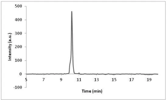
Figure 3.2. Liquid chromatogram of C12-VVAGK-Am after subtraction of water, the change of response units with respect to time at 220 nm. Purity is calculated as 89%.
Figure 3.3. Mass spectrum of C12-VVAGK-Am. Mass data [M+H]+ (calculated): 654.48, [M+H]+ (observed): 654.49, [2M+H]+ (calculated): 1307.96, [2M+H]+ (observed): 1307.98, [2M+Na]+ (calculated): 1329.96, [2M+Na]+ (observed): 1329.98.
43
Figure 3.4. Liquid chromatogram of C12-VVAGKS(Glc)-Am after subtraction of water, the change of response units with respect to time at 220 nm.
Figure 3.5. Mass spectrum of C12-VVAGKS(Glc)-Am. Mass data [M+H]+ (calculated): 903.57, [M+H]+ (observed): 903.58, 2[M+H]+ (calculated): 1807.14, 2[M+H]+ (observed): 1807.16.
44
Figure 3.6. Liquid chromatogram of C12-P3GK-Am after subtraction of water, the change of response units with respect to time at 220 nm. Purity is calculated as 98%.
Figure 3.7. Mass spectrum of C12-P3GK-Am. Mass data [M+H]+ (calculated): 676.47, [M+H]+ (observed): 676.48, [2M+H]+ (calculated): 1351.94, [2M+H]+ observed): 1351.95 , [M+2H]+/2 (calculated): 338.73, [M+2H]+/2 (observed): 338.74.
45
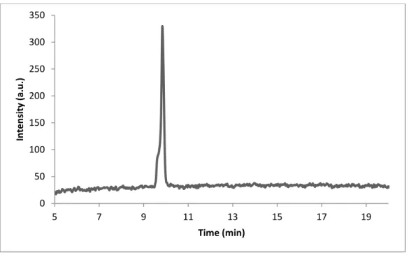
Figure 3.8. Liquid chromatogram of C12-P3GKS(Glc)-Am after subtraction of water, the change of response units with respect to time at 220 nm. Purity is calculated as 99%.
Figure 3.9. Mass spectrum of C12-P3GKS(Glc)-Am. Mass data [M+H]+ (calculated): 925.55, [M+H]+ (observed): 925.56, 2[M+H]+ (calculated): 1851.10, 2[M+H]+ (observed): 1851.12 , [M+2H]+/2 (calculated): 463.27, [M+2H]+/2 (observed): 463.28. 0 50 100 150 200 250 300 350 5 7 9 11 13 15 17 19 In te n si ty (a.u .) Time (min)