IKKs and tumor cell plasticity
Serkan I. G€oktuna1,2 , Michaela A. Diamanti3 and Tieu Lan Chau1
1 Department of Molecular Biology and Genetics, Bilkent University, Ankara, Turkey 2 National Nanotechnology Research Center (UNAM), Bilkent University, Ankara, Turkey
3 Georg-Speyer-Haus, Institute for Tumor Biology and Experimental Therapy, Frankfurt am Main, Germany
Keywords
EMT; IkappaB kinases; IKK; inflammation; metastasis; Stemness; tumor cell plasticity Correspondence
S. I. G€oktuna, Department of Molecular Biology and Genetics, Bilkent University, 06800 Bilkent, Ankara, Turkey
Fax: +90 3122665097 Tel: +90 3122902418
E-mail: [email protected] (Received 28 November 2017, revised 22 February 2018, accepted 21 March 2018) doi:10.1111/febs.14444
Nuclear factor jB (NF-jB) transcription factors are the central hubs of signaling pathways connecting proinflammatory signals to cell survival, proliferation and cytokine production. In cancers, NF-jB signaling influ-ences many aspects of tumor development, from initiation to metastasis. These functions are mediated by tumor-induced plasticity that allows tumor cells to adapt and survive in changing conditions within the tumor microenvironment. Tumor cell plasticity is shaped by the inflammatory microenvironment in tumors. This review focuses on inhibitor of NF-jB kinases, the direct upstream elements of NF-jB regulation, specifically on their conventional and non-conventional functions in animal models of tumorigenesis from the recent literature.
Introduction
A relationship between inflammation and cancer has been proposed since Rudolph Virchow observed an increase in the number of infiltrating leukocytes in tumors in 1863 [1]. Since then, a large body of evi-dence has accumulated supporting such a link, and the underlying molecular mechanisms have gradually been uncovered. Today we know that a history of chronic inflammation can be attributed to more than 17% of malignancies worldwide [2]. Prolonged intake of
non-steroidal anti-inflammatory drugs significantly lowers the risk of certain cancers, such as breast and colon cancer [3], suggesting that therapies targeting inflam-matory processes rather than directly killing tumor cells may overcome complications due to resistance of tumors to current therapeutics.
Drug resistance is acquired from cellular heterogene-ity within tumors, which in turn arises from tumor cell plasticity. The mechanisms leading to tumor cell
Abbreviations
ATM1, ataxia telangiectasia mutated serine/threonine kinase 1; BAFF, B-cell activating factor; CAC, colitis-associated colorectal cancer; CAF, cancer-associated fibroblast; CaP, prostate cancer; CRC, colorectal cancer; CYLD, cylindromatosis lysine 63 deubiquitinase; DEN,
diethylnitrosamine; DMBA, 7,12-dimethylbenzanthracene; E2F1, E2F transcription factor 1; EGFR, epithelial growth factor receptor; EMT, epithelial–mesenchymal transition; ER, endoplasmic reticulum; ERK, extracellular signal-regulated kinase; FOXA2, forkhead box protein A2; FOXO3a, forkhead box protein O3a; HCC, hepatocellular carcinoma; IFN, interferon; IKK, inhibitor ofjB kinase; IL, interleukin; IjB, inhibitor of nuclear factorjB; JNK, c-Jun N-terminal kinase; LT, lymphotoxin; MAPK, mitogen-activated protein kinase; mTOR, mechanistic target of rapamycin; NEMO, NF-jB essential modulator; NF-jB, nuclear factor jB; NMSC, non-melanoma skin cancer; NPM, nucleophosmin; Rac1, Rac Family Small GTPase 1; RANK, receptor activator of nuclear factorjB; RIPK1, receptor-interacting serine/threonine-protein kinase 1; ROS, reactive oxygen species; SCC, squamous cell carcinoma; SMRT, silencing mediator of retinoic acid and thyroid hormone receptor; SOD, superoxide dismutase; STAT3, signal transducer and activator of transcription 3; TAX1BP1, Tax1 binding protein 1; TGFb, tumor growth factor beta; TNF, tumor necrosis factor; TNFR, tumor necrosis factor receptor; TPA, 12-O-tetradecanoylphorbol-13-acetate; TSC, tuberous sclerosis complex subunit; VEGF-A, vascular endothelial growth factor A; XBP1, X-box-binding protein 1.
plasticity include mutations, epithelial–mesenchymal transition (EMT), dedifferentiation and inflammation, which help cancer cells adapt to changes and threats within the tumor microenvironment. Among these mechanisms, inflammation plays a central role. As will be explained in detail, the inflammatory microenviron-ment provides all the conditions for cells to accumu-late further mutations, to dedifferentiate to gain stem cell-like properties and to go through the EMT for invasion and formation of distant metastases. There-fore, we need a better understanding of inflammatory pathways and their regulators to be able to develop superior therapeutic options that target tumor cell plasticity and tumor development. This review particu-larly focuses on inhibitor of nuclear factor jB (IjB) kinases (IKKs) as the master regulators of inflamma-tory signaling mechanisms that shape tumor cell plas-ticity in various cancer models.
Tumor cell plasticity
Plasticity by definition means the ability to change or adapt to varying conditions. Cellular plasticity is required for tumor cells to adapt to an ever-changing tumor microenvironment for growth and survival amidst various environmental threats. Most of the tumors arising from a single cell have an unlimited pro-liferative ability yet differ in many other capabilities such as self-renewal, handling stress to avoid death, escaping immune surveillance and invading to form dis-tant metastases. All these differences within tumors are the result of a wide-ranging cellular heterogeneity [4–6]. In addition to heterogeneity, the great level of complex-ity within tumors is achieved by elaborate interaction of various cell types in response to alteration of the tumor microenvironment [7]. Altogether, cellular plasticity-driven heterogeneity accounts for the development of resistance to chemotherapeutic reagents.
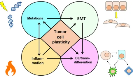
How is tumor cell plasticity achieved? There are basically four fundamental mechanisms, namely muta-tion, dedifferentiation/transdifferentiamuta-tion, EMT and inflammation (Fig. 1). Together with epigenetic inter-actions, tumor cell fusion and exocytic vesicles, these shape the cellular plasticity of the tumors [8]. How-ever, this review focuses only on the fundamental mechanisms that lead to plasticity during the initia-tion, progression and metastasis of tumors.
The first source of tumor cell plasticity is mutations. As seen in the process of evolution, heterogeneity– vari-ation– must pre-exist in tumor cell populations before any change in the tumor microenvironment occurs. Mutations are key players in cellular heterogeneity within tumors [9]. Driver mutations, such as
chromosomal or microsatellite instability and loss of DNA proofreading or repair machinery, increase tumor cell susceptibility to further mutations to escape cell death or to gain new features required for malignant transformation [10]. Besides, mutations within a solid tumor can give rise to different populations that take part in invasion, metastasis, drug resistance and tumor recurrence [11]. Mutations are not only a component of tumor cell plasticity, but also the basis of all the other mechanisms leading to it. Therefore, every fundamental feature of tumor cell plasticity is based on acquiring suf-ficient mutations to achieve heterogeneity within the tumors for the growth and expansion of cancer cells.
The second fundamental mechanism that drives tumor cell plasticity is dedifferentiation and/or transd-ifferentiation, respectively the ability of tumor cells to acquire stem cell-like characteristics supporting con-tinuous proliferation of growing tumors and to shift back to differentiated states [8]. In a given tissue, stem cells are the only cell type that maintains tissue homeostasis following damage or renews the tissue by transforming into any other cell type [8,11,12]. Nor-mally, differentiated cells lose this capability, but dif-ferentiated cells within tumors can dedifferentiate, adopting a stem cell-like phenotype [11], and there-fore cancer stem cells can arise from the tissue stem cells or from differentiated quiescent cells to sustain tumor growth. This ability is essential to repopulate the cancer stem cell niche against any cellular stress leading to the loss of stem cells [13], thus making stemness and dedifferentiation major assets for tumor cell plasticity.
The third important mechanistic insight into tumor cell plasticity is related to EMT, which grants tumor cells the ability to invade deeper into the tissue or to form distant metastases [14]. When tumors grow to their natural boundaries limited by the surrounding tissues and extracellular matrix, it is a challenge for their metabolically active cells to maintain a nutrient supply sufficient for their high demands [15]. To do so, some tumor cells remodel the extracellular matrix to open up extra space for new tumor cells to invade by coordinating fibroblasts and leukocytes, while others induce angiogenesis to initiate a new blood supply together with endothelial cells, and others still intrava-sate into the blood circulation to find new places to grow without competition [14,15]. Since only mes-enchymal-like cells can activate molecular pathways related to motility, almost all these processes require EMT for cells to gain motility to leave the tumor stroma and form new colonies [16]. However, the mes-enchymal state is not as proficient as the epithelial state in terms of proliferative ability [17,18]. Epithelial
cells have a basal attachment for efficient proliferation and additionally paracrine growth signals become more effective when the cells are closely packed. Although mesenchymal cells lose these advantages, metastasizing mesenchymal cells still have the capacity to return to a highly proliferative epithelial state to form distant colonies [19–21]. Therefore, epithelial– mesenchymal plasticity is a two-way process helping the tumor cells reprogram their physiology according to the changing demands of the tumor microenvironment.
The last but not least important mechanism of tumor cell plasticity is the ability to create a chronic inflammatory environment within solid tumors. An inflammatory microenvironment can promote tumor growth by triggering the release of growth hormones from surrounding stromal or immune cells, leading to altered cytokine profiles within the tumor, and thus evasion of immunosurveillance. Chronic inflam-mation also induces a wound healing response, angiogenesis or EMT to enable tumor cells to invade and eventually escape to form metastases [22,23]. Recently, it has been found that a microenvironment that favors growth and increased mutational rates due to reactive oxygen species (ROS) stress and compensatory proliferation upon excessive cell death can initiate tumorigenesis as well [24]. Taken together, an inflammatory environment can be the driving force for tumor cell plasticity. Eventually, the success of tumor cells not only results from their intrinsic ability to alter their own physiology but also depends on how they transform the whole tumor microenvironment to promote tumor growth.
Inflammation as the driving force for
tumor cell plasticity
Inflammation is the natural response, elicited through concerted activity of infiltrating leukocytes, cytokine secretion and tissue regeneration, when the host is faced with tissue injury or pathogens, in an attempt to heal a wound or eliminate a pathogen [25]. Normally, inflam-mation is resolved as soon as the threat to tissue integ-rity has been eliminated. However, incomplete elimination of a pathogen or failure to properly shut down the inflammatory pathways will result in chronic inflammation. This is one of the hallmarks of cancer, leading to continuous cytokine and protease secretion, and production of ROS and nitric oxides that eventually create an inflammatory microenvironment favoring tumor initiation [24,26]. This microenvironment has many cellular components, namely tumor cells, stromal cells, and innate and adaptive immune cells. It is the intricate signaling among these cell types that tilts the balance towards protumorigenic or antitumorigenic immunity [27].
Among all the molecular pathways that bridge inflammatory regulation of tumor microenvironment and cellular plasticity, nuclear factor jB (NF-jB) sig-naling represents a central hub due to its versatile roles in inflammation and tissue regeneration [28–31]. While chronic inflammation increases susceptibility to cancer development, NF-jB signaling can also contribute to tumor promotion in cancers not associated with pre-existing inflammation by creating an inflammatory tumor microenvironment [32]. The NF-jB family com-prises proteins that regulate expression of genes
Fig. 1. Mechanisms of tumor cell plasticity. Mutations, EMT, dedifferentiation/transdifferentiation and inflammation are the main mechanisms that gives rise to tumor heterogeneity. Heterogeneity is important in enabling tumor cells to adapt to and survive environmental threats.
involved in inflammation, innate and adaptive immune responses, cell survival, and cancer [28,33–35]. Although both NF-jB (e.g. RelA, RelB, cRel, p50, p52) and its regulatory IjB subunits (e.g. IjBa, IjBb, IjBe, Bcl3, p100, p105) are numerous, little has been revealed of the mutations that affect the functions of these molecules in different diseases [36]. Consequently, our knowledge about their regulation has come mainly from genetic models of upstream regulatory elements, such as IKKs, in cancers of hematopoietic origin [37].
A core element of NF-jB activation is the IKK com-plex built from two catalytic subunits, IKKa and IKKb kinases, and a regulatory subunit, NF-jB essen-tial modulator (NEMO)/IKKc [29,31]. We know that NF-jB subunits are regulated by the IKK complex, which is in turn regulated by diverse upstream signaling mechanisms comprising growth, death, stress and pathogen recognition receptors. Activation of NF-jB transcription factors is highly dependent on both classi-cal and alternative pathways that mediate degradation of IjB and IjB-related proteins [29,30]. The classical (or canonical) pathway utilizes IKKb- and IKKc-dependent IjBa degradation in response to proinflam-matory stimuli (such as interleukin (IL)-1, tumor necrosis factor (TNF)-a and toll-like receptor ligands) releasing active NF-jB dimers into the nucleus, where they bind to DNA and mediate gene expression [38,39]. In contrast, the alternative (or non-canonical) pathway can be activated by ligands (such as B-cell activating factor (BAFF), lymphotoxin (LT) -a1b2 and CD40L)
acting on their specific receptors (BAFFR, LTbR and CD40, respectively) and depends exclusively on IKKa and its upstream kinase, NF-kB-inducing kinase [40– 43]. Throughout the text we will use the term ‘conven-tional’ to refer to the NF-jB-dependent (comprising both classical and alternative pathways) functions of IKKs and ‘non-conventional’ for the NF-jB-independent functions or targets of IKKs. Still, it cannot be ruled out that IKKa/b activity on non-conventional targets or pathways may also be induced through similar upstream elements to those inducing conventional (classical or alternative) NF-jB signaling pathways [44]; nonetheless, IKKa/b activity on non-conventional targets may not require any of the downstream elements activated by NF-jB transcription factors [45,46]. To better illustrate conventional and non-conventional targets of IKKs in different cancers, Tables 1 and 2.
Despite the fact that canonical IKKs have structural and functional similarities, they have distinct roles, and diverse phenotypes have been reported for IKKa and IKKb knockout mice [32]. Many studies have addressed the role of IKK subunits in tumor develop-ment because of their therapeutic potential, which is
largely absent for NF-jB or IjB subunits. Although there is a potential use of specific peptides blocking the interaction of non-kinase subunits of signaling complexes, such as IKKc in IKK complex, to termi-nate downstream signaling [47], the number of such studies is still limited compared with studies addressing catalytic subunits of IKK complexes [48–50]. There-fore, in the next section, we concentrate on IjB kinases as the key mediators of cytokine signaling that are able to orchestrate inflammation in the tumor microenvironment. The focus on both conventional and non-conventional functions, with specific examples of in vivo animal models of solid tumors, will explain how these molecules affect tumor cell plasticity by shaping the tumor microenvironment.
Specific roles of IKKs in tumor cell
plasticity
Though the roles of canonical NF-jB signaling have long been well described in many cancers, specific functions of IKKb or IKKa in tumor cell plasticity were poorly characterized until recently. Accumulating evidence suggests that in their non-conventional roles, these kinases possess a greater level of complexity in the way they regulate inflammation, proliferation and metastasis due to their ability to phosphorylate diverse substrates [51]. Table 1 summarizes conventional and non-conventional targets or signaling pathways con-trolled by IKKs and their effects on tumor cell plastic-ity in different cancers (also see Fig. 2). Detailed information on the non-conventional IKKa/b targets in different signaling pathways can be found in Table 2. In contrast to IKKb, which spends most of its time in the cytoplasm chasing canonical and non-conventional targets, IKKa translocates into the nucleus where it regulates non-conventional substrates such as p65, histone H3, p27, forkhead box protein A2 (FOXA2) and silencing mediator of retinoic acid and thyroid hormone receptor (SMRT) to direct vari-ous mechanisms in inflammation and cancer (Table 2 and Fig. 3) [32,51–53].
IKKs are involved in tumor-elicited inflammation through their indispensable roles in regulating inflam-matory cells and their interactions with tumor cells within the tumor microenvironment. Specifically, IKKs regulate proinflammatory or anti-inflammatory signal-ing machinery through phosphorylation of numerous targets, such as NF-jB, protein inhibitor of activated STAT1 (PIAS), interferon regulatory factor 3/7 and Tax1 binding protein 1 (TAX1BP1). These targets in turn regulate downstream factors such as TNF, IL-1, IL-6, IL-18, interferon (IFN)-a/b, IFNc, various other
cytokines and signal transducer and activator of tran-scription 3 (STAT3) [45,54,55], which also have pleio-tropic effects on proliferation, oxidative stress, endoplasmic reticulum (ER) stress and anti-apoptotic machinery, challenging genomic integrity and facilitat-ing mutations, another important mechanism in tumor cell plasticity.
Besides these inflammatory targets, IKKs can also directly or indirectly regulate many proliferative fac-tors, such as (a) cyclin D1, epidermal growth factor receptor (EGFR), tumor growth factor b (TGFb), c-Myc and FOXO3a, which in turn favor mutations due to increased proliferation [46]; (b) many components in survival pathways activated upon stress such as CYLD Lysine 63 Deubiquitinase, X-box-binding pro-tein 1 (XBP1), superoxide dismutase (SOD), Rac Family Small GTPase 1 (Rac1), ATG16 l1, receptor-interacting serine/threonine-protein kinase 1 (RIPK1), extracellular signal-regulated kinase (ERK) and c-Jun
N-terminal kinase (JNK) [45,56–59]; and (c) various controlling factors such as p53, Aurora A, ATM Ser-ine/Threonine Kinase 1 or nucleophosmin (NPM) that orchestrate factors to preserve genomic integrity in other cancers [45,51]. IKKs also control factors such as SMRT, FOXA2, histone deacetylase 3, histone H3a, Smad2/3 or E2F transcription factor 1 (E2F1) partici-pating in NOTCH, Wnt or TGFb pathways [46,51]. The latter is important signaling machinery as highly dividing tumor cells require stemness and dedifferentia-tion/transdifferentiation to maintain the ability to regenerate or adapt to various stressful conditions within the tumor microenvironment. Last but not least, the specific functions of IKKs in tumor cell plasticity include their abundant functions in controlling EMT through factors such as Wnt, Maspin1, Twist and Snail [8,60]. Formation of tumor cell plasticity highly depends on the regulation of canonical and non-canoni-cal targets by the tissue- and context-specific roles of
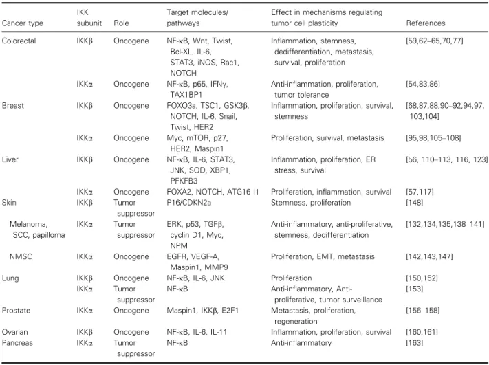
Table 1. Conventional and non-conventional targets/signaling pathways controlled by IKKs and their effects in tumor cell plasticity in different cancers. Gsk3b, glycogen synthase kinase 3b; iNOS, inducible nitric oxide synthase; NMSC, non-melanoma skin cancer; PFKFB3, 6-phosphofructo-2-kinase/fructose-2,6-biphosphatase 3; SCC, squamous cell carcinoma.
Cancer type
IKK
subunit Role
Target molecules/ pathways
Effect in mechanisms regulating
tumor cell plasticity References
Colorectal IKKb Oncogene NF-jB, Wnt, Twist,
Bcl-XL, IL-6, STAT3, iNOS, Rac1, NOTCH
Inflammation, stemness, dedifferentiation, metastasis, survival, proliferation
[59,62–65,70,77]
IKKa Oncogene NF-jB, p65, IFNc, TAX1BP1
Anti-inflammation, proliferation, tumor tolerance
[54,83,86]
Breast IKKb Oncogene FOXO3a, TSC1, GSK3b,
NOTCH, IL-6, Snail, Twist, HER2
Inflammation, proliferation, survival, stemness
[68,87,88,90–92,94,97, 103,104]
IKKa Oncogene Myc, mTOR, p27, HER2, Maspin1
Proliferation, survival, metastasis [95,98,105–108]
Liver IKKb Oncogene NF-jB, IL-6, STAT3,
JNK, SOD, XBP1, PFKFB3
Inflammation, proliferation, ER stress, survival
[56, 110–113, 116, 123]
IKKa Oncogene FOXA2, NOTCH, ATG16 l1 Proliferation, inflammation, survival [57,117]
Skin IKKb Tumor
suppressor
P16/CDKN2a Stemness, proliferation [148]
Melanoma, SCC, papilloma IKKa Tumor suppressor ERK, p53, TGFb, cyclin D1, Myc, NPM Anti-inflammatory, anti-proliferative, stemness, dedifferentiation [132,134,135,138–141]
NMSC IKKa Oncogene EGFR, VEGF-A,
Maspin1, MMP9
Proliferation, EMT, metastasis [142,143,147]
Lung IKKb Oncogene NF-jB, IL-6, JNK Proliferation [150,152]
IKKa Tumor
suppressor
NF-jB inflammatory,
Anti-proliferative, tumor surveillance
[153] Prostate IKKa Oncogene Maspin1, IKKb, E2F1 Metastasis, proliferation,
regeneration
[156–158] Ovarian IKKb Oncogene NF-jB, IL-6, IL-11 Inflammation, proliferation, survival [160,161]
Pancreas IKKa Tumor
suppressor
the IKK kinases. To better illustrate this, we give sev-eral examples of the pivotal roles IKKs play in shaping tumor cell plasticity in what follows.
Among many studies associating NF-jB signaling with almost any cancer in the past two decades, there have been only a limited number providing mechanistic insights, and even fewer describing the roles of IKKs in animal models of tumorigenesis. Since we believe that the complex regulations and interactions within the tumor microenvironment can be best understood through the use of genetic disease models and trans-genic animal models, here we select those studies, clas-sified by individual cancers, to demonstrate how canonical or non-canonical IKK functions affect tumor cell plasticity. Of note, however, due to the genetic and physiological differences between these species, there is a possibility that not all the information derived from animal models can be translated into human disease. Recent literature has witnessed an increased interest in patient-derived xenografts or patient-derived organoid models to better approximate and model human dis-eases. Although these are powerful tools for rapid and
reliable drug screening, toxicological assessment and combinatorial targeting, they fail to establish complex interactions within the tumor microenvironment. To resolve such problems, advances in cell-based co-cultures of multiple origin may be a good alternative. Develop-ments in this area or in related cell-based techniques will eventually eliminate the requirement for animal use in cancer or any other disease research, yet the information derived from such studies about the specific functions of IKKs in various solid malignancies is very limited at the present.
Cancer specific functions of IKKs in
tumor cell plasticity
Colorectal cancer
Constitutive NF-jB activation, a common theme in most cancers, is one of the leading reasons that tumors develop resistance against chemotherapy or radiation. NF-jB activity is crucial for the activation of anti-apoptotic signaling that protects tumor cells from
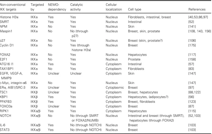
Table 2. Detailed information about non-conventional IKKa/b targets in different signaling pathways. The information in this table is compiled mostly from studies with animal tumor models. For more exhaustive information about non-conventional IKKa/b targets in various signaling machinery, not necessarily related to tumor cell plasticity, refer to previous reviews [45,46,51]. AIB1/SRC-3 alliases for NCOA3, nuclear receptor coactivator 3; ERa, esterogen receptor alpha; MMP9, matrix metalloprotease 9; PFKFB3, 6-phosphofructo-2-kinase/ fructose-2,6-biphosphatase 3. Non-conventional IKK targets Targeted by NEMO-dependency Catalytic activity Cellular
localization Cell type References
Histone H3a IKKa Yes Yes Nucleus Fibroblasts, intestinal, breast [40,53,86,97]
SMRT IKKa Yes Yes Nucleus Intestinal [52]
NPM IKKa No Yes Nucleus Skin [141]
Maspin1 IKKa No No (through
p27)
Nucleus Breast, skin, prostate [108, 143, 156]
p27 IKKa No Yes Nucleus Breast (skin, prostate?) [106]
Cyclin D1 IKKa No Yes (through
histone H3a)
Nucleus Breast [175]
FOXA2 IKKa No Yes Nucleus Hepatocytes [117]
E2F1 IKKa No Yes Nucleus Prostate [158]
ATG16 l1 IKKa Yes Yes Cytoplasm Intestinal [57]
TAX1BP1 IKKa No Yes Cytoplasm Fibroblasts [83]
EGFR, VEGF-A, MMP9
IKKa Unclear Unclear Cytoplasm Skin [147]
c-Myc, integrin-a6 IKKa No Yes Nucleus Skin [147]
ERa, AIB1/SRC-3 IKKa Unclear Yes Cytoplasmic Breast [97]
TSC1 IKKb Unclear Yes Cytoplasm Breast, hepatocytes [88,122]
XBP1 IKKb Yes Yes Cytoplasm Hepatocytes, (adipocytes?) [56]
PFKFB3 IKKb Yes Yes Cytoplasm Breast, fibroblasts [123]
FOXO3a IKKb Unclear Yes Cytoplasm Breast [87]
RIPK1 IKKa/b Yes Yes Cytoplasm Hepatocytes [115]
NOTCH IKKa/b No No (through SMRT
or FOXA2/NUMB)
Nucleus Intestinal and breast (through SMRT), hepatocytes (through FOXA2)
[52,103]
IL-6 IKKa/b Yes No (through NOTCH) Nucleus Breast [103]
death [61]. Activation of the anti-apoptotic machinery in most cancers, especially in colorectal cancer (CRC), through induction of NF-jB-dependent gene expres-sion requires IKKb catalytic activity. In a colitis-associated colorectal cancer (CAC) model for tumorigenesis, IkkbDIEC (Ikbkb) mice were found to have reduced tumor burden due to increased apoptosis through downregulation of Bcl-xL [62]. On the other
hand, IkkbDMΦ mice in a CAC model were found to have reduced tumor number and size due to decreased proliferation resulting from diminished macrophage-derived IL-6 synthesis [62]. Later, it was suggested that macrophage-derived IL-6 and inducible nitric oxide synthase promote tumor growth via inflammatory STAT3 [63,64], activation of which has been shown to regulate proliferation, survival and NF-jB signaling in a CAC tumor model [65,66].
IKKb-driven inflammation not only provides prolif-erative or survival advantage but may also influence stemness and differentiation. As suggested in previous studies on different tissues, IKKb-driven inflammation blocks the differentiation of adipocytes, osteoclasts
and vascular endothelia to lock them in a more stem cell-like phenotype through various mechanisms involving regulation of Wnt signaling [67–69]. In our recent work, we have shown that non-stem cells from intestinal epithelia can convert to a stem cell-like phe-notype in the presence of constitutive Wnt and NF-jB activation in spontaneous mouse models of colorectal tumorigenesis [70]. In the same work, activated NF-jB could induce Wnt signaling or vice versa through the cooperation of their transcription factors at related gene promoters [70]. However, the latter was not mechanistically illustrated and the proof for this mech-anism came from a related study in which it was found that both NF-jB activation and ROS production are controlled by Rac1 in a Wnt-driven colorectal carcino-genesis model by loss of the Apc gene [59]. Accord-ingly, Lgr5+ stem cell expansion in Wnt-driven tumors mechanistically required the activation of Rac1 to induce ROS production, and as a result, a marked activation of NF-jB to protect stem cells from apopto-sis [59]. Other studies showed that IKKb expression in cancer-associated fibroblasts (CAFs), also known as
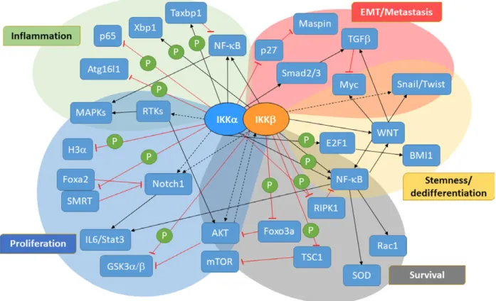
Fig. 2. Signaling pathways governed by IKKs in tumor development and tumor cell plasticity. A summary of the combination of signaling molecules and pathways targeted by IKKs in different cancers. It should be noted that not all pathways are active at the same time or work in the same way in each cancer. Most of these pathways are differentially regulated in different cancers and even in different subtypes. Tables 1 and 2 give more detailed information about IKK target molecules/pathways and how they affect tumor development in various genetic cancer models. GSK3a/b, glycogen synthase kinase 3a/b; H3a, histone H3 alpha; RTK, receptor tyrosine kinase.
intestinal mesenchymal cells, may have potential roles in colorectal tumor cell plasticity [71,72]. Despite the fact that contrasting results were obtained in these studies, the oncogenic potential of IKKb in CRC may be due to differences in tissue-specific ablation strate-gies that were constitutive [71] rather than inducible [72]. Although there are some critics requesting further proof on IKKb-specific roles in CAFs and CRC [73], it is noteworthy that while germline mutations are often compensated during the course of development, studies with inducible models can illustrate better the sporadic nature of cancer.
It remains unknown how IKKb-driven NF-jB acti-vation triggers the metastatic shift in colorectal tumors. It was first shown that constitutively induced cytokines and a chronic inflammatory phenotype were not sufficient to drive inflammation in the intestine without mitogen-activated protein kinase (MAPK) sig-naling [74]. MAPKs are required for c-Myc stabiliza-tion upon activastabiliza-tion of toll-like receptor signaling in Wnt-driven tumor models [75]. Hence, MAPKs and their downstream transcription factors regulate the expression of various cytokines and proliferative fac-tors by co-operating with NF-jB at promoter regions
of target genes [74,76]. However, in later studies, our colleagues have proven that this proinflammatory envi-ronment created by constitutive IKKb activation is not only required for tumor initiation and progression but also for invasion and metastasis upon p53 loss [77]. Consequently, Twist1, an EMT marker, was tran-scriptionally activated leading to tumor invasion and lymph node metastasis [77].
IKKa regulates intestinal tumor cell plasticity through modulation of inflammation and cell to cell interactions within the tumor microenvironment. It is widely known that immune cells can suppress, tolerate or promote the growth of tumor cells in various can-cers, yet the regulation of immune cells in the early stages of tumor development has been poorly described until recently. A recent study on the V12RAS-driven zebrafish melanoma model clearly demonstrated that myeloid cells were recruited to the tumor-initiating cells as soon as the oncogenic trans-formation occurred [78]. Interaction of both cell types is required for the tumor progression parallel to the wound healing response [78]. Similar to these observa-tions, we have identified a unique intraepithelial local-ization of myeloid cells in the intestines of mice devoid
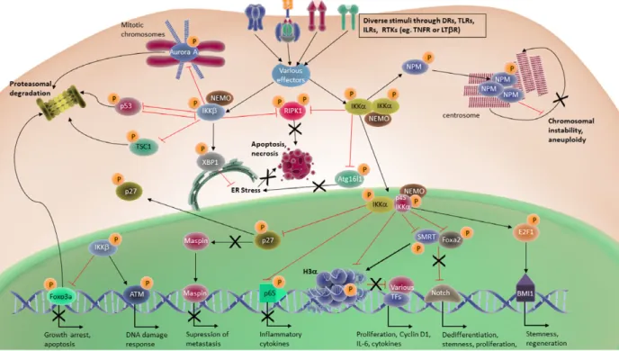
Fig. 3. NF-jB-independent functions of IKKs in tumor development. This figure summarizes some of the most common non-conventional IKK targets in different tumors. Again the regulation on these molecules is determined according to tissue type, tumor stage and other factors within the tumor microenvironment. Therefore, the outcome of any given regulation may show dramatic differences from one cancer to another. Table 2 gives more detailed information about non-conventional IKK targets in various malignancies. ATM1, ATM Serine/ Threonine Kinase 1; BMI1, B cell-specific Moloney murine leukemia virus integration site 1; DR, death receptor; IL1R, interleukin 1 receptor; RTK, receptor tyrosine kinase; TF, transcription factor; TLR, toll-like receptor.
of IKKa activation (IkbkaS181A/S185A or simply IKKaAA/AA) upon b-catenin-induced transformation
[54]. Mechanistically, IKKa represses an NF-jB-dependent cytokine expression, required for enhanced recruitment of myeloid cells to the site of tumors [54]. In this model, IKKa-deficient myeloid cells showed M1-like polarization and actively produced IFNc upon tumorigenesis. Additionally, the tumor suppressive function of myeloid cells in the absence of IKKa acti-vation may not be limited to enhanced paracrine IFNc secretion, which has been shown to be important in the anti-tumor response [79,80], and they may also phagocytize tumor cells [81] more efficiently, as their specific localization may suggest. Prior to our observa-tions, it was reported that IKKa limits macrophage activation and contributes to the resolution of inflam-mation via preventing extended promoter binding of RelA and c-Rel [82]. IKKa was found to be crucial in limiting macrophage or dendritic cell activity [82–85]. Given that proinflammatory signaling governed by enhanced NF-jB activation may exacerbate inflamma-tion in different diseases, IKKa-dependent feedback inhibition may prove to be indispensable for preven-tion of tissue damage in various pathologies [54]. Pre-viously, it had been reported that nuclear localization of IKKa results in phosphorylation and release of the repressor protein SMRT from NOTCH1 target genes, leading to their aberrant expression upon malignant transformation in CRC [52]. In a recent report, Margalef and colleagues [86] showed that phospho-activated IKKa is first cleaved to a shorter form (p45-IKKa), and then this active and truncated IKKa is translocated to the nucleus where it phosphorylates and release the transcriptional repressor SMRT from the promoters of NOTCH signaling targets, preventing cancer cells from differentiating. The existence of cleaved-nuclear IKKa, a primary form in human patient samples, correlates with tumor stage and prog-nostic outcome in patients [86]. Thus, IKKa can gov-ern stemness and proliferation of intestinal tumor cells through these non-conventional functions to further enhance their plasticity to tumor microenvironment changes.
Breast cancer
Breast cancer is another example where canonical and non-canonical functions of IKKs affect tumor cell plasticity in various settings related to tumor develop-ment and metastasis. One of the first pieces of mecha-nistic evidence showing that IKKb can regulate non-canonical targets to promote tumorigenesis came from work with animal models of breast cancer [87]. This
study demonstrated a physical FOXO3a–IKKb inter-action, followed by FOXO3a phosphorylation, ubiqui-tination and degradation [87], all of which occur independently of AKT. FOXO3a-depleted tumor cells grow better in mouse xenografts due to extensive NF-jB activation leading to survival of tumor cells. As a result, FOXO3a-positive breast cancer patients have improved survival due to activation of proapoptotic signaling while FOXO3a IKKb+ patients had worse prognosis [87]. In another study, IKKb was found to connect tumor-elicited inflammation to angiogenesis by directly phosphorylating the mechanistic target of rapamycin (mTOR) repressor TSC complex subunit 1 (TSC1)–TSC2 tumor suppressor complex to initiate angiogenesis within breast tumors [88]. Tumor xeno-grafts with mutant TSC1 lacking a phosphoregulatory site grew poorly, whereas human patients positive for both phospho-TSC1 and phospho-S6K had worse prognosis [88]. Hence, IKKb activation upon hypoxia-induced proinflammatory TNF expression is crucial in angiogenesis induction for further tumor growth and invasion in breast cancer. Additionally, IKKb and NF-jB were shown to be essential for EMT and metastasis in breast cancer cells [89]. It subsequently became evident that NF-jB is required for stabiliza-tion of Snail or Twist1 in breast cancer cell lines to ini-tiate EMT, invasion and metastasis of breast cancer [90–92]. IKKb was also shown to be crucial for endothelial cell permeability, which is important for the extravasation of metastatic cells [68]. Park and col-leagues [93] described that NF-jB-dependent granulo-cyte-colony stimulating factor secretion by metastatic breast cancer cells was directing osteoclast differentia-tion and osteolytic bone metastasis into skeletal tissue, which is frequently observed in breast cancer patients. Similarly, Chen and colleagues [94] observed that IKKb implements a LIN28B/TCF7L2 feedback loop that promotes breast cancer cell stemness and metasta-sis with the help of in vivo xenograft and pharmaco-logical inhibition studies. Briefly, IKKb regulates tumor cell plasticity in breast cancer by regulating many facets of related mechanisms.
In the signaling believed to be involved in breast carcinogenesis, the non-canonical roles of IKK kinases, IKKa in particular, have not been fully inves-tigated until recently. We now know that primary human breast cancer specimens and breast cancer cell lines exhibit aberrant IKKa and IKKb kinase activi-ties that may not involve NF-jB [95] or its down-stream targets [96] to enhance transformation of breast cancer cells. IKKa/b kinases have been also shown to promote mammary tumorigenesis through inhibition of FOXO3a [97] and glycogen synthase kinase 3b by
the activation of mTORC2, which further regulates the phosphatidylinositol-3-kinase–AKT pathway [98]. Moreover, implication of a role for constitutively active IKK complex and NF-jB activation in breast cancer cell migration and metastasis has also been reported [99]. Given the important role of NOTCH1 [100,101] and IL-6 signaling in breast tumorigenesis [102], it was recently reported that IKKa and IKKb, independent of NF-jB, control NOTCH-mediated IL-6/Janus kinase/STAT signaling in breast tumors cells [103]. IKKa was then discovered to be essential for oncogenic transformation in in vivo models of Her2/ neu-induced breast tumorigenesis [104]. In this study, using two independent mouse models of mammary tumorigenesis, the authors provided evidence support-ing the importance of IKKa kinase activity for the increased incidence and advanced onset of ErbB2/ Her2-induced breast tumors [104]. This is reinforced with more recent findings as well [105]. In these stud-ies, the enhanced, rather than totally inhibited, tumori-genesis was attributed to the kinase domain of IKKa, which maintains signals essential for the self-renewal potential of tumor-initiating cells. Currently, we know that IKKa regulates the expansion of these cells, in Her2-initiated tumors through a mechanism that involved NF-jB inducing kinase/IKKa axis induced p27/Kip1 phosphorylation and nuclear exclusion, con-tributing to augmented tumorigenesis [106]. On the other hand, IKKa was shown to be more efficient than IKKb in activating NF-jB and the expression of downstream genes involved in cell invasion [107] in Her2-overexpressing breast cancer cells. The prometa-static activity of IKKa was stimulated by receptor activator of NF-jB (RANK) ligand (RANKL)/RANK to repress Maspin expression [108] and correlated inversely with nuclear p27/Kip1 in human invasive breast carcinoma specimens [106]. The studies summa-rized above indicate a protumorigenic and prometa-static role of IKKa in the mammary gland in which the kinase activity of IKKa stimulates proliferation of breast cancer cells and protects them from being killed by anti-cancer drugs. Consequently, IKKa can be con-sidered an important regulator of breast cancer pro-gression and a notable target for the development of therapeutic strategies targeting inhibition proliferation, metastasis and drug resistance in breast cancer cells.
Hepatocellular carcinoma
IKKb plays opposing roles in different cells at different time points in hepatic cancer development. Since there were no hepatitis B virus or hepatitis C virus equivalents for generating a hepatitis-induced
carcinogenesis in mice, Mdr2 knockout mice, which develop cholestatic hepatitis followed by hepatic tumorigenesis, were used in early research [109]. By the use of this model, inflammatory cell-derived TNF is found to promote hepatocyte survival via activation of canonical anti-apoptotic NF-jB signaling [110]. Interestingly, using an IjB super-repressor model to silence NF-jB specifically in hepatocytes, no difference was observed in the tumor development in normal and mutant mice [110]. Only when the same model was used to inactivate NF-jB conditionally at a later point or when anti-TNF antibody was applied, was tumor growth seriously prevented due to apoptosis induction in the transformed hepatocytes [110]. In sharp contrast to these observations, in a mouse model of chemical carcinogen (diethylnitrosamine; DEN)-induced hepato-cellular carcinoma (HCC), it was shown that IKKb deletion in hepatocytes dramatically increased tumor burden while its deletion in macrophages inhibited tumor formation [111]. The observed differences in these two studies may be due to high regenerative abil-ities of hepatocytes in which IKKb or NF-jB plays an important protective role against damage-induced cell death and resulting compensatory proliferation upon liver injury by DEN [111]. As shown in another study, NF-jB activates SOD to counteract ROS-induced stress in hepatocytes [112], whereas in the absence of NF-jB, ROS induces the activation of JNK and acti-vator protein 1, which eventually leads to hepatocyte death via apoptosis [113]. Further work has clarified that ROS-damage-induced compensatory proliferation in HCC was driven by STAT3 activation in hepato-cytes [114]. The contradictory NF-jB involvement in early and late stages of hepatocellular cancer was fur-ther supported by IKKc/NEMO mutant mice, bearing IKKc-deficient liver parenchymal cells. These mice spontaneously developed steatosis or multiple tumors in their livers [115]. The absence of IKKc halts kinase complex formation and causes excessive cell death due to uncontrolled RIPK1 activation. Thus, RIPK1 abla-tion or RIPK1 kinase inactive knock-in models could prevent hepatocyte apoptosis and HCC in mice [115]. Later studies proposed that the control of RIPK1 to prevent hepatocellular necrosis or apoptosis relies on non-conventional functions of IKKs and differs from their roles in TNF-driven canonical NF-jB activation-related anti-apoptosis [58]. This observation partially explains the different observations between NF-jB super-repressor and IKKb-deficient mouse models.
Aside from hepatocytes in HCC, the resident liver macrophages, Kupffer cells, when induced by apoptotic hepatocytes, secrete inflammatory cytokine IL-6, which in turn drives proliferation in hepatocytes through
activation of STAT3 [111,116]. Therefore, when tumor cell plasticity relies rather on myeloid cell-dependent activation of IKKb in HCC, IKKb in myeolid cells does not display a protective role in the late hepatic tumorigenesis as it does in hepatocytes at the earlier stage. In contrast, elevated IKKa levels have been asso-ciated with increased oncogenic potential in HCC mod-els of tumorigenesis since IKKa has been found to be activated with elevated TNF expression during hepati-tis [117]. Following this, IKKa translocates into the nucleus where it phosphorylates FOXA2 to suppress its downstream targets such as NUMB which in turn is a NOTCH1, an important transcription factor that regu-lates cell fate and differentiation during various devel-opmental processes, inhibitor [117]. Therefore, this specific role for IKKa helps the proliferation of hepa-tocytes in an inflammation-driven and NOTCH1-dependent manner [117].
IKKa/b activation in HCC can be also induced through other pathways than tumor necrosis factor receptor (TNFR) signaling. Indeed, more recent stud-ies with hepatitis-driven cancer models indicated that TNFR can be dispensable for NF-jB activation and its downstream effects in chronic hepatitis-driven HCC [118–120]. Upon observing that LTs and their receptor LTbR were highly upregulated in HCC, Haybaeck and colleagues [118] developed a chronic HCC mouse model (to simulate hepatitis B virus- and hepatitis C virus-driven chronic hepatitis and HCC) with LT-over-expressing hepatocytes. They showed that only an LT (a and b) response of hepatocytes leads to IKKa/b activation in tumor cells. Therefore, only LTbR activa-tion is required for oncogenic transformaactiva-tion upon chronic hepatitis while TNFR1 was found to be dis-pensable. Later studies, with mouse models of high-fat-diet-induced steatohepatitis-driven HCC, showed that LTs or LIGHT (TNFSF14) produced by natural killer T cells or CD8+ cells specifically signaled through LTbR on hepatocytes to induce steatohepati-tis and hepatocyte damage through the activation of the canonical NF-jB pathway for hepatocellular tumorigenesis [119,120]. Hence, the differences in the observed results are probably due to differences in experimental models. More satisfactory conclusions about the upstream factors leading to IKK activation in hepatocellular tumorigenesis may be derived from advanced mouse models that better mimic the develop-ment of human disease.
As with other mechanisms, ER stress can alter the tumor microenvironment and tumor cell plasticity through modulation of immune responses, fueling tumor development [121]. Earlier studies proposed that IKKb control on TSC1 and the mTOR signaling
pathway may regulate glucose metabolism and insulin resistance through the regulation of insulin receptor substrate 1 in hepatocytes [122]. Similarly IKKb has been shown to regulate glutamine deprivation, result-ing from increased anabolic activity in highly dividresult-ing cancer cells, through its direct control on 6-phospho-fructo-2-kinase/fructose-2,6-biphosphatase isoform 3 in the glycolytic pathway [123]. Recent studies have pro-vided solid connections between ER stress, inflamma-tion and cancer in certain malignancies [124]. Especially in liver and colorectal cancers, elements of ER stress, nutrition and metabolism have been shown to be connected with tumor-elicited inflammation in tumor suppression and maintenance of the stem cell niche [125,126]. The important actors in ER stress, such as XBP1 and ATG16L1, have been shown to be associated with IKKa/b in the modulation of inflam-mation through protection from ER stress-induced apoptosis [57] and glucose homeostasis through their interaction [56]. All these observations indicate that IKKs can be the key regulators of ER stress- and tumor-elicited inflammation. Therefore, better under-standing of IKK kinases in the mechanism of ER stress and cancer development is required to develop better treatment options for these cancers.
Skin cancers
IKKa has emerged as an innate surveillant in the skin controlling homeostasis and suppressing tumor forma-tion. Mice with IKKa deletion exhibit numerous mor-phological abnormalities and die soon after birth, displaying defects in keratinocyte terminal differentia-tion and epidermal hyperplasia [127–129]. Reintroduc-tion of an IKKa transgene in IKKa-deficient keratinocytes in mice induces terminal differentiation and rescues the phenotype of Ikka / (Ikbka) mice [130–132]. This function of IKKa in the formation of epidermis was independent of the kinase domain and NF-jB activities [130,131,133]. Decreased IKKa expression has been reported in several mouse [132– 137] and human squamous cell carcinomas (SCCs) [133–136] suggesting that IKKa has a key role as a tumor suppressor in the skin. It has been proposed that IKKa expression level is important for skin tumor development. Liu et al. [132] highlighted the impor-tance of IKKa in the development of squamous cell carcinoma in mice and humans. They showed that overexpression of an IKKa transgene in the epidermis (Lori-IKKa-Tg mice) enhanced terminal differentiation and inhibited carcinoma development and tumor metastasis in a 7,12-dimethylbenzanthracene (DMBA) and 12-O-tetradecanoylphorbol-13-acetate (TPA)
chemical model of skin carcinogenesis [132]. In the same study they identified novel IKKa mutations in human SCC samples, suggesting that the aggressive-ness of SCC is inversely correlated with IKKa expres-sion [132]. Moreover, elevated IKKa expression suppressed epidermal thickness and skin inflammation in a dose-dependent manner. In a follow-up study, the authors used two transgenic mouse lines with different expression levels of IKKa in the basal epidermis and demonstrated that higher IKKa levels are critical for mouse embryonic development and suppression of excessive keratinocyte mitosis [137]. Also, in DMBA/ TPA-induced skin carcinogenesis IKKa hemizygotes developed more papilloma and carcinoma than the control group. Most of the carcinoma and some papil-loma of Ikka+/ mice displayed reduced IKKa
expres-sion [138]. Furthermore, Ikka deletion in basal epidermal keratinocytes (using keratin promoter 5) caused the same phenotype observed in Ikka / mice
and was reversed by introduction of a keratinocyte-specific IKKa transgene [139]. Therefore, during SCC development the levels of IKKa are critically reduced, yet a sufficient dose is required for skin development and protection. In UVB-triggered carcinogenesis, IKKa reduction induced increased ERK activity and proliferation in the skin resulting in enhanced tumor numbers and p53 mutations [140]. Lastly, other studies suggested that inflammation-activated IKKa helps to protect skin cells against genomic instability by phos-pho-activation (at Ser125) of NPM oligomerization, activity of which blocks centrosome doubling [141]. As a result, in human SCC samples both IKKa and NPM are often observed to be decreased. Therefore, IKKa and NPM have profound effects in preventing aneu-ploidy and protecting genomic integrity in squamous skin epithelia [141].
In contrast, implication of IKKa in the malignant potential of skin tumors in the DMBA/TPA model has also been reported [142]. Although the essential role of IKKa in keratinocyte differentiation and strati-fication has been confirmed, an IKKa-overexpressing three-dimensional human skin equivalent (HaCaT and fibroblast cultures) has shown marked increase in ker-atinocyte proliferation and invasiveness [143]. In a chemically induced skin carcinogenesis model initiated by DMBA/TPA [144–146], IKKa reduced ERK acti-vation affecting carcinoma development and vascular endothelial growth factor A (VEGF-A) expression impeding tumor invasion, independent of kinase activity and NF-jB activation [132,138]. Similarly, in non-melanoma skin cancer (NMSC) IKKa has shown protumorigenic roles depending on its subcellular localization [147]. NMSC mouse models were
developed by the use of Tg.AC mice, expressing v-Ha-Rasoncogene, and these mice were mated to cytoplas-mic or nuclear expressing human-IKKa transgenic mice. As a result, cytoplasmic IKKa-induced tumori-genesis was dependent on EGFR, VEGF-A and matrix metalloprotease 9, while nuclear IKKa transgene-induced oncogenicity was dependent on c-Myc, Mas-pin and integrin-a6 expression [147]. Consequently, IKKa exerts its oncogenicity in NMSCs while acting as a tumor suppressor in melanoma and papilloma, which further demonstrates its versatility in tumor cell plasticity, depending both on cell type and cellular localization. In contrast to numerous studies revealing the role IKKa, there are only a few associating IKKb with skin tumorigenesis. A recent work showed IKKb to act as a tumor suppressor in basal skin cancer mod-els through regulation of p16-Ink4a (CDKN2a) [148]. The authors argued that IKKb mediates resistance towards tumor development in epidermal cells but not in other cell types. Therefore, both IKKb and IKKa may play a dual role in skin cancer depending on the cell type and interactions within the tumor microenvi-ronment.
Lung, prostate and other cancers
Nicotine-derived nitrosamine ketone, a tobacco smoke byproduct, is a chemical carcinogen frequently used to generate lung cancer models in rodents. Although it was reported to induce inflammation and proliferation in bronchial epithelial cells through activation of NF-jB and cyclin D1 [149], previous studies using these models failed to report an association of tobacco smoke with increased tumor progression in vivo. How-ever, a modified regime of tobacco exposure has been recently utilized to prevent desensitization of pul-monary cells to tobacco smoke [150] in a K-Ras (K-RasG12D)-driven pulmonary carcinogenesis model [151]. It indicated that chronic inflammation due to repeated tobacco smoke exposure promoted tumor growth in an IKKb- and JNK-dependent manner. Consequently, tobacco smoke-driven chronic inflam-mation utilizes IKKb for the activation of inflamma-tory signaling in macrophages leading to tissue damage and, following MAPK activation, to promote tumor growth via increased proliferation [150]. Later IL-6, a target of NF-jB activation in myeloid cells, was found to be a mediator of increased proliferation in inflammation-driven lung cancer [152].
The role of IKKa in macrophage regulation of lung tumorigenesis strikingly differs from that of colorectal cancer. These differences became more apparent in a study where IKKa kinase dead (KD) mutant animals
were generated [153] instead of the use of K-RasG12D-driven lung adenocarcinoma model. These mice produced sporadic lung tumors resembling human small cell carcinoma, marked by the upregula-tion of markers such as K5, p63 and Trim29 [153]. Reintroduction of transgenic K5 and wild-type bone marrow transplantation reversed the SCC-like pheno-type in IKKa-KD mice [153]. Therefore, IKKa down-regulation in bronchial epithelia triggers an inflammatory response that causes polarization of macrophages towards the tumor-promoting M2 phe-notype. While both in colon and lung cancers IKKa plays an anti-inflammatory role to augment activation of NF-jB-driven inflammation, in lung cancer the absence of IKKa leads to excessive inflammation that arguably involves T-cell activation. As a result, the latter eventually leads to the recruitment of anti-inflammatory M2 macrophages into lung tumors.
Although most prostate cancers (CaP) are gen-dependent and responds quite efficiently to andro-gen ablation, they become androandro-gen-independent later on [154]. Therefore, the major risk for CaP patients is the development of drug resistance. While tumor initi-ation and progression clearly involve inflammatory processes, such as NF-jB and STAT3 activation, the role of inflammation in metastatic progression still awaits more evidence. In one study, IKKa was found to be an important mediator of CaP metastasis in a mouse model of prostate adenocarcinoma (transgenic adenocarcinoma of the mouse prostate, in which the SV40 large T antigen is specifically expressed in the prostate epithelium) that closely reflects the pathogene-sis of human prostate cancer [155]. Using these mice, Luo et al. [156], demonstrated that IKKa kinase activ-ity regulates the interplay between inflammation and prostate metastatogenesis. They suggested that IKKa activation and nuclear translocation are the critical events at the onset of metastasis since it is required for the repression of the metastasis suppressor Maspin. IKKa activation relies on the interaction of myeloid and lymphoid cells that are attracted into the tumors and secreted cytokines such as RANKL and LT [156]. The activation of IKKa in CaP cells was shown to be IKKb-dependent but NF-jB-independent [156,157]. Leukocytes and B cells displaying IKKb activation were recruited to the vanishing tumor and produced cytokines, such as LT, which in turn activated IKKa on the surviving CaP cells resulting in increased metas-tasis [157]. However, the effects of infiltrating B cells on IKKa activation were not limited to the metastasis but also involved in castration resistance and tumor recurrence in CaP through the regeneration of tumor cells [158]. In IKKa-deficient (IKKaAA/AA) CaP mice,
tumor recurrence was considerably diminished. Mecha-nistically, upon activation by B-cell-derived LTs in CaP tumors, IKKa was localized to the nucleus where it associated with transcription factor E2F1 on the Bmi1 promoter to activate a cancer stem cell renewal pathway [158].
In other cancers, such as ovarian and pancreatic, it has also been reported that myeloid-derived IL-6 is important in inflammation-induced tumorigenesis models through activation of STAT3 [159–161]. There-fore, IKKb mostly, but not necessarily, exerts its pro-tumorigenic function through canonical NF-jB signaling via activation of proinflammatory cytokine signaling in myeloid cells or anti-apoptotic pathways to protect tumor cells from stress-induced cell death. Indeed, proinflammatory cytokines such as TNF, IL-6, IL-11 and IL-22 activate proliferative STAT3 signaling that converges to canonical NF-jB signaling to orches-trate diverse mechanisms leading to tumor cell plastic-ity in solid tumors [37]. IKKa has tumor suppressive roles through suppression of destructive inflammation in skin, lung [139,153,162] and possibly in pancreatic cancer [163], but the recent literature has shown onco-genic roles of IKKa in driving tumor initiation, pro-gression and metastasis in various other cancers [54,106,117,156]. The differential involvement of IKKa in various cancers can depend on upstream signals in a spatiotemporal manner and on variations in tumor microenvironment such as the microbiota or the cyto-kine milieu in different organs [54,164].
Perspectives
Since NF-jB signaling is constitutively activated in numerous cancers including hematological and solid malignancies, the pharmaceutical industry has aggres-sively sought to develop therapeutic strategies to target the NF-jB pathway [37]. There are as yet many con-straints in developing effective IKK inhibitors that could make an ideal drug. They should (a) be very specific to NF-jB signaling (but not to other signaling pathways); (b) selectively target specific NF-jB compo-nents in a given disease, (c) be more effective on tumor cells than on any other cells; (d) have an effect that is transient and highly reversible to prevent harmful side effects on innate immunity and extended immunosup-pression; and (e) have efficacy, dose, delivery method and delivery schedules that are carefully determined for a single agent or in combination with others [165]. With much effort put in by pharmaceutical companies, inhibitors targeting NF-jB, NEMO and IKKb have been developed [165]. Early success with these was reached in a wide array of preclinical studies in
hematological malignancies, such as multiple myeloma, and some other solid malignancies where tumor devel-opment was largely dependent on NF-jB signaling [37]. However, none of this preclinical knowledge could be translated into clinical outcomes and none of these inhibitors could get clinical approval due to toxi-city linked with systemic NF-jB suppression [166]. Due to the complexity of NF-jB regulation in various cells within the tumor microenvironment, systemic inhibition of any of the IjB kinases would lead to potential development of septic shock, as seen in many different animal cancer models [82,167,168]. Even indi-rect inhibitors, such as the proteasomal inhibitor bortezomib that targets multiple signaling pathways, could not avoid dose-limiting toxicities as their efficacy was significantly limited due to their interference with systemic NF-jB activities [169].
Although therapeutic approaches targeting the core elements of NF-jB signaling, such as IKKs, in princi-ple have the potential to block cancer-promoting activ-ity, they would also challenge the essential functions of this pathway in immunity, inflammation and anti-apoptosis [165]. It gets even more complicated when the effects of NF-jB signaling, being vastly tissue- and context-specific, have to be considered. That is the rea-son that many new studies are carried out with more specific strategies to only intervene with the NF-jB pathway in a cancer- and tissue-selective manner with-out affecting NF-jB’s essential functions in other tissues [166]. Such approaches seem promising and indispens-able to preserve non-conventional functions of specific IKK subunits. However, the biggest challenge is still how to develop IKKa- or IKKb-specific inhibitors due to the great similarity in their catalytic domains. Over the past two decades, no such inhibitors have been developed [37] by the pharmaceutical industry.
Regardless of the pitfalls in developing effective tar-geting of the NF-kB pathway, the studies we presented in this review on IKKa and IKKb involvement in tumor cell plasticity still support the idea that these proteins are potential therapeutic targets for many dif-ferent malignancies. There are yet challenges in putting the theoretical knowledge into practice because of tumor cell plasticity. Problems include recurrence of tumors due to acquired resistance, and non-cancer stem cells gaining a stem cell-like phenotype [170]. Given that heterogeneity stemming from tumor cell plasticity makes the latter harder to control, the direct killing of cells by chemotherapeutic agents may lead to greater tumor stem cell propagation as a side effect [171]. Moreover, tumor cells cannot be targeted by one or more drugs efficiently due to coexistence of differ-ent subpopulations of them within tumors. On the
other hand, intervention on EMT, as seen with many drugs, seems to increase colonization efficiency of cir-culating metastatic cells [20]. Due to the aforemen-tioned obstacles, therapies aiming at immune cells within the tumor microenvironment may reverse drug resistance. We suggest that it could be essential for the future of clinical cancer research, such as reprogram-ing blood cells for the initiation of anti-tumor activity [172] or cell-specific inactivation of inflammatory mole-cules like IKKs by means of gene therapy. In particu-lar, to circumvent associated problems with the development of a specific inhibitor, one may propose the design of non-ATP competitive kinase inhibitors (for example small molecule inhibitors), which may offer better specificity and prevention of off-target effects [173,174]. For the design of such inhibitors, dif-ferences in structural information on catalytic domains of individual IKKs or structural differences in phos-phorylation or docking sites of non-overlapping sub-strates targeted by these kinases can be used. Such information will be instrumental in identifying novel non-canonical targets. Since we have pointed out here that inflammatory pathways are the very source of plasticity within tumors, it is important to have a bet-ter understanding of regulatory pathways in inflamma-tion-driven tumorigenesis to develop more advanced strategies to tackle cancer.
Acknowledgements
SIG is supported by B_IDEB 2232, T €UBA-GEB_IP and BAGEP fellowships.
Conflict of interest
The authors declare no conflict of interest.
Author contributions
SIG and MAD drafted the manuscript. TLC checked the manuscript and made critical changes and contri-butions. All authors read and approved the final manuscript.
References
1 Balkwill F & Mantovani A (2001) Inflammation and cancer: back to Virchow? Lancet357, 539–545. 2 Parkin DM (2006) The global health burden of
infection-associated cancers in the year 2002. Int J Cancer118, 3030–3044.
3 Mantovani A, Allavena P, Sica A & Balkwill F (2008) Cancer-related inflammation. Nature454, 436–444.
4 Marusyk A, Almendro V & Polyak K (2012) Intra-tumour heterogeneity: a looking glass for cancer? Nat Rev Cancer12, 323–334.
5 Clevers H (2011) The cancer stem cell: premises, promises and challenges. Nat Med17, 313–319. 6 Magee JA, Piskounova E & Morrison SJ (2012)
Cancer stem cells: impact, heterogeneity, and uncertainty. Cancer Cell21, 283–296.
7 Schwitalla S (2014) Tumor cell plasticity: the challenge to catch a moving target. J Gastroenterol49, 618–627. 8 Varga J, De Oliveira T & Greten FR (2014) The
architect who never sleeps: tumor-induced plasticity. FEBS Lett588, 2422–2427.
9 Meacham CE & Morrison SJ (2013) Tumour heterogeneity and cancer cell plasticity. Nature501, 328–337.
10 Fodde R, Smits R & Clevers H (2001) APC, signal transduction and genetic instability in colorectal cancer. Nat Rev Cancer1, 55–67.
11 Beck B & Blanpain C (2013) Unravelling cancer stem cell potential. Nat Rev Cancer13, 727–738.
12 Welling M & Geijsen N (2013) Uncovering the true identity of naive pluripotent stem cells. Trends Cell Biol23, 442–448.
13 Shoshani O & Zipori D (2015) Stress as a fundamental theme in cell plasticity. Biochim Biophys Acta1849, 371–377.
14 Kalluri R & Weinberg RA (2009) The basics of epithelial-mesenchymal transition. J Clin Invest119, 1420–1428.
15 Kalluri R (2009) EMT: when epithelial cells decide to become mesenchymal-like cells. J Clin Invest119, 1417–1419.
16 Friedl P & Alexander S (2011) Cancer invasion and the microenvironment: plasticity and reciprocity. Cell 147, 992–1009.
17 Vega S, Morales AV, Ocana OH, Valdes F, Fabregat I & Nieto MA (2004) Snail blocks the cell cycle and confers resistance to cell death. Genes Dev18, 1131– 1143.
18 Mejlvang J, Kriajevska M, Vandewalle C, Chernova T, Sayan AE, Berx G, Mellon JK & Tulchinsky E (2007) Direct repression of cyclin D1 by SIP1
attenuates cell cycle progression in cells undergoing an epithelial mesenchymal transition. Mol Biol Cell18, 4615–4624.
19 Gao D, Joshi N, Choi H, Ryu S, Hahn M, Catena R, Sadik H, Argani P, Wagner P, Vahdat LT et al. (2012) Myeloid progenitor cells in the premetastatic lung promote metastases by inducing mesenchymal to epithelial transition. Cancer Res72, 1384–1394. 20 Ocana OH, Corcoles R, Fabra A, Moreno-Bueno G,
Acloque H, Vega S, Barrallo-Gimeno A, Cano A & Nieto MA (2012) Metastatic colonization requires the
repression of the epithelial-mesenchymal transition inducer Prrx1. Cancer Cell22, 709–724.
21 Polyak K & Weinberg RA (2009) Transitions between epithelial and mesenchymal states: acquisition of malignant and stem cell traits. Nat Rev Cancer9, 265– 273.
22 Colotta F, Allavena P, Sica A, Garlanda C & Mantovani A (2009) Cancer-related inflammation, the seventh hallmark of cancer: links to genetic instability. Carcinogenesis30, 1073–1081.
23 Hanahan D & Coussens LM (2012) Accessories to the crime: functions of cells recruited to the tumor microenvironment. Cancer Cell21, 309–322. 24 Grivennikov SI, Greten FR & Karin M (2010)
Immunity, inflammation, and cancer. Cell140, 883– 899.
25 Coussens LM & Werb Z (2002) Inflammation and cancer. Nature420, 860–867.
26 Hanahan D & Weinberg RA (2011) Hallmarks of cancer: the next generation. Cell144, 646–674. 27 Lin WW & Karin M (2007) A cytokine-mediated link
between innate immunity, inflammation, and cancer. J Clin Invest117, 1175–1183.
28 Hayden MS & Ghosh S (2011) NF-kappaB in immunobiology. Cell Res21, 223–244.
29 Solt LA & May MJ (2008) The IkappaB kinase complex: master regulator of NF-kappaB signaling. Immunol Res42, 3–18.
30 Beinke S & Ley SC (2004) Functions of NF-kappaB1 and NF-kappaB2 in immune cell biology. Biochem J 382, 393–409.
31 Karin M & Ben-Neriah Y (2000) Phosphorylation meets ubiquitination: the control of NF-[kappa]B activity. Annu Rev Immunol18, 621–663.
32 Liu F, Xia Y, Parker AS & Verma IM (2012) IKK biology. Immunol Rev246, 239–253.
33 Bonizzi G & Karin M (2004) The two NF-kappaB activation pathways and their role in innate and adaptive immunity. Trends Immunol25, 280–288. 34 Hu MC & Hung MC (2005) Role of IkappaB kinase
in tumorigenesis. Future Oncol1, 67–78.
35 Ghosh S & Hayden MS (2008) New regulators of NF-kappaB in inflammation. Nat Rev Immunol8, 837–848. 36 Hacker H & Karin M (2006) Regulation and function
of IKK and IKK-related kinases. Sci STKE2006, re13.
37 DiDonato JA, Mercurio F & Karin M (2012) NF-kappaB and the link between inflammation and cancer. Immunol Rev246, 379–400.
38 Li ZW, Chu W, Hu Y, Delhase M, Deerinck T, Ellisman M, Johnson R & Karin M (1999) The IKKbeta subunit of IkappaB kinase (IKK) is essential for nuclear factor kappaB activation and prevention of apoptosis. J Exp Med189, 1839–1845.
39 Ghosh S & Karin M (2002) Missing pieces in the NF-kappaB puzzle. Cell109 (Suppl), S81–S96.
40 Park GY, Wang X, Hu N, Pedchenko TV, Blackwell TS & Christman JW (2006) NIK is involved in nucleosomal regulation by enhancing histone H3 phosphorylation by IKKalpha. J Biol Chem281, 18684–18690.
41 Senftleben U, Cao Y, Xiao G, Greten FR, Krahn G, Bonizzi G, Chen Y, Hu Y, Fong A, Sun SC et al. (2001) Activation by IKKalpha of a second, evolutionary conserved, NF-kappa B signaling pathway. Science293, 1495–1499.
42 Sun SC (2012) The noncanonical NF-kappaB pathway. Immunol Rev246, 125–140.
43 Sun SC (2017) The non-canonical NF-kappaB pathway in immunity and inflammation. Nat Rev Immunol17, 545–558.
44 Hinz M & Scheidereit C (2014) The IjB kinase complex in NF-jB regulation and beyond. EMBO Rep 15, 46–61.
45 Chariot A (2009) The NF-kappaB-independent functions of IKK subunits in immunity and cancer. Trends Cell Biol19, 404–413.
46 Colomer C, Marruecos L, Vert A, Bigas A & Espinosa L (2017) NF-kappaB members left home: NF-kappaB-independent roles in cancer. Biomedicines 5, E26.
47 May MJ, D’Acquisto F, Madge LA, Glockner J, Pober JS & Ghosh S (2000) Selective inhibition of NF-kappaB activation by a peptide that blocks the interaction of NEMO with the IkappaB kinase complex. Science289, 1550–1554.
48 Carvalho G, Fabre C, Braun T, Grosjean J, Ades L, Agou F, Tasdemir E, Boehrer S, Israel A, Veron M et al.(2007) Inhibition of NEMO, the regulatory subunit of the IKK complex, induces apoptosis in high-risk myelodysplastic syndrome and acute myeloid leukemia. Oncogene26, 2299–2307.
49 Solt LA, Madge LA & May MJ (2009) NEMO-binding domains of both IKKalpha and IKKbeta regulate IkappaB kinase complex assembly and classical NF-kappaB activation. J Biol Chem284, 27596–27608.
50 Tian F, Zhou P, Kang W, Luo L, Fan X, Yan J & Liang H (2015) The small-molecule inhibitor selectivity between IKKalpha and IKKbeta kinases in NF-kappaB signaling pathway. J Recept Signal Transduct Res35, 307–318.
51 Espinosa L, Margalef P & Bigas A (2015) Non-conventional functions for NF-kappaB members: the dark side of NF-kappaB. Oncogene34, 2279–2287. 52 Fernandez-Majada V, Aguilera C, Villanueva A,
Vilardell F, Robert-Moreno A, Aytes A, Real FX, Capella G, Mayo MW, Espinosa L et al. (2007) Nuclear IKK activity leads to dysregulated
notch-dependent gene expression in colorectal cancer. Proc Natl Acad Sci USA104, 276–281.
53 Yamamoto Y, Verma UN, Prajapati S, Kwak YT & Gaynor RB (2003) Histone H3 phosphorylation by IKK-alpha is critical for cytokine-induced gene expression. Nature423, 655–659.
54 Goktuna SI, Canli O, Bollrath J, Fingerle AA, Horst D, Diamanti MA, Pallangyo C, Bennecke M, Nebelsiek T, Mankan AK et al. (2014) IKKalpha promotes intestinal tumorigenesis by limiting recruitment of M1-like polarized myeloid cells. Cell Rep7, 1914–1925.
55 Grivennikov SI & Karin M (2010) Dangerous liaisons: STAT3 and NF-kappaB collaboration and crosstalk in cancer. Cytokine Growth Factor Rev21, 11–19. 56 Liu J, Ibi D, Taniguchi K, Lee J, Herrema H,
Akosman B, Mucka P, Salazar Hernandez MA, Uyar MF, Park SW et al. (2016) Inflammation improves glucose homeostasis through IKKbeta-XBP1s interaction. Cell167, 1052–1066. e18
57 Diamanti MA, Gupta J, Bennecke M, De Oliveira T, Ramakrishnan M, Braczynski AK, Richter B, Beli P, Hu Y, Saleh M et al. (2017) IKKalpha controls ATG16L1 degradation to prevent ER stress during inflammation. J Exp Med214, 423–437.
58 Dondelinger Y, Jouan-Lanhouet S, Divert T, Theatre E, Bertin J, Gough PJ, Giansanti P, Heck AJ, Dejardin E, Vandenabeele P et al. (2015) NF-kappaB-independent role of IKKalpha/IKKbeta in preventing RIPK1 kinase-dependent apoptotic and necroptotic cell death during TNF signaling. Mol Cell60, 63–76. 59 Myant KB, Cammareri P, McGhee EJ, Ridgway RA,
Huels DJ, Cordero JB, Schwitalla S, Kalna G, Ogg EL, Athineos D et al. (2013) ROS production and NF-kappaB activation triggered by RAC1 facilitate WNT-driven intestinal stem cell proliferation and colorectal cancer initiation. Cell Stem Cell12, 761– 773.
60 Affara NI & Coussens LM (2007) IKKalpha at the crossroads of inflammation and metastasis. Cell129, 25–26.
61 Karin M (2008) The IkappaB kinase - a bridge between inflammation and cancer. Cell Res18, 334– 342.
62 Greten FR, Eckmann L, Greten TF, Park JM, Li ZW, Egan LJ, Kagnoff MF & Karin M (2004) IKKbeta links inflammation and tumorigenesis in a mouse model of colitis-associated cancer. Cell118, 285–296.
63 Grivennikov S, Karin E, Terzic J, Mucida D, Yu GY, Vallabhapurapu S, Scheller J, Rose-John S, Cheroutre H, Eckmann L et al. (2009) IL-6 and Stat3 are required for survival of intestinal epithelial cells and development of colitis-associated cancer. Cancer Cell 15, 103–113.