Effect of Parenteral Administration of Vitamin B to Goats on
Performance, Lice (Phthiraptera) Infestations and Cellular Immunity
[1]Uğur USLU
1
Tahir BALEVİ
2Uçkun Sait UÇAN
3Onur CEYLAN
1Ali USLU
3[1] This study was supported by Selcuk University Scientific Research Project Coordination Unit (Project Number: 15401069)
1 Department of Parasitology, Faculty of Veterinary Medicine, University of Selcuk , TR-42250 Konya - TURKEY
2 Department of Animal Nutrition and Nutritional Diseases, Faculty of Veterinary Medicine, University of Selcuk , 42250 Konya - TURKEY
3 Department of Microbiology, Faculty of Veterinary Medicine, University of Selcuk , TR-42250 Konya - TURKEY
Article Code: KVFD-2017-17592 Received: 17.02.2017 Accepted: 13.04.2017 Published Online: 14.04.2017 Citation of This Article
Uslu U, Balevi T, Uçan US, Ceylan O, Uslu A: Effect of Parenteral Administration of Vitamin Bto Goats on Performance, Lice (Phthiraptera) Infestations
and Cellular Immunity. Kafkas Univ Vet Fak Derg, 23 (5): 715-720, 2017. DOI: 10.9775/kvfd.2017.17592
Abstract
This study was carried out on 20 of 8-10 months old Anatolian black goats which were healthy or Linognathus africanus infested (n=10 each). Each goat was housed individually all along the study. Five of ten louse-infested goats were administered vitamin B complex (Benefor enj) intramuscularly at a dose of 40mL/goat.Remaining goats (n=5) were not given vitamin B complex. The other goats that have not been infested with L. africanus were grouped exactly the same as above. All the animals (n=20) were first sensitized and then measured for hypersensitivity reaction in terms of cellular immune response. Infested goats given vitamin B complex were shown a decrease in L. africanus infestation. The other 5 number of infested goats that not given vitamin B complex were detected with an increase in the louse infestation. At the end of the trial, the higher live weight was recorded in healthy goats than those not administered vitamin B complex.
Keywords: Linognathus africanus, Vitamin B, Performance, Cellular immunity, Goat
Keçilerde Parenteral Verilen B Vitamininin Performans, Bit (Phthiraptera)
Enfestasyonları ve Hücresel İmmünite Üzerine Etkisi
Özet
Bu araştırma 20 adet 8-10 aylık, Linognathus africanus ile enfeste (n=10) ve sağlıklı (n=10) erkek kıl keçilerinde yapıldı. Her bir keçi çalışma süresince bireysel bölmelerdeayrı ayrı beslendi. Bitli olan 10 adet keçinin 5 tanesinevitamin B kompleksi (Benefor enj) 40mL/keçi dozunda kas içi yolla uygulandı. Diğer 5 adet bitli hayvana vitamin B kompleksi uygulanmadı. Bitsiz 10 adet keçi ise aynı dozda vitamin B komleksi uygulanan ve uygulanmayan olmak üzere iki gruba ayrıldı. Tüm hayvanlar (n=20) önce duyarlılaştırıldı ve ardından aşırı duyarlılık reaksiyonu ile hücresel immün cevap yönünden değerlendirildi. Çalışma sonunda, bitli olup vitamin Bkompleksi uygulanan 5 adet keçide L. africanus enfestasyonunda önemli derecede azalma görülürken, vitamin B kompleksi uygulanmayan diğer 5 adet keçide ise bit enfestasyonunda artış gözlendi. Deneme sonunda en yüksek canlı ağırlık bit enfestasyonu olmayan ve B vitamini verilmeyen gruptaki keçilerden elde edildi.
Anahtar sözcükler: Linognathus africanus, B Vitamini, Performans, Hücresel immünite, Keçi
INTRODUCTION
Infestation of louse is called pediculosis or phthirapteriasis [1]. Louse infestation is the most common in winter and early spring. The rate of infestation is higher in animals with poor body structure than animals with good body composition [2-9]. Lice cause significant decline in productivity and major economic losses worldwıde [1,10-13]. It has been reported that inTurkey there are 109 lice
species belonging to a large number of genus that morphologically diagnosed in birds and mammals up to day. Among these mammalian lice, 20 lice species belonging to 8 genera in the Anoplura, 8 lice species belonging to 3 genera in Ischnocera and 2 lice species belonging to 2 genera in Amblycera were identified [14].
Konya is a pioneering region of national goat breeding with the number of 260.577 goats [15]. Huge economic
İletişim (Correspondence)
+90 332 2233621loses in the yields of meat, milk and wool are caused by lice infestations in goats every year. Lice are known to cause weakening in animals by diminishing body conditions of animals. This causes great damage not only to the country’s economy but also to its animal health. Millions of dollars of drugs are used every year depending on
pediculosis throughout the World [12,16,17].
Despite not enough, studies have been conducted on lice infestations in various regions of the country [18-20]. In a study from the Region Van, a positive lose rate of 57.60% was found in examined goats. Infestations caused by the species of Bovicola caprae, B. crassipes, B. limbata,
L. africanus, L. stenopsis were reported as being 52.51%,
7.56%, 6.39%, 17.81%, 15.71%, respectively. Additionally, it was emphasized that the infestation was more common in winter months with the rate of 40.16%. By some other studies carried out in Turkey,the rate of lice infestations in goats was found to vary between 57.60% and 82.10% [2,21,22]. The lice on sheep can cause itching because of intense irritation, secondary bristle and wool lossby destroying nerve endigs in the skin. However, small ruminants carrying few lice do not show clinical signs. This parasite also causes restlessness, weak condition, low efficiency and leads to friction and thickening of the skin caused by the itching [16,17]. Blood-sucking lice may penetrate through the mouth organelles and bleed. Serum leaked from wound cause the wool to become dirty. These wounds attract flies and predispose the infested goats to myiasis [23,24].
The purpose of this research is to measure relationship between the lice infestation, cellular immune response and performance in goats whichvitamin B complex were administered parenterally and thus indirectly determine the effect of vitamin B complex on host resistance to infestation.
MATERIAL and METHODS
The ethic committee document related to this study was taken from the Ethics Committee of the Experimental Animal Production and Research Center of the Veterinary Faculty of Selcuk University with the number of Decision 2016/16.
This research was carried out between 21 January and 21 March 2016. The research material consisted of 20 goats obtained from a private flock in Konya region and from which lice collected. All the goats from the same breed (hair goat) and gentle (male) were placed to individual compartments one by one in sheep unit of Prof. Dr. Hümeyra Özgen Research and Application Farm, Faculty of Veterinary Medicine, Selcuk University.
Firstly, animals were weighed in the experiment, then were divided into two groups in which each group has 10 goats. Infested animals were put into the first group. The first group were further divided into two groups, consisting
of one given the vitamin B complex via parenteral route (n=5), and the other (n=5) not given vitamin B complex. The same trial procedure was also applied to the second group including 10 animals.
Parasitological Examination
Flotation, Sedimentation and Barmen-Wetzeltechniques were applied to all animals before starting to trial. Positive animals for the presence of other parasites were only included in the study provided that treatment completed. At first, 10 goats infested with lice were macroscopically examined. Later, the hairs on the back, neck, tail and foot parts were collected by scraping into a plastic cuvette with an appropiate comb. White-barrel-shaped eggs were identified by appearing on the bristles of dark hairy goats. Identification of lice, nymphs and ovaries was performed by macroscopic and microscopic examinations [16]. In addition, the hair in the form of pinches was taken from these areas by using scissors. These bristles were put in nylon bags separately and the protocol numbers were noted. Afterwards all samples were brought to the laboratory of the Parasitology, Veterinary Faculty of Selcuk University. Lice and eggs fallen into a white plastic cuvette were collected and taken into bottles containing 70% alcohol. The lice were fixed on slides with Canadian Balsam followed by a treatmentin 10% KOH to make them transparent.Lice were identified by examining in binocular light micro-scopein accordance with the relevant literature. Data for performance, condition and parasite presence were evaluated in goat groups with and without vitamin B complex. Vitamin B complex usedin this study includes100 mg Thiamin HCl (Vit. B), 5 mg Riboflavin (Vit. B2), 10 mg Pridoxin HCl (Vit. B6), 100 mcg Cyanocobalamin (Vit B]2), 100 mg Niasin and 10 mg D-Panthenol per 1 mL. Five goats in first group infested with lice and other 5 in second group without infestation were injected with vitamin B complex intramuscularly (dose 5 mL/goat/week; 8 times, totally 40 ml/goat) and performance values were evaluated.
Determination of Nutrition Performance
At the beginning of the study,all the goats, taken to the experimentwere weighed usingan electronic weighing machine on an empty stomachin the morning. During the experiment, weights were measured four times for two months, once an every two weeks. The live weights as well as live weight gains were determined.
Individual feeding program was applied in the trial. Every goat was nourished with 4% of its weight concen-trated feed. Roughage was given to the animals as ad libitum.
Immunologicalexamination
For the immune measurements, all the goats were first sensitized and then measurements for delayed type allergy
reactionwere performed. For this purpose,a method were used with modifications [25]. On the 50th day of the experiment, one side of necks of all the goats were shaved 15 cm x 15 cm in dimensions and topical application of 0.2 mL 90% (in alcohol) DMSO (Dimethylsulfoxide; Sigma D2650) was applied to the center of this field. Three minutes later, the same area was again topically applied with 0.2 mL of 5% DNCB (1-Chloro-2,4-dinitrobenzene; Sigma-Aldrich-237329) (wt/vol solvent alcohol). This sensitization was repeated in the same manner and to the same spots after 2 days (the 52nd day of the experiment). The skin thicknesses of the two points (10 cm apart from each other) in sensitized area were measuredat 57th day of trial. 0.1 mL as control to first spot (4:1acetone:oil) of 0.1% acetone (Sigma Aldrich 320110) + olive oil (Sigma C8392) and 5% DNCB as test to second spot were injected. Sensitivity determination was recorded by measuring skin thickness at 24 h.
Statistical Analysis
Anderson Darling normality test was first applied to analyze distribution of the samples then Paired sample t test was used. P values of weight gain and skin thickness for all groups less than 0.05 (P<0,05) were considered as statistically significant.
RESULTS
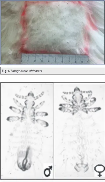
By the parasitological examination of the lice from infested group, all the lice collected from all parts of the body were found to be L. africanus, a blood-sucking louse species (Fig.1, Fig. 2). In the second subgroup of infested group and the goats from which the vitamin B complex has not been applied to, lice infestation was observed to increase at the end of the study. At the beginning of the work, the average number of lice
counted was 25 in the 10 cm2 area, while at the end it was 50 from the same area.
Significant losses of wool amounts and levels of irritations were observed due to severe infestations in goats without vitamin B complex of the infested group.
Skin thicknesses were recorded before and after sensitization as well as following testing by using digital calipers (Fig. 3).
Based on groups, degrees of hypersensitivity reactions and values measured afterwards are shown in Table 1. When skin thick-ness of control site compared with
Fig 3. Normal, sensitized and test skin thickness measurements, A- Shaved skin, skin ready for
sensitization, B- Sensitized skin, C- Control and test injections, D- Test reading at 24th h
Fig 1. Linognathus africanus
those of first (for sensitization) or second (for trial) injections all determined to be statistically significant (P<0.05). However, no differences were seen between the
groups with or without louse infestations (P>0.05). This is also the same case for the groups of vitamine supplemantation (P>0.05) (Fig. 4).
Tablo 1. Effect of vitamin B application on hypersensitivity reaction in healthy and lice-infested goats
Skin Thickness (ST) Groups*
1 2 3 4 Before sensitization ST 3.89±0.63 4.13±0.45 3.49±0.36 3.78±0.75 Before ID application ST 6.71±0.77 8.55±1.09 7.62±2.78 8.31±2.36 24 h after ID application ST Control ST 9.72±1.80 9.89±1.79 7.55±2.02 7.85±1.58 Test ST 17.98±1.93 19.51±2.26 19.89±1.96 21.43±4.19 Test ST-Control ST 8.26±2.41 9.62±1.94 12.34±1.77 13.58±4.02
* 1: Lice infested, vitamin B complex applied, 2: Lice infested, vitamin B complex unapplied, 3: Non-lice infestation, vitamin B complex applied, 4: Non-lice infestation, vitamin B complex unapplied
Fig 4. Statistically comparison of each group skin thickness
Table 2. The live weight averages obtained at the end of the study
Days Lousy Not Lousy
1 (Vitamined, Kg) 2 (Without Vitamin, Kg) 3 (Vitamined, Kg) 4 (Without Vitamin, Kg)
26.01.2016 19.50 19.92 20.02 21.68 09.02.2016 21.62 20.14 22.22 24.20 23.02.2016 23.48 22.80 23.66 28.44 12.03.2016 24.16 24.38 24.70 29.26 23.03.2016 26.08 26.86 26.72 33.72 Average 22.97 22.82 23.46 27.46
Fig 5. Statistically comparison of
each group at the beginning and end of the study
The measurements of nutrition performances are presented in Table 2 and Fig. 5.
DISCUSSION
Single-sex goats were used and no gender comparisons were made in this study, sincetype of goat genderhas been reported as not one of the determining factors for spreading of lice [2,9,22,26,27].
L. africanus toxins cause paralysis in hair roots and lead
to alopecia resulting in development of suppurative lesions by disruping skin integrity. However, no dermatitis occurrence was not detected in this study.
Two sides of neck are the body regions recommended for the measurement of cellular immunity using allergic skin tests [28]. In addition, as the primary region where the lice are localized on the host and in cases where the number of lice is low, both sides are suggested as suitable sides. In this trial, these regions were preferred for intra-dermal injection.
The delayed type of hypersensitivity reactions are used to assess cellular immune response. IVth type of hypersensitivity reactions is an in vivo test and has found clinical use in animals [29-31]. It has long been known that the vitamin B complex is one of the major factors in the development of resistance to ectoparasites in animals [32,33]. Various studies have been made to explain this effect of vitamin B [34,35]. Atpresent study, the development of the hypersensitivity reaction induced by DNCB was examined in both healthy and L. africanus infected goats. The adaptation of the method to the goats was clinically determined to develop a hypersensitivity reaction in the goats. Of vitamin B complex applied goats, the skin thickness of L. africanus infested goats (17.98±1.93 mm) was found to be lower than that measured by healthy goats (19.89±1.96 mm) (P>0.05).
A similarity was the case for the groups which did not receive vitamin B complex (19.51±2.26, 21.43±4.19 mm) (P>0.05) (Fig. 4). These findings may led to the conclusion that L. africanus infestation had an effect on suppressing the development of cellular immune response. On the other hand, with the planning of new trials with more goats and different types of lice infestations in the future, it would then be possible to determine statistically meaningful conclusions.
It is known that various toxins of blood-sucking lice cause local paralysis and alopeciaby reaching the hair roots [16]. Although the L. africanus lice are not deeply established on surface skin, it is suggested that migration of cells which affect the skin and subcutaneous tissuescontributing to the formation of cellular granulomas (primarily T cells) to the region was inhibited by lice.Lice may cause this effect either locally via their secretions on wool to reach the skin
or by a systemic wayfollowing absorption of secretions leading to T cell differentiation or even by both ways. At the beginning of the trial, live weights were determined as 19.50, 19.92, 20.02 and 21.68 kg in the groups. At the end of the experiment, the average live weights obtained from the same groups were as 22.97, 22.82, 23.46 and 27.46 kg, respectively. At this time of the study, the highest live weight was obtained from a group which no lice infestation occurred and no vitamin applied (P<0.05). In this particular group, the increase in live weight can be due to the absence of lice-infestation and injection stress.
In the first group of the infested animals applied with vitamin B complex, there was a decrease in the number of lice at the end of the experiment while the lice had already been removed in the goat number 3. This was inter preted as the possibility that the application of the vitamin B complex would likely remove the stress factors in the goat and reduce the intensity of the lice infestation.
It was concluded that the DNCB hypersensitivity reaction can be used when cellular immunity measurementis needed in L. africanusinfested goats, but this should be interpreted with caution and the recommendation of applying alternative tests or methods should be taken into account. In addition, when the vitamin B complex was administered in dose of 40 mL/goat by parenteral route (IM) in L. africanus infested goats, cellular immune system of the host and development of lice are negatively influenced. Vitamin B complex in goat breedingis concluded to be recomended parenterally at the dose used in this study. REFERENCES
1. Soulsby EJL: Helminths, Arthropods and Protozoa of Domesticated
Animals. Bailliere & Tindall, London, UK, 1986.
2. Taşcı S, Topcu A: Van yöresi keçilerinde Phthiraptera (bit) türleri ve
bunların mevsimsel aktiviteleri. Ankara Üniv Vet Fak Derg, 36, 503-525, 1989.
3. Taşcı S, Topcu A: Van yöresi koyunlarında bit (Mallophaga ve Anoplura)
türleri ve bunların mevsimsel aktiviteleri. Ankara Üniv Vet Fak Derg, 36, 467-476, 1989.
4. Urquhart GM, Armour J, Duncan JL, Dunn AM, Jennings FW:
Veterinary Parasitology, Second Edn, 141-205, Blackwell Sci. Ltd, UK, 1996.
5. Durden LA: Lice (Phthiraptera). In, Durden LA, Mullen GR (Eds):
Medicaland Veterinary Entomology. 45-65, Academic Press, San Diego, 2002.
6. Price RD, Hellenthal RA, Palma RL, Johnson KP, Clayton DH: The
Chewing Lice: World checklist and biological overview. Illinois Natural History Survey Special Publication, 24. x + 501 p, 2003.
7. Sisay A, Yilkal A, Yacob TH: Ectoparasites of sheep and goats in north-
west Amhara Regional State Ethiopia. Ethiop Vet J, 17, 55-67, 2013. DOI: 10.4314/evj.v17i1.5
8. Tamerat Beyene N, Zeryehun T: Prevalence and identification of
ectoparasites fauna in small ruminants in selected areas of Eastern Ethiopia.
Afr J Basic Appl Sci, 7, 240-246, 2015.
9. Tamerat N, Korso L, Mengistu S, Keffale M: Prevalence and
identification of ectoparasites fauna in small ruminants in and around Adami Tulu, east Shawa Zone of Oromia, Ethiopia. Livest Res Rural
Develop, 28 (11), 2016.
10. Fekadu A, Tolosso YH, Ashenafi H: Ectoparasites of small ruminants
in three agro-ecological districts of Southern Ethiopia. Afr J Basic Appl
Sci, 581, 47-54, 2013.
11. Tolosso YH: Ectoparasitism: Threat to Ethiopian small ruminant
populations and tanning industry. J Vet Med Anim Health, 6, 25-33, 2014. DOI: 10.1155/2015/216085
12. Cornall K, Wall R: Ectoparasites of goats in the UK. Vet Parasitol,
207, 176-179, 2015. DOI: 10.1016/j.vetpar.2014.11.005
13. Israel Y, Abera T, Wakayo BU: Epidemiological study on ecto-
parasites infestation of small ruminants in Sodo Zuria District, Southern Ethiopia. J Vet Med Anim Health, 7, 140-144, 2015. DOI: 10.5897/ JVMAH2014.0358
14. İnci A, Yıldırım A, Dik B, Düzlü Ö: Current knowledge of Turkey›s
louse fauna. Türkiye Parazitol Derg, 34, 174-178, 2010. DOI: 10.5152/ tpd.2010.17
15. TUİK: Türkiye İstatistik Kurumu. İllere göre keçi sayısı, 2015. http://
www.tuik.gov.tr/HbGetir.do?id=18852&tb_id=5; Erişim Tarihi: 06.01.2017.
16. Wall R, Shearer D: Veterinary Ectoparasites. Biology, Pathologyand
Control. Second Edn. BlackwellScienceLtd, OsneyMead, Oxford OX2 0EL, 25 John Street, London WCIN 2BS, UK, 2001.
17. Otter A, Twomey DF, Crawshaw TR, Bates P: Anaemia and mortality
in calves infested with the long-nosed sucking louse (Linognathus vituli).
Vet Rec,153, 176-179, 2003. DOI: 10.1136/vr.153.6.176
18. Dumanlı N, Köroğlu E, Erdoğmuş Z, Angın M, Yılmaz H: Elazığ
yöresi keçilerinde bulunan bit (Phthiraptera) türleri. Tr J Vet Anim Sci, 19, 291-295, 1995.
19. Topcu A, Ulu İ: Niğde Yöresi keçilerinde bulunan Bit (Anoplura
ve Mallophaga) türleri. Ankara Üniv Vet Fak Derg, 45, 201-205, 1998. DOI: 10.1501/Vetfak_0000000598
20. Akdemir C, Biçek K, Değer S: Van ve yöresi koyun ve keçilerinde
bit (Phthiraptera) enfestasyonları. YYÜ Vet Fak Derg, 11, 5-7, 2000.
21. Değer S, Taşçı S, Akgül Y, Alkan İ: Van ve yöresinde evcil hayvanlarda
ektoparaziter dermatitisler. YYÜ Vet Fak Derg, 5, 155-161, 1994.
22. Dumanlı N, Erdoğmuş Z, Köroğlu E, Angın M, Yılmaz H: Elazığ
yöresi koyunlarında bulunan bit (Mallophaga ve Anoplura) türleri. F Ü
Sağlık Bil Vet Derg, 6, 67-73, 1992.
23. Wall R, Shearer D: Veterinary entomology: Arthropod ectoparasites
of veterinary importance, 1st edn., Champan and Hall, United Kingdom, 1-439, 1997.
24. Durden LA: Lice (Phthiraptera). In, Williams ES, Barker IK (Eds):
Parasitic Diseases of Wild Mammals. Third edn., 3-17, Iowa State University Press/Ames, Iowa, 2001.
25. Burton JL, Kennedy BW, Burnside EB, Wilkie BN, Burton JH:
Dinitrochlorobenzene contact hypersensitivity as a marker trait for selection to ımprove disease resistance in calves. J Dairy Sci, 72, 2351-2361, 1989. DOI: 10.3168/jds.S0022-0302(89)79368-1
26. Tesfaye D, Assefa M, Demissie T, Taye M: Ectoparasites of small
ruminants presented at Bahir Dar Veterinary Clinic, Northwest Ethiopia.
Afr J Agric Res, 7, 4669-4674, 2012. DOI: 10.5897/AJAR12.599
27. Seyoum Z, Tadesse T, Addisu A: Ectoparasites prevalence
in small ruminants in and around Sekela, Amhara Regional State, Northwest Ethiopia. J Vet Med, 2015 (2015), Article ID: 216085. DOI: 10.1155/2015/216085
28. Erganiş O, İstanbulluoğlu E: İmmünoloji. Mimoza Yayınevi, Konya,
1993.
29. Uçan US, Kuyucuoğlu Y, Kuyucuoğlu EÖ: İmmün sistemi
baskılanmış ve uyarılmış broiler civcivlerde kutanöz bazofil aşırı duyarlılık reaksiyonu. Tr J Vet Anim Sci, 23, 585-589, 1999.
30. Sayın Z, Erganiş O: Sığır tübekülozunun teşhisinde kullanılan
metotlar. Kocatepe Vet J, 3, 77-82, 2010.
31. Lydyard P, Whelan A, Fanger M: İmmünoloji. 3. Basımdan Çeviri.
Çeviri Ed. Erganiş O, Uçan US. Nobel Akademik Yayıncılık Eğ. Dan. Tic. Ltd. Şti., Ankara, 2013.
32. Kartman L: A note on vitamins in relation to ectoparasite resistance. J Parasit, 28, 170-171, 1942.
33. Ely DG, Harvey TL: Relation of ration to short-nosed cattle louse
infestations. J Econ Entomol, 62, 341-344, 1969. DOI: 10.1093/jee/62.2.341
34. Gibney VJ, Campbell JB: Effects of Ariboflavinosis on population of
Polyplaxspinulosa (Burmeister) (Anoplura: Haematopinidae) in artificially Infested albino rats. J Kansas Entomol Soc, 54, 531-536, 1981.
35. Tamura J, Kubota K, Murakami H, Sawamura M, MatsushimaTamura T, Saitoh T, Kurabayshi H, Naruse T: Immunomodulation by vitamin B12:
Augmentation of CD8+ T lymphocytesandnatural killer (NK) cellactivity in vitamin B12-deficient patientsby methyl-B12 treatment. Clin Exp