Yazışma Adresi/Address for Correspondence: Dr. Sema Polat, Cukurova University Faculty of Medicine, Department of Anatomy, Adana,Turkey E-mail: [email protected]
ARAŞTIRMA / RESEARCH
Determination of lower extremity anthropometric measurements in
adult healthy women wearing and non-wearing narrow toed high
heeled shoes
Dar burunlu yüksek topuklu ayakkabı giyen ve giymeyen yetişkin sağlıklı kadınlarda
alt ekstremite antropometrik ölçümlerin belirlenmesi
Sema Polat
1, Ahmet Hilmi Yücel
11Cukurova University Faculty of Medicine, Department of Anatomy, Adana, Turkey
Cukurova Medical Journal 2018;43(3):545-551
Abstract Öz
Purpose: The present study was aimed to analyze
morphometric measurements of lower extremity dimensions in healthy females wearing and non-wearing high heeled shoes with narrow toe box.
Materials and Methods: Lower extremity length
measurements were taken from 121 healthy adult females (64 females non-wearing high heel shoes with narrow toe box, 57 females wearing high heeled shoes with narrow toe box) aged 20 to 40 years using an anthropometer and caliper. Additionally, demographic data (age, height, weight and body mass index-BMI) were taken and tibia-femur ratio (the ratio between tibia length and tibia-femur length-T/F) were calculated.
Results: The mean values of Tibia - femur length ratio
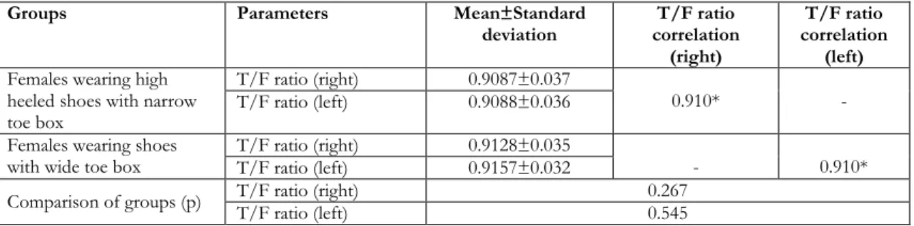
were found as 0.9087±0.038 (right) and 0.9088±0.036 (left) in females wearing high heeled shoes with narrow toe box, whereas the same values were established as 0.9128±0.035 (right) and 0.9157±0.032 (left) in females non-wearing high heeled shoes with narrow toe box. Furthermore, 53.12% of females non-wearing high heeled shoes with narrow toe box suffer from foot, knee and back pain, whereas 78.94% of females wearing shoes with narrow toe box suffer from foot, knee and back pain.
Conclusion: The observations presented in this report
have defined anatomic parameters and morphometric changes that need to be taken into consideration when wearing narrow high heeled shoes and shoes having wide toe box in females.
Amaç: Bu çalışma dar burunlu yüksek topuklu ayakkabı
giyen ve giymeyen yetişkin sağlıklı kadınlarda alt ekstremite ile ilgili morfometrik ölçümleri analiz etmek için yapılmıştır.
Gereç ve Yöntem: Alt ekstremite uzunluk ölçümleri
yaşları 20 ile 40 arası 121 yetişkin sağlıklı (geniş burunlu ayakkabı giyen 64 kadın; dar yüksek topuklu ayakkabı giyen 57 kadın) kadından antropometre ve kaliper kullanılarak yapıldı. Ayrıca, demografik veriler (yaş, vücut ağırlığı, boy uzunluğu, vücut kitle indeksi-BKI) alındı ve tibia-femur oranı hesaplandı.
Bulgular: Dar burunlu yüksek topuklu ayakkabı giyen
kadınlarda tibia’nın femur uzunluğuna oranı 0.9087±0.038 (sağ) ve 0.9088±0.036 (sol) iken, dar burunlu yüksek topuklu ayakkabı giymeyen kadınlarda aynı ölçümler sırasıyla 0.9128±0.035 (sağ) ve 0.9157±0.032 (sol) olarak hesaplandı. Son olarak, dar burunlu yüksek topuklu ayakkabı giymeyen kadınların %53.12’i ayak, diz ve bel ağrısı çekerken, dar burunlu yüksek topuklu ayakkabı giyen kadınların %78.94’si ayak, diz ve bel ağrısı problemi yaşamaktadır.
Sonuç: Çalışmada elde edilen değerler, kadın
populasyonunda dar burunlu yüksek topuklu ayakkabı ve geniş burunlu ayakkabı giyildiğinde dikkat edilmesi gereken anatomik parametreleri ve morfometrik değişimleri içermektedir.
Key words: Femur, morphometry, the ratio of tibia to
femur, lower extremity anthropometry. Anahtar kelimeler: Femur, morfometrisi, Tibia uzunluğunun femur uzunluğuna oranı, Alt ekstremite antropometrisi.
546
INTRODUCTION
The design and popularity of high heeled shoes (HHS) have always been a topic of interest to Today’s fashion trends1. HHS are not only foot
accessories but also an important part of female fashion2. Females wear high-heeled shoes due to
some reasons3. Especially, these shoes are preferred
by all females due to enhancing beauty and providing authority source4,5. Millions of females use
high heeled shoes3. Moreover, walking with HHS
shifts body weight toward medial with respect to the foot and alters the normal function of the ankle. Compensations occur at the knee and hip to maintain stability and progression during walking3.
Furthermore, studies in literature have shown that wearing HHS leads to smaller and unstable supporting surface and alters lower extremity structure, reduces the shock absorbing ability and weight bearing functions6,7.
The weight bearing bone of lower limb is the femur. It is the longest and strongest bone of the body. While the proximal part of femur constitutes the hip joint with the pelvis and femur distal part constitutes knee joint with tibia8. Tibia is the second longest
bone and the proximal part joins in knee joint structure while the distal part constitutes ankle joint8. The fibula is usually used for bone grafts.
Also, fractional surgical resection of this bone is performed in limb lengthening9. The fibula plays a
role in stabilization of the talocrural joint in spite of deficiency of weight-bearing function and it is not directly a part of the knee joint10.
Lower extremity that have been studied in various scientific fields including anatomy, anthropology, forensic medicine, ergonomics and orthopedics is well-adjusted bipedal locomotion and weight bearing as both structurally and functionally. This adaptation affects bone features such as length or strength11. Moreover, in forensic investigations long
bones like tibia and fibula play a critical function for stature estimation. These present more reliable estimates of stature than the upper extremity bones12. Femoral anthropometric measurements
show differences due to racial variations in nutrition, heredity, climate and geographical factors13. In a
surgical correction of lower extremity limb deficiency, the surgeon must decide whether the tibia or femur should lengthen or shorthen in orthopaedic clinics14.
In the literature, little is known about the ideal T/F ratio. The study declares the mean ideal T/F ratio to be between 0.78 and 0.85 and it is stated that increase in the ratio of T/F leads to hip and knee arthritis. Moreover, the shorter femur requires increased motion at the hip and knee joints. Furthermore, it is stated that this condition leads to develop speedy of degenerative joint disease. Briefly, longer femur and shorter tibia or decrease in the ratio of tibia to femur can prevent development of osteoarthritis (OA)14. OA of the hip and knee is an
important public health subject15. The prevelance
studies both in Turkey and in international literature show that women are more inclined to OA according to men15-17. Additionally, it is declared the
ratio of body measurements to each other is affected from demographic data, geographical condition, genetic and enviromental factors and these features can change according to race and country. In anatomical studies made in the human body especially it is aimed to find the ideal beauty from the artistic point18,19.
Footwear is more important for prevent foot from some problems. The main task of footwear began to change due to fashion from 15th.century on and fashion caused the changes of footwear features such as high heel or extended forward toe box20.
Especially, it is declared that forefoot pain is often related to wear inappropriate shoes and wearing shoe with diminished toe box induces the health problems about foot21. High heeled shoes is
commonly contain narrow toe box, rigit heel cap and curves plantar region. So, high heeled shoes vary from normal footwear22.
As a result, we believe that the data obtained in this study can provide principal information for lower extremity morphometry and may help the orthopaedic surgeons and rheumatologists design for having a succesful surgery and minimize the related problems. Additionally, we think that our study will make a significant contribution to the literature about the ratio of tibia/femur studies in females wearing two type shoes what these length must be aesthetically.
MATERIALS AND METHODS
The study was approved by our institutional review board and ethics committee approval also was obtained. Bilateral lower extremity length measurements were obtained from 121 adult
females (64 females non-wearing high heeled shoes wide narrow toe box, 57 females wearing high heeled shoes with narrow toe box) between 20 and 40 years of age with no history of trauma or congenital anomalies. Each subject was asked to gently place her/his body in suitable position to each measure over a platform. In this study, 57 healthy females wearing high heel shoes with narrow toe box at least 5 cm height, at least one year and three days in a week were recruited, whereas 64 females non-wearing high heeled shoes with narrow toe box, less than 5 cm height and shoes having wide toe box were included.
Measurements and reference points
The distance between umblicus and malleolus medialis (MM), The distance between spina iliaca anterior superior (SIAS) and malleolus medialis (MM), the distance between trochanter major and malleolus medialis (MM) were measured. The femur length is the distance from the greater trochanter to the lateral condyle8. The tibia length measurement
was taken from the medial condyle of the tibia to the medial malleolus (spherion)12. The fibula length
was performed from the head of fibula to the lateral malleolus12. Tibia/femur ratio was calculated14.
After these measurements, the body mass index (BMI) was calculated with kg/m2 formula. All these
measurements were done with antropometer (Anthropometry 01290 Lafayette Instrument) and caliper [Mitutoyo Vernier Kaliper (0-150 mm)] measurement tools.
Statistical analysis
After these calculations statistical analysis was performed with SPSS 21.0. From these measurements, minimum (min.), maximum (max.), means and standard deviations (SD) values were evaluated. Moreover, the correlation analysis between parts of the lower limb were performed. Furthermore, females were asked whether they suffer from pain or not. After, the pain ratio calculated in two groups.
RESULTS
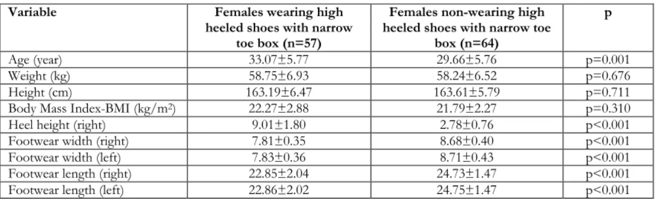
The records of 121 adult females wearing and non-wearing high heeled shoes with narrow toe box were assessed. The mean and standard deviation values of age, weight, height and BMI measurements were found to be 33.07±5.77 years, 58.75±6.93 kg,
163.19±6.47 and 22.27±2.88 kg/m2 in females
wearing high heeled shoes with narrow toe box respectively, whereas the same values were 29.66±5.76 years, 58.24±6.52 kg, 163.61±5.79 cm and 21.79±2.27 kg/m2 in females non-wearing high
heeled shoes with narrow toe box (Table 1). Moreover, the mean and standard deviation values of heel height, footwear length and footwear width were measured as 2.78±0.76 cm; 24.73±1.47 mm (right) and 24.75±1.47 mm (left); 8.68±0.40 cm (right) and 8.71±0.43 (left) in non-wearing high heeled shoes with narrow toe box, whereas, corresponding values were determined as 9.01±1.80 cm; 7.81±0.35 cm (right) and 7.83±0.36 (left); and 22.85±2.04 mm (right) and 22.86±2.02 mm (left) in wearing high heeled shoes with narrow toe box (Table 1). There were found a significiant difference in heel height, footwear length and foot wear width measurements (p<0.05).
Statistical analysis of lower extremity length measurements of females wearing shoes having two type were shown in Table 2. There are no statistically differences in measurements including distance between umbilicus and malleolus medialis, spina iliaca anterior superior (SIAS) and malleolus medialis and trochanter major and malleolus medialis in females wearing and nonwearing high heeled shoes with narrow toe box. Moreover, in comparing the mean values of the right extremity with that of the left extremity, there was no difference between them for two groups. Furthermore, the correlation analysis of lower extremity measurements were shown in Table 3. The tibia/femur length ratio (T/F) were found as 0.9087±0.038 (right), 0.9088±0.036 (left) in females wearing high heeled shoes with narrow toe box, whereas the same values were established as 0.9128±0.035 (right) and 0.9157±0.032 (left) in females wearing shoes with wide toe box (Table 4). In addition, 53.125% (34) of females non-wearing high heeled shoes with narrow toe box suffer from foot, knee and back pain, whereas 78.947% (45) of females wearing high heeled shoes with narrow toe box have foot, knee and back pain.
DISCUSSION
Anthropometry is a reliable technique to evaluate various body parts. Body proportions show change between population11. Lower extremity is
well-adjusted bipedal locomotion and weight bearing as both structurally and functionally11. This adaptation
548 affects musculoskeletal system including the length and strength of the bone, muscle origin, insertion and mass11. The measurements of body are affected
from gender, age, socio-economic condition, nutrition and geographic factors18-20. Moreover, the
long bones play an important role in forensic studies12. Furthermore, the information about the
tibia dimensions is critical in forensic, anatomic, orthopaedic clinic and archeological studies to determine unknown bodies and stature21.
Table 1. Demographic data in females wearing and non-wearing high heeled shoes with narrow toe box
Variable Females wearing high
heeled shoes with narrow toe box (n=57)
Females non-wearing high heeled shoes with narrow toe
box (n=64)
p
Age (year) 33.07±5.77 29.66±5.76 p=0.001
Weight (kg) 58.75±6.93 58.24±6.52 p=0.676
Height (cm) 163.19±6.47 163.61±5.79 p=0.711
Body Mass Index-BMI (kg/m2) 22.27±2.88 21.79±2.27 p=0.310
Heel height (right) 9.01±1.80 2.78±0.76 p<0.001
Footwear width (right) 7.81±0.35 8.68±0.40 p<0.001
Footwear width (left) 7.83±0.36 8.71±0.43 p<0.001
Footwear length (right) 22.85±2.04 24.73±1.47 p<0.001
Footwear length (left) 22.86±2.02 24.75±1.47 p<0.001
p=significant value
Table 2. Statistical analysis of lower extremity length measurements in females wearing and non-wearing high heeled shoes with narrow toe box
Length Measurements (cm) Females wearing high
heeled shoes with narrow toe box (n=57)
Females non-wearing high heeled shoes with narrow
toe box (n=64)
P
Femur length (right) 38.32±2.43 37.94±1.99 p=0.352
Femur length (left) 38.28±2.45 37.98±2.02 p=0.464
Tibia length (right) 34.79±2.17 34.62±2.00 p=0.647
Tibia length (left) 34.76±2.09 34.76±1.83 p=0.995
Fibula length (right) 37.79±2.27 36.96±1.92 p=0.031
Fibula length (left) 37.71±2.27 36.98±1.95 p=0.060
Umbilicus- Malleolus medialis (right) 89.36±4.91 88.73±4.17 p=0.447 Umbilicus – Malleolus medialis (left) 89.42±4.94 88.95±4.28 p=0.583 Spina Iliaca anterior superior- malleolus
medialis (SIAS-right) 82.76±4.60 82.09±4.35 p=0.410
Spina Iliaca anterior superior- malleolus
medialis (left) 82.89±4.48 82.32±4.35 p=0.478
Trochanter major - malleolus medialis
(right) 74.01±4.09 74.41±4.19 p=0.590
Trochanter major - malleolus medialis (left) 73.96±4.03 74.45±4.29 p=0.523
p=significant value
Table 3. Correlation analysis of lower extremity measurements according to height and weight measurements
Measurements Height Weight
Femur length (right) 0.671 p<0.001 0.682 p<0.001
Femur length (left) 0.376 p<0.001 0.388 p<0.001
Tibia length (right) 0.680 p<0.001 0.326 p<0.001
Tibia length (left) 0.694 p<0.001 0.323 p<0.001
Fibula length (right) 0.707 p<0.001 0.322 p<0.001
Fibula length (left) 0.713 p<0.001 0.339 p<0.001
SIAS-MM distance (right) 0.746 p<0.001 0.300 p<0.001
SIAS-MM distance (left) 0.741 p<0.001 0.303 p<0.001
Umblicus-MM (right) 0.801 p<0.001 0.339 p<0.001
Umblicus-MM (left) 0.800 p<0.001 0.338 p<0.001
Table 4. Ratio of tibia to femur length (T/F) and correlation analysis between right and left ratio in two groups.
Groups Parameters Mean±Standard
deviation correlation T/F ratio (right)
T/F ratio correlation
(left)
Females wearing high heeled shoes with narrow toe box
T/F ratio (right) 0.9087±0.037
0.910* -
T/F ratio (left) 0.9088±0.036 Females wearing shoes
with wide toe box T/F ratio (right) T/F ratio (left) 0.9128±0.035 0.9157±0.032 - 0.910* Comparison of groups (p) T/F ratio (right) T/F ratio (left) 0.267 0.545
r≥0.900*: Very highly correlated
The femur is the weight bearing lower extremity bone8 and is one of the most crucial bones in
identifying gender, race, sport-culture habits23. The
anatomical knowledge of different dimensions of femur can provide very important data for radiologist, rheumatologists and orthopaedic surgeons for treatment planning and diagnosis8. In
Indians, the mean value of femur length was found as 43.98±2.15 cm (right) and 44.15±2.15 cm (left)8.
Whereas, in Iran the mean value of the femur length were declared as 40.81±2.18 cm24. In this paper, the
mean value of femur length was established as 38.32±2.43 cm (right) and 38.28±2.45 cm (left) in wearing high heeled shoes with narrow toe box. The same measurements were found as 37.94±1.99 cm (right) and 37.98±2.02 cm (left) in females non-wearing high heeled shoes with narrow toe box. There was no significant differences in the mean values of the femur lengths for our two groups. Moreover, the differences in literature can result from racial variations, genetic, climate and geographical conditions14. In Thai Cadaveric study
performed by Pureepatpong et al, it was declared that the longest bones were femur, fibula and tibia respectively25.
In Sudanese, the mean value of tibia length was found as 38.21±1.93 cm11, whereas in Indians, the
same values were 35.26±1.69 cm (right) and 35.43±1.71 cm (left)12. In our study, this
measurement was 34.79±2.17 cm (right) and 34.76±2.09 cm (left) in wearing high heeled shoes with narrow toe box and the same values were 34.62±2.00 cm (right) and 34.76±1.83 cm in females non-wearing high heeled shoes with narrow toe box, respectively. Due to these reports, we found differences in mean values of Indian and Sudanese populations compared with our result: they have higher values than us. We think that these
discrepancies can result from race variation, climate, geographical condition, genetic features.
Gaur et al reported the mean value of fibula were established as 36.39±1.95 cm (right) and 36.35±1.85 cm (left) in Indians12. In this study, the same value
was declared as 37.79±2.27 cm (right) and 37.71±2.27 cm (left) in females wearing high heeled shoes with narrow toe box whereas, in females non-wearing high heeled shoes with narrow toe box, these were found as 36.96±1.92 cm (right) and 36.98±1.95 cm (left) respectively. Also, this study showed that the fibula is longer than tibia in a similarly to our study.
After these measurements, the tibia- femur ratio was estimated. The advantages of this ratio include that it is easy and it does not require the use of investigation such as magnetic resonance. In a surgical correction of lower extremity limb deficiency, the orthopedist must decide whether lengthening or shortening of the tibia or femur should occur, because of preventing osteoarthritis hip and knee and decreasing the tibia femur ratio which is reported the tibia femur ideal ratio as 0.78 and 0.8514. In this investigation, the tibia femur ratio
value was found as 0.9087±0.038 (right), 0.9088±0.036 (left) in females wearing narrow high heeled shoes, whereas the same measurement was established as 0.9128±0.035 (right) and 0.9157±0.032 (left) in females non wearing high heeled shoes with narrow toe box. It was found no significant differences between our two groups. According to the literature data, our findings were higher than this study. However, we think that this situation results from especially climate and racial differences and this results from both narrow high heeled shoes and inappropriate wide shoes or unsuitable heel height which may predispose to OA in the joint. Kerrigan et al declared during walking in
550 high heeled shoes than barefoot, increased force across the patellofemoral joint and a greater compressive force on the medial compartment of the knee3. Moreover, this increase in compressive
forces the tibiofemoral region for knee OA (26). It could be important because osteoarthtitic changes are more common in the medial than in the lateral aspect of the knee. Both prolonged strain and pressures may lead to degenerative joint changes3.
Additionally, It was shown that wearing wide heeled shoes and narrow heeled shoes increased peak knee varus torque by 26% and 22%, respectively and wide heeled shoes have the same effect on knee torque as narrow heeled shoes3. Whereas, other
study showed that mildly high-heels (1.5 inch=3.81) cause changes in knee joint torques that are similar to that caused by females’ wear shoes with heel heights mean 2.5 (6.5 cm) and 2.8 inch (7.11 cm) 26.
We consider that these diversities may be a result of such factors like individual constitution, genetic variables, climate type and the using of high heeled shoe or unsuitable heel height as well. We can say both wearing narrow high heeled shoes and low heeled shoes may not cause the differences in right and left side about lower extremity lengths including Umblicus-medial malleolus (MM), Spina iliaca anterior superior (SIAS)-Medial malleolus (MM) and trochanter major-Medial malleolus (MM) distances. As a result, we believe that the data obtained in this study can provide principal information for lower extremity morphometry and may help the orthopaedic surgeons, rheumatologists for minimize the problems connected with ill-fitting shoes. We think that the T/F ratio is as one of the important guide for determining knee osteoartrit. Moreover, normally the ratio of tibia length to femur length is known as between 0.78 and 0.85, whereas in knee dislocation and hip - knee osteoarthritis, this value shows deviation from ideal values. Additionally, we believe that our study values will contribute to the literature about lower limb anatomy and the ratio of tibia/femur studies regarding what these length must be aesthetically. But, further researches are needed to determine whether high heeled shoes affect the osteoarthritis or not and moreover, we think that shoes that are fit and suitable subjects’feet structure should be prefer. So, the foot injuries can be decreased.
REFERENCES
1. Curran SA, Holliday JL, Watkeys L. Influence of
high heeled footwear and pre-fabricated foot orthoses on energy efficiency in ambulation. The Foot and Ankle Online Journal. 2010;3(3):1. 2. Xiong S, Hapsari VD. Effects of heel height and
wearing experience on human standing balance. J Foot Ankle Res. 2014;7:A97.
3. Kerrigan DC, Todd MK, O Riley P. Knee osteoarthritis and high heeled shoes. Lancet. 1998;351:1399-401.
4. Iqbal R, De A, Mishra W, Maulik S, Am C. Study on lumbar kinematics and the risk of low back disorder in female university students by using shoes of different heel heights. Work. 2012;41:2521-6. 5. Park S, Lee K. Simulation of biomechanical influence
of high heels on musculuskeletal system of foot and ankle. Proceedings of ISB2009, Cape Town, South Africa, Jul. 2009.
6. Hong WH, Lee YH, Lin YH, Tang SFT, Chen HC. Effect of shoe heel height and total contact insert on muscle loading and foot stability while walking. Foot Ankle Int. 2013;34:273-81.
7. Srivastava A, Mishra A, Tewari RP. Electromyography analysis of high heel walking. International Journal of Electronics & Communication Technology. 2012;3:166-9.
8. Khan SM, Saheb AH. Anthropometric study of femur in South India. Int J Anat Res. 2014;2:630-2. 9. Uchiyama E, Suzuki D, Kura H, Yamashita T,
Murakami G. Distal fibular length needed for ankle stability. Foot Ankle Int. 2006;27:185-9.
10. Naidoo N, Ishwarkumar S, Lazarus L, Pillay P, Satyapal KS. Osteometry and morphology of the human fibula: a South African study. Int J Morphol. 2015;33:1071-7.
11. Ahmed AA. A study of correlations within the dimensions of lower limb parts for personal identification in a Sudanese population. The Scientific World Journal. 2014;2014:541408. 12. Gaur R, Kaur K, Airi R, Jarodia K. Estimation of
stature from percutaneous lengths of tibia and fibulaa of scheduled castes of hayrana state, India. Ann Forensic Res Anal. 2016;3:1025-30.
13. Bokariya P, Kothari R, Waghmare J, Tarnekar A, Ingole IV. Anthropometric study of femur in central Indian population. Journal of Mahatma Gandhi Institute of Medical Sciences. 2009;14:47-9. 14. Weinberg DS, Liu RW. The association of tibia
femur ratio and degenerative disease of the spine, hips, and knees. J Pediatr Orthop. 2015;1-6. 15. O’Connor MI. Osteoarthritis of the hip and knee:
sex and gender differences. Orthop Clin North Am. 2006;37:559-68.
16. Uysal FG, Başaran S. Diz osterartriti. Türkiye Fiziksel Tıp ve Rehabilitasyon Dergisi. 2009;55:1-7. 17. Kerrigan DC, Todd MK, O’Riley P. Knee
osteoarthritis and high-heeled shoes. Lancet. 1998;351:1399-1401.
yaş grubu kız öğrencilerde boy ve alt ekstremite ölçümleri. Ondokuz Mayıs Üniversitesi Tıp Dergisi. 1995;12;207-13.
19. Güleç E, Akın G,Sağır M, Özer BK, Gültekin T, Bektaş Y. Anadolu insanının antropometrik
boyutları: 2005 yılı Türkiye antropometri anketi genel sonuçları. Ankara Üniversitesi Dil ve Tarih Coğrafya Fakültesi Dergisi. 2009;49:187-201.
20. Menz HB, Roddy E, Marshall M, Thomas MJ, Rathod T, Peat GM, Croft PR. Epidemiology of shoe wearing patterns over time in older women: Associations with foot pain and hallux valgus. J Gerontol A Biol Sci Med Sci. 2016;71:1682-7. 21. Hurst B, Branthwaite H, Greenhalgh A,
Chockalingam N. Medical-grade footwear: the impact of fit and comfort. J Foot Ankle Res. 2017;10:1-7.
22. Bae YH, Ko M, Park YS, Lee SM. Effect of revised high heeled shoes on foot pressure and static balance
during standing. J Phys Ther Sci. 2015;27:1129-31. 23. Vaghefi SHE, Elyasi L, Amirian SR, Reigan P,
Akbari H, Sheikhshoaiee M et al. Evaluating antropometric dimensions of the femur using direct and indirect methods. Anatomical Sciences. 2015;12;89-92.
24. Bokariya P, Sontakke B, Waghmare JE, Tarnekar A. The anthropometric measurements of tibia. J Indian Acad Forensic Med. 2012;4:322-3.
25. Pureepatpong N, Sangiampongsa A, Lerdpipatworakul T, Sangvichien S. Stature estimation of modern Thais from long bones: A cadaveric study. Siriraj Medical Journal. 2012;64;22-5. 26. Kerrigan C, Johansson JL, Bryant MG, Boxer JA, Croce UD, Riley PO. Moderate heeled shoes and knee joint torques relevant to the development and progression of knee osteoarthritis. Arch Phys Med Rehabil. 2005;86:871-5.