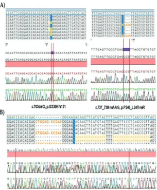
©Copyright 2020 by Turkish Society of Hematology
Turkish Journal of Hematology, Published by Galenos Publishing House Turk J Hematol 2020;37:98-103
Received/Geliş tarihi: July 24, 2019 Accepted/Kabul tarihi: November 19, 2019 Address for Correspondence/Yazışma Adresi: Müge Sayitoğlu, Ph.D., İstanbul University, Aziz Sancar Institute of
Experimental Medicine, Department of Genetics, İstanbul, Turkey Phone : +90 212 414 20 00 -33314
E-mail : mugeay@istanbul.edu.tr ORCID: orcid.org/0000-0002-8648-213X Objective: PTEN/AKT pathway deregulations have been reported to
be associated with treatment response in acute leukemia. This study examined pediatric T-cell acute lymphoblastic leukemia (T-ALL) samples for PTEN and AKT1 gene variations and evaluated the clinical findings.
Materials and Methods: Fifty diagnostic bone marrow samples of
childhood T-ALL cases were investigated for the hotspot regions of the PTEN and AKT1 genes by targeted next-generation sequencing.
Results: A total of five PTEN variations were found in three of the 50
T-ALL cases (6%). Three of the PTEN variations were first reported in this study. Furthermore, one patient clearly had two different mutant clones for PTEN. Two intronic single-nucleotide variations were found in AKT1 and none of the patients carried pathogenic AKT1 variations.
Conclusion: Targeted deep sequencing allowed us to detect both
low-level variations and clonal diversity. Low-low-level PTEN/AKT1 variation frequency makes it harder to investigate the clinical associations of the variants. On the other hand, characterization of the PTEN/ AKT signaling members is important for improving case-specific therapeutic strategies.
Keywords: T-ALL, PTEN, AKT1, Next-generation sequencing
Amaç: PTEN/AKT yolak düzensizliklerinin akut lösemide tedavi yanıtı
ile ilişkili olduğu bildirilmiştir. Çalışmanın kapsamı, pediatrik T-ALL hastalarının PTEN ve AKT1 genlerinin sıcak bölge varyasyonları için incelenmesi ve klinik bulgularla değerlendirilmesidir.
Gereç ve Yöntemler: Elli pediatrik T-ALL olgusunun tanı zamanı
kemik iliği örnekleri, PTEN ve AKT1 genlerinin sıcak bölgeleri için hedefe yönelik yeni nesil dizileme ile dizilenmiştir.
Bulgular: Elli T-ALL olgusunun %6’sında PTEN varyasyonu
saptanmıştır. Tespit edilen varyasyonlardan üçü ilk defa bu çalışmada gösterilmiştir. Ayrıca bir hastanın PTEN açısından iki farklı mutant klon taşıdığı belirlenmiştir. AKT1 geninde iki intronik tek nükleotid polimorfizmi tespit edilirken hiçbir olguda patojenik AKT1 varyasyonu saptanmamıştır.
Sonuç: Derin dizileme, hem düşük düzeydeki varyasyonların hem
de klonal çeşitliliğin belirlenmesine olanak sağlamıştır. T-ALL hastalarındaki düşük düzey PTEN/AKT1 varyasyon sıklığı, varyantların klinikle ilişkisinin ortaya çıkarılmasını zorlaştırmaktadır. Diğer yandan, PTEN/AKT sinyal yolağının karakterizasyonu hasta spesifik terapötik stratejilerin uygulanabilirliği için önemlidir.
Anahtar Sözcükler: T-ALL, PTEN, AKT1, Yeni nesil dizileme
Öz
Abstract
1İstanbul University, Aziz Sancar Institute of Experimental Medicine, Department of Genetics, İstanbul, Turkey 2Altınbaş University Faculty of Medicine, İstanbul, Turkey
3İstinye University Faculty of Art and Science, Department of Molecular Biology and Genetics, İstanbul, Turkey 4Acıbadem Mehmet Ali Aydınlar University Faculty of Medicine, Department of Medical Biology, İstanbul, Turkey 5İstanbul University Faculty of Medicine, Department of Pediatrics Hematology, İstanbul, Turkey
6İstanbul University-Cerrahpaşa Cerrahpaşa Faculty of Medicine, Department of Pediatric Hematology, İstanbul, Turkey 7Acıbadem Mehmet Ali Aydınlar University Faculty of Medicine, Department of Medical Genetics, İstanbul, Turkey aF.K. and Y.E. contributed equally to this work.
Fulya Küçükcankurt1,2,a, Yücel Erbilgin1,a, Sinem Fırtına1,3, Özden Hatırnaz Ng1,4, Zeynep Karakaş5, Tiraje Celkan6,
Ayşegül Ünüvar5, Uğur Özbek1,7, Müge Sayitoğlu1
Çocukluk Çağı T-hücreli Akut Lenfoblastik Lösemi Hastalarında PTEN ve AKT1 Varyasyonlar
PTEN and AKT1 Variations in Childhood T-Cell Acute
Lymphoblastic Leukemia
DOI: 10.4274/tjh.galenos.2019.2019.0282Introduction
One of the key signal transduction pathways involved in malignant transformation is the PTEN/PI3K/AKT pathway, which regulates cellular metabolism, cell growth, translation,
chromosome stability, and cell survival [1]. Phosphatase and tensin homolog deleted on chromosome ten (PTEN) is a lipid and dual function phosphatase that antagonizes the PI3K/ AKT pathway; by dephosphorylating phosphoinositide 3-kinase
(PI3K) it produces PIP2 (phosphatidylinositol 4,5-bisphosphate) and PIP3 (phosphatidylinositol (3,4,5)-triphosphate) and so terminates the transmission of the signal to AKT and other PIP3-effector targets [2]. AKT1 is a serine threonine kinase that modulates the cell cycle checkpoint [3]. AKT1 is activated by platelet-derived growth factor and its activation is deregulated by mutations in the pleckstrin homology domain of
AKT1. Survival factors can suppress apoptosis in a
transcription-independent manner by activating the serine/threonine kinase
AKT1, which then phosphorylates and inactivates components
of the apoptotic machinery [4].
PTEN as a tumor suppressor is frequently mutated in cancers
and its inactivation results in constitutive activation of the PI3K/AKT pathway. PTEN is a regulatory key to prevent the malignant transformation of T-cells [5]. The PTEN/AKT pathway has an important role in the β-selection checkpoint in T-cell development and lymphocyte homeostasis [6]. PTEN-deficient T-cells are found to be highly proliferative as a cause of increased phosphorylation of AKT [7]. AKT1 is highly expressed in thymus tissue and knockout studies showed that terminal differentiation in CD8+ T-cells failed, with increased proliferation, cytokine secretion, and prolonged survival [8,9]. PTEN/AKT abnormalities resulting in deletion, insertion, or missense mutations lead to differential regulation in different hematologic malignancies [10,11,12,13,14]. Genomic resequencing results showed that PI3K/AKT pathway genes are commonly mutated in pediatric and young adult T-cell acute lymphoblastic leukemia (T-ALL) cases [11,15]. In this study, PTEN and AKT1 variations and their clinical associations were analyzed in a group of childhood T-ALL cases.
Materials and Methods
Childhood T-ALL cases (n=50) diagnosed at the İstanbul University Faculty of Medicine and Cerrahpaşa University Faculty of Medicine were included in this study. Patients were treated according to the BFM-ALL protocol. Diagnostic bone marrow samples with a blast count of >80% were included in the study. The T-cell origin of ALL was defined by the expression of immunophenotype markers that included CD1a, CD2, cytoplasmic CD3, surface CD3, CD4, CD5, CD7, and CD8. T-ALL cases were evaluated according to the European Group for the Immunological Characterization of Leukemia classification scale as immature (n=20), cortical (n=17), or mature (n=4); however, nine cases were not able to be further classified due to limited immunological marker information [11]. Median age was 8 (range=0.9-17) years and other clinical features of the T-ALL cases are summarized in Table 1. Written and oral informed consent was obtained from the legal representatives of the pediatric patients.
Identification of PTEN and AKT1 variations
The mononuclear cells of the bone marrow samples were isolated by the Ficoll density gradient procedure [16]. Genomic DNA was isolated with the QIAamp DNA Mini Kit (QIAGEN GmbH, Hilden, Germany) according to the manufacturer’s protocol. DNA quality and quantity were checked with a spectrophotometer (NanoDrop 100, Thermo Scientific, USA). The hotspot regions of
PTEN (exons 7 and 8) and AKT1 (exon 2) were covered by primer
pairs, which were designed and validated by the ALL package of the IRON-II (Interlaboratory Robustness of Next-Generation Sequencing) study (Table 2). Exons were amplified using the FastStart High Fidelity PCR System and GC-RICH PCR System kits (Roche Applied Science, Penzberg, Germany). Amplicons were purified with Ampure XP beads (Beckman Coulter, Krefeld, Germany) and libraries were quantified by Quant-iT PicoGreen dsDNA Reagent (Invitrogen, Carlsbad, CA, USA). Deep sequencing was performed on a Roche FLX GS Junior (454-Life Sciences, Branford, CT, USA) according to the manufacturer’s instructions. The minimum read depth threshold per amplicon per sample was set to 500x. Sanger sequencing was used to confirm the variations, and low-level variants (variant calling was <20%) were re-sequenced by using a different MID. After the data quality assessment, variant detection analyses were done by AVA software (GS Amplicon Variant Analyzer software version 2.5.3, Roche Applied Science). The in silico prediction tools MutationTaster [17] and SIFT [18] were used to evaluate the functional effects of identified variants in PTEN (NM_000314.4) and AKT1 (NM_005163.2).
Results
A total of 50 childhood T-ALL patients were screened for hotspot regions of PTEN and AKT1 by targeted deep sequencing. All detected variations are listed in Table 3. A total of five PTEN variations were found in three of the 50 T-ALL cases (6%) and all the variations occurred in exon 7, truncating PTEN in the C2-domain.
A nonsense c.781C>T, p.Q261* (rs730882131) pathogenic variant was found in one patient (P#7) with a low frequency (2.1%), and this somatic variation was evaluated as a small background clone without any clinical significance. P#7 is a 3.5-year-old boy who was classified in the medium-risk group (MRG), a responder to induction therapy who was followed for 27 months.
Three novel variants including insertions and deletions were detected in two T-ALL cases. One patient (P#48) had two different mutant clones for PTEN; the first clone carried c.700_701insCTGGAGCCGAC p.R234Pfs*26 with 40% frequency and the second clone harbored c.707_720delACAAGTTCATGTAC and c.724_740delGAGTTCCCTCAGCCGTT deletions that cause p.D236Vfs*6 with 16% frequency, which are classified as “deleterious” by SIFT. The deletion area was able to be detected by conventional sequencing; however, it was not possible to
distinguish the clones (Figure 1B). P#48 is a 12-year-old girl who had high white blood cell count at diagnosis (170x109/L)
with lymphadenopathy, splenomegaly, and hepatomegaly; she was a responder to induction therapy and has been followed in remission for 90 months.
One patient (P#27) also had two variations in the PTEN gene: a likely pathogenic deletion c.703delG, p.G235Kfs*21 with 10% frequency and a novel insertion c.737_738insAAG, p.P246_ L247insR with 4.6% frequency (Figure 1A). She is 7 years old and classified in the MRG, a responder to induction therapy. She
Table 1. Clinical features of childhood T-ALL patients.
Clinical features All cases
(n=50) PTEN variation (+) (n=3) PTEN variation (-) (n=47) Sex Male:Female 39:11 1:2 37:10 Platelets, 109/L Median (min-max) 43000 (5400-450000) 60000 (51000-72000) 44000 (5400-450000) WBC, 109/L Median (min-max) 86000 (1300-603000) 90000 (10300-170000) 76400 (1300-603000) Hemoglobin, g/dL Median (min-max) 10 (1.2-13.5) 10 (9.3-12) 8.6 (1.2-13.5) CNS involvement, n (%) Yes No NA 12 (24) 26 (52) 12 (24) 0 (0) 2 (67) 1 (33) 12 (25.5) 24 (51.1) 11 (23.4) Risk group, n (%) MRG HRG SRG 13 (26) 20 (40) 17 (34) 1 (33) 2 (67) 0 (0) 12 (25.6) 18 (38.2) 17 (36.2) Steroid response, n (%) Yes No NA 17 (34) 2 (4) 31 (62) 1 (33) 0 (0) 2 (67) 16 (34) 2 (4) 29 (62) Day 33 BM, n (%) Remission No remission NA 31 (62) 8 (16) 11 (22) 3 (100) 0 (0) 0 (0) 28 (59.6) 8 (17) 11 (23.4) Relapse, n (%) Yes No NA 11 (22) 29 (58) 10 (20) 1 (33) 2 (67) 0 (0) 10 (21.3) 27 (57.4) 10 (21.3) Last status, n (%) Live Dead NA 20 (40) 20 (40) 10 (20) 3 (100) 0 (0) 0 (0) 17 (36.2) 20 (42.5) 10 (21.3) NOTCH1/FBXW7 mutation, n (%) Yes No NA 7 (14) 17 (34) 26 (52) 0 (0) 1 (33) 2 (67) 7 (14.9) 16 (34) 24 (51.1) t(9;22), n (%) Yes No 0 (0) 50 (100) 0 (0) 0 (0) 0 (0) 50 (100) t(4;11), n (%) Yes No 8 (16) 42 (84) 0 (0) 0 (0) 8 (17) 42 (83)
BM: Bone marrow, WBC: white blood cells, Hb: hemoglobin, CNS: eentral nervous system, SRG: standard risk group, MRG: medium risk group, HRG: high risk group, NA: not available, t: translocation, min: minumum, max: maximum.
presented with lymphadenopathy, splenomegaly, hepatomegaly, and mediastinum involvements. She had early relapse and has now been in remission for 80 months. Furthermore, two common intronic single-nucleotide variations, rs2494749 (8%) and rs2494748 (6%), were found in the AKT1 gene. However, none of the patients carried the diseased-linked variation in exon 2 of the AKT1 gene.
NOTCH1/FBXW7 mutation data were available for 24 of the
patients [19]. None of the patients who had NOTCH1/FBXW7 variations carried PTEN or AKT1 mutations for the respective exons. Furthermore, patients who had PTEN or AKT1 variations did not carry t(9;22), t(12;21), or t(4;11).
Discussion
PTEN has an important role in the proliferation and survival of T-cell progenitors, and its loss may sustain leukemic T-cell viability in T-ALL [20]. PTEN function is often inactivated by different mechanisms such as mutations, epigenetic alterations, gene silencing, and post-translational modifications in cancers where it can be associated with reduced chemotherapy response and poor prognosis [21,22].
Table 2. Gene-specific primer sets for deep sequencing.
Gene Exon Forward primer 5’-3’ Reverse primer 5’ -3’
PTEN 7 GCATTTCCTGTGAAATAATACTGG CACCAATGCCAGAGTAAGCA
PTEN 8 TGTTTAACATAGGTGACAGATTTTCTT AAGTCAACAACCCCCACAAA
AKT1 2 GGTCAGAGAGCTTAGAGGGATG CACAGACCCTGGGGCTACTA
Table 3. Pathogenic PTEN variations in childhood T-ALL patients.
Patient ID HGVSc Protein Variation dbSNP MutationTaster SIFT
P#48 c.700_701insCTGGAGCCGAC p.R234Pfs*26 Insertion Novel DC DM
P#48 c.707_720delACAAGTTCATGTAC c.724_740delGAGTTCCCTCAGCCGTT p.D236Vfs*6 Deletion Novel DC DM
P#7 c.781C>T p.Q261* Nonsense rs730882131 DC DM
P#27 c.703delG p.G235Kfs*21 Deletion
ExAc-likely
pathogenic DC DM
P#27 c.737_738insAAG p.P246_L247insR Insertion Novel DC DM
HGVS: Human Genome Variation Society, SIFT: sorting Intolerant from Tolerant, where SIFT scores predict the effect of variants on protein function and ≤0.05 is predicted to have damaging effects on protein function; dbSNP: database for Single Nucleotide Polymorphisms; MutationTaster indicates that the amino acid sequence changed and protein features (might have) affected splice site changes, ExAc: Exome Aggregation Consortium, DC: Disease-causing, DM: damaging, DL: Deleterious (NM_000314.4 reference for PTEN).
Figure 1. Novel variants including insertions and deletions were
The frequency of PTEN variation was previously reported as 5%-27% in different studies of T-ALL patients. Different methodologies, numbers of analyzed cases, and whole exome or hot spot region examinations may explain this diversity. In our study, exon 7 and exon 8, which are the hot spot regions for PTEN gene variations, were screened with targeted amplicon sequencing. Three patients had PTEN mutations in our cohort; on the other hand, two of the patients harbored multiple
PTEN mutant clones that we were able to distinguish by deep
sequencing. Furthermore, two patients showed low-level PTEN variations; we may consider that PTEN mutations were not the first to be hit for the oncogenic behavior in these T-ALL patients. In common with other studies, all the mutations were located in exon 7 and two novel frameshift mutations were detected in one patient, predicted to cause truncated protein. Truncating mutations located within the first eight exons of the PTEN gene lead to mono-allelic expression by nonsense mediated decay [23]. Furthermore, a nonsense PTEN variation was found in a T-ALL patient that resulted in the loss of PTEN protein levels [10]. All the patients with mutations for PTEN achieved remission after induction therapy and one patient developed early relapse. Furthermore, all patients were alive during the follow-up. PTEN is implicated in regulating downstream effects of NOTCH1 signaling such as proliferation and survival of T-cell progenitors.
PTEN mutations were also suggested to be secondary mutations
following NOTCH1-activating mutations, rendering cells insensitive to γ-secretase inhibitors. On the other hand, other studies suggested that NOTCH1-activating mutations and PTEN mutations were two different hits in different T-ALL subgroups [21,24]. Patients with PTEN mutations were particularly associated with the TAL-1-expressing group in T-ALL cases. In our cohort, 30% of T-ALL patients harbored NOTCH1/FBXW7 mutations and none of the PTEN mutant samples carried
NOTCH1/FBXW7 aberrations [19].
Previous studies have reported controversial prognostic effects of PTEN variations in childhood T-ALL [11,13,25]. In the BFM (n=301) and GBTL1 ALL-99 (Brazilian) (n=62) pediatric ALL cohort studies, it was shown that in the absence of NOTCH1 mutations PTEN gene variations were associated with poor prognosis, while the DCOG/COALL (German) (n=146) cohort study reported PTEN variations as independent high-risk factors for relapse [10]. However, the UKALL2003 (n=145) pediatric cohort could not find any association between PTEN variations and clinical findings [11]. An Italian study group examined 257 children with T-ALL treated with AIEOP-BFM protocols. They found an association between increased risk of relapse and PTEN mutations in pediatric T-ALL [26]. In another study, Szarzynska-Zawadzka et al. [27] screened 162 patients with T-ALL for
PTEN aberrations (mutations, copy number variations, and
deletions) and found that PTEN deletions were more common than mutations (16% vs. 9%) in the patients. Additionally,
bi-allelic inactivation of PTEN (co-occurrence of deletions and mutations) was detected in 8% of patients. PTEN deletions were associated with worse survival and increased risk of relapse. However, PTEN mutations were associated with poor survival but not with relapse. These findings suggest the existence of multiple leukemic sub-clones displaying various PTEN anomalies, with each of these subsets possibly having different biological and clinical features. Detailed analysis of the type of genetic anomaly would be useful to refine risk stratification based on PTEN status.
Study Limitations
This study has some limitations. The number of patients in the study is limited and the patients had only been screened for the hot spot regions of the genes, although variation frequencies are similar to those of other studies.
Conclusion
PTEN tumor suppressor gene inactivation is a frequent event in T-ALL, but its effect on patient therapy response is debatable. Herein, only a small proportion of T-ALL patients had PTEN and AKT1 variations. Therefore, it is not possible to reach a meaningful conclusion about the prognostic value of
PTEN mutations in T-ALL. In our cohort, screening for PTEN
abnormalities at diagnosis did not add further information about patients’ risk groups. However, the PTEN genotype may serve as a potential biomarker for targeted therapy in later perspective studies. Furthermore, PTEN mutations are not the only aberrations that contribute to the loss of PTEN protein in T-ALL patient samples. Other PTEN aberrations (copy number variations, deletions), different molecular mechanisms like effective PTEN-splicing, long noncoding RNAs, and epigenetic modulations that also lead to PTEN inactivation should also be evaluated in the future. The PTEN/AKT pathway has a critical role in cell growth and survival and has become a target pathway for pharmacological studies due to its frequent activation in various types of tumors [28,29,30,31,32]. In order to identify patients who may benefit from novel developed therapeutics, it is important to characterize the molecular background of the patients.
Ethics
Ethics Committee Approval: The ethical committee of
the İstanbul Medical Faculty (reference number and date: 1298/22.08.2014) approved this study.
Informed Consent: It was obtained from the parents or legal
guardians before patients’ enrollment in the study.
Authorship Contributions
Concept: M.S, U.Ö., O.H.; Design: S.F., M.S., O.H., Y.E.; Data Collection or Processing: F.K., Y.E., M.S., Z.K, T.C., A.Ü; Analysis
or Interpretation: F.K., Y.E., M.S, S.F; Literature Search: F.K., Y.E., M.S.; Writing: F.K., Y.E., M.S.
Acknowledgments: We highly appreciate the efforts of Monica
Ann Malt, MSN, RN, and CPAN (Bezmialem Vakıf University, Turkey), for language editing of this paper.
Conflict of Interest: No conflict of interest was declared by the
authors.
Financial Disclosure: The present work was supported by the
Research Fund of İstanbul University (Project No. 48185) and the İstanbul Development Agency, Investment in the Future: Project of BIOBANK (Project No: TR10/15/YNK/0093).
References
1. Yin Y, Shen WH. PTEN: A new guardian of the genome. Oncogene 2008;27:5443-5453.
2. Hopkins BD, Hodakoski C, Barrows D, Mense SM, Parsons RE. PTEN function: The long and the short of it. Trends Biochem Sci 2014;39:183-190. 3. Cai J, Xu L, Tang H, Yang Q, Yi X, Fang Y, Zhu Y, Wang Z. The role of the
PTEN/PI3K/Akt pathway on prognosis in epithelial ovarian cancer: a meta-analysis. Oncologist 2014;19:528-535.
4. Mahajan K, Mahajan NP. PI3K-independent AKT activation in cancers: a treasure trove for novel therapeutics. J Cell Physiol 2012;227:3178-3184. 5. Maehama T. PTEN: Its deregulation and tumorigenesis. Biol Pharm Bull
2007;30:1624-1627.
6. Suzuki A, Yamaguchi MT, Ohteki T, Sasaki T, Kaisho T, Kimura Y, Yoshida R, Wakeham A, Higuchi T, Fukumoto M, Tsubata T, Ohashi PS, Koyasu S, Penninger JM, Nakano T, Mak TW. T cell-specific loss of Pten leads to defects in central and peripheral tolerance. Immunity 2001;14:523-534.
7. Carnero A, Paramio JM. The PTEN/PI3K/AKT pathway in vivo, cancer mouse models. Front Oncol 2014;4:252.
8. Le Page C, Koumakpayi IH, Alam-Fahmy M, Mes-Masson AM, Saad F. Expression and localisation of Akt-1, Akt-2 and Akt-3 correlate with clinical outcome of prostate cancer patients. Br J Cancer 2006;94:1906-1912. 9. Sauer S, Bruno L, Hertweck A, Finlay D, Leleu M, Spivakov M, Knight ZA,
Cobb BS, Cantrell D, O’Connor E, Shokat KM, Fisher AG, Merkenschlager M. T cell receptor signaling controls Foxp3 expression via PI3K, Akt, and mTOR. Proc Natl Acad Sci U S A 2008;105:7797-7802.
10. Zuurbier L, Petricoin EF 3rd, Vuerhard MJ, Calvert V, Kooi C, Buijs-Gladdines JG, Smits WK, Sonneveld E, Veerman AJ, Kamps WA, Horstmann M, Pieters R, Meijerink JP. The significance of PTEN and AKT aberrations in pediatric T-cell acute lymphoblastic leukemia. Haematologica 2012;97:1405-1413. 11. Jenkinson S, Kirkwood AA, Goulden N, Vora A, Linch DC, Gale RE. Impact
of PTEN abnormalities on outcome in pediatric patients with T-cell acute lymphoblastic leukemia treated on the MRC UKALL2003 trial. Leukemia 2016;30:39-47.
12. Larson Gedman A, Chen Q, Kugel Desmoulin S, Ge Y, LaFiura K, Haska CL, Cherian C, Devidas M, Linda SB, Taub JW, Matherly LH. The impact of NOTCH1, FBW7 and PTEN mutations on prognosis and downstream signaling in pediatric T-cell acute lymphoblastic leukemia: a report from the Children’s Oncology Group. Leukemia 2009;23:1417-1425.
13. Gutierrez A, Sanda T, Grebliunaite R, Carracedo A, Salmena L, Ahn Y, Dahlberg S, Neuberg D, Moreau LA, Winter SS, Larson R, Zhang J, Protopopov A, Chin L, Pandolfi PP, Silverman LB, Hunger SP, Sallan SE, Look AT. High frequency of PTEN, PI3K and AKT abnormalities in T cell acute lymphoblastic leukemia. Blood 2009;114:647-650.
14. Morotti A. The role of the tumor suppressor PTEN in chronic myeloid leukemia pathogenesis. Science Proceedings 2015;2:e638.
15. Liu Y, Easton J, Shao Y, Maciaszek J, Wang Z, Wilkinson MR, McCastlain K, Edmonson M, Pounds SB, Shi L, Zhou X, Ma X, Sioson E, Li Y, Rusch M, Gupta P, Pei D, Cheng C, Smith MA, Auvil JG, Gerhard DS, Relling MV, Winick NJ, Carroll AJ, Heerema NA, Raetz E, Devidas M, Willman CL, Harvey RC, Carroll WL, Dunsmore KP, Winter SS, Wood BL, Sorrentino BP, Downing JR, Loh ML, Hunger SP, Zhang J, Mullighan CG. The genomic landscape of pediatric and young adult T-lineage acute lymphoblastic leukemia. Nat Genet 2017;49:1211-1218.
16. Panda SK, Ravindran B. Isolation of human PBMCs. Bio-Protocol 2013;3:4-6. 17. Schwarz JM, Cooper DN, Schuelke M, Seelow D. MutationTaster2: Mutation
prediction for the deep-sequencing age. Nat Methods 2014;11:361-362. 18. Choi Y, Sims GE, Murphy S, Miller JR, Chan AP. Predicting the functional
effect of amino acid substitutions and indels. PLoS One 2012;7:e46688. 19. Erbilgin Y, Sayitoglu M, Hatirnaz O, Dogru O, Akcay A, Tuysuz G, Celkan T,
Aydogan G, Salcioglu Z, Timur C, Yuksel-Soycan L, Ure U, Anak S, Agaoglu L, Devecioglu O, Yildiz I, Ozbek U. Prognostic significance of NOTCH1 and FBXW7 mutations in pediatric T-ALL. Dis Markers 2010;28:353-360. 20. Tesio M, Trinquand A, Macintyre E, Asnafi V. Oncogenic PTEN functions and
models in T-cell malignancies. Oncogene 2016;35:3887-3896.
21. Mendes RD, Canté-Barrett K, Pieters R, Meijerink JP. The relevance of PTEN-AKT in relation to NOTCH1-directed treatment strategies in T-cell acute lymphoblastic leukemia. Haematologica 2016;101:1010-1017.
22. Milella M, Falcone I, Conciatori F, Cesta Incani U, Del Curatolo A, Inzerilli N, Nuzzo CM, Vaccaro V, Vari S, Cognetti F, Ciuffreda L. PTEN: Multiple functions in human malignant tumors. Front Oncol 2015;16;5:24. 23. Tétreault MP. Esophageal cancer: insights from mouse models. Cancer
Growth Metastasis 2015;8(Suppl 1):37-46.
24. Palomero T, Sulis ML, Cortina M, Real PJ, Barnes K, Ciofani M, Caparros E, Buteau J, Brown K, Perkins SL, Bhagat G, Agarwal AM, Basso G, Castillo M, Nagase S, Cordon-Cardo C, Parsons R, Zúñiga-Pflücker JC, Dominguez M, Ferrando AA. Mutational loss of PTEN induces resistance to NOTCH1 inhibition in T-cell leukemia. Nat Med 2007;13:1203-1210.
25. Jotta PY, Ganazza MA, Silva A, Viana MB, da Silva MJ, Zambaldi LJ, Barata JT, Brandalise SR, Yunes JA. Negative prognostic impact of PTEN mutation in pediatric T-cell acute lymphoblastic leukemia. Leukemia 2010;24:239-242. 26. Paganin M, Grillo MF, Silvestri D, Scapinello G, Buldini B, Cazzaniga G,
Biondi A, Valsecchi MG, Conter V, Te Kronnie G, Basso G. The presence of mutated and deleted PTEN is associated with an increased risk of relapse in childhood T cell acute lymphoblastic leukaemia treated with AIEOP-BFM ALL protocols. Br J Haematol 2018;182:705-711.
27. Szarzynska-Zawadzka B, Kunz JB, Sedek Ł, Kosmalska M, Zdon K, Biecek P, Bandapalli OR, Kraszewska-Hamilton M, Jaksik R, Drobna M, Kowalczyk JR, Szczepanski T, Van Vlierberghe P, Kulozik AE, Witt M, Dawidowska M. PTEN abnormalities predict poor outcome in children with T-cell acute lymphoblastic leukemia treated according to ALL IC-BFM protocols. Am J Hematol 2019;94:93-96.
28. Dillon L, Miller T. Therapeutic targeting of cancers with loss of PTEN function. Curr Drug Targets 2014;15:65-79.
29. Hall CP, Reynolds CP, Kang MH. Modulation of glucocorticoid resistance in pediatric T-cell acute lymphoblastic leukemia by increasing BIM expression with the PI3K/mTOR inhibitor BEZ235. Clin Cancer Res 2016;22:621-632. 30. Azimi A, Azimi A. Hypothesis: ROBOPHERA, a phosphatase and tensin
homolog-targeted antineoplastic therapy. Anticancer Drugs 2017;28:369-375. 31. Zhang X, Park JS, Park KH, Kim KH, Jung M, Chung HC, Rha SY, Kim HS. PTEN deficiency as a predictive biomarker of resistance to HER2-targeted therapy in advanced gastric cancer. Oncology 2015;88:76-85.
32. Moses C, Nugent F, Waryah CB, Garcia-Bloj B, Harvey AR, Blancafort P. Activating PTEN tumor suppressor expression with the CRISPR/dCas9 system. Mol Ther Nucleic Acids 2019;14:287-300.