Non-Hematologic Malignancies Metastasing to the
Bone Marrow: A Record-Based Descriptive Study
From A Tertiary Center
Pelin AYTAN1, Nazım Emrah KOCER2, Mahmut YERAL1, Cigdem GEREKLIOGLU3,
Mutlu KASAR1, Nur Hilal BUYUKKURT1, Suheyl ASMA3, Hakan OZDOGU1, Can BOGA1 1 Adana Baskent University, Faculty of Medicine, Department of Hematology, Adult Bone Marrow Transplantation Unit
2 Adana Baskent University, Faculty of Medicine, Department of Pathology
3 Adana Baskent University, Faculty of Medicine, Department of Family Medicine, Adana, TURKEY
ABSTRACT
The aim of this study is to assess the cases of nonhematologic malignancies that had bone marrow (BM) metastasis with respect to hematologic abnormalities, radiologic findings and pathologic findings. All of the patients with BM investigation were retrospectively evaluated. The patients with BM metastasis by a non-hematologic malignancy were assessed. Data regarding patient characteristics including peripheral blood evaluation findings, imaging findings, BM evaluation results and survival were obtained from patient files and computer based electronic database. 30 cases were detected among 1831 BM aspirations and biopsies. The most common malignancies were breast (36.7%), prostate (13.3%), gastric(13.3%) and lung (13.3%) adenocarcinomas. 90.9% and 75% of the cases had positive radiologic findings with PET/CT and CT respectively. 43.3% of the patients died during the study period and the median time from BM assessment to death was 2 months. Anemia, thrombocytopenia and leukopenia were present in 90%, 73.3% and 20% respectively. Lactate dehydrogenase and alkaline phosphatase were elevated in 90% and 80% respectively. In 76.2% a leu-koerythroblastic blood picture was present. All the cases were diagnosed with biopsy and aspiration detected infiltration in 40% and in 4 metastatic patients (13.3%) the aspiration was false negative. In 46.7% the aspiration resulted with dry tap. Grade 3 fibrosis was present in 76.7%. BM assessment is a minimally invasive technique and provides very beneficial clinical data, however, because the survival is very short after BM assessment and the PET/CT has a considerable sensitivity it is not necessary to confirm BM metastasis in patients whose tumor stage is already known.
Keywords: Non-hematologic malignancy, Bone marrow, Biopsy, Aspiration ÖZET
Kemik İliğine Metastaz Yapan Non-Hematolojik Malignansiler: Üçüncü Basamak Bir Merkezden Kayıt Tabanlı Tanımlayıcı Çalışma
Bu çalışmanın amacı, kemik iliğine metastaz yapmış olan nonhematolojik malignansilerin hematolojik anormallikler, radyolojik ve pa-tolojik bulgular açısından değerlendirilmesidir. Ocak 2014 ile Ağustos 2018 arasında kemik iliği aspirasyonu ve biyopsisi yapılan tüm hastalar retrospektif olarak incelendi. Nonhematolojik bir malignansi tarafından kemik iliği metastazı olan hastalar değerlendirildi. 18 yaşından küçük hastalar, hematolojik malignansisi olan hastalar, supresif etki altında kemoterapi tedavisi alan hastalar çalışma dışı bırakıldı. Perifer kan değerlendirme, görüntüleme ve kemik iliği değerlendirme sonuçları ve sağkalım verilerini da içeren hasta özellikleri hasta dosyalarından,ve elektronik bilgisayar veri tabanından elde edildi. 1831 tane kemik iliği biyopsi ve aspirasyon içinden 30 vaka tespit edildi. En sık görülen malignansiler meme (%36.7), prostat (%13.3), mide (%13.3) ve akciğer (%13.3) adenokanserleri idi. PET/ CT ve CT ile vakaların sırasıyla %90.9 ve %75’inde radyolojik bulgular tespit edildi. Hastaların %43.3’ü çalışma süresinde öldüler ve kemik iliğinden ölüme kadar geçen ortanca süre 2 ay idi.
ORCIDs: Pelin Aytan: 0000-0002-4213-1565 Nazim E. Kocer: 0000-0003-3636-8109 Mahmut Yeral: 0000-0002-9580-628X
Cigdem Gereklioglu: 0000-0002-0886-9750 Mutlu Kasar: 0000-0003-3856-7005 Nurhilal Buyukkurt: 0000-0002-0895-4787 Suheyl Asma: 0000-0001-5335-7976 Hakan Ozdogu: 0000-0002-8902-1283 Can Boga: 0000-0002-9680-1958
UHOD
INTRODUCTION
Metastasis to bone marrow is a rare but important finding in non-hematologic tumors as it is related to advanced stage and poor prognosis. Carcinoma of the prostate, breast and lung are most common non-hematologic tumors that have tendency to me-tastasize to the bone marrow1; however, any
malig-nancy that metastasize via blood may involve bone marrow.
Assessment of bone marrow consists of aspiration which allows assessment of cellular morphology and biopsy which provides samples for optimal evaluation of cellularity, fibrosis and infiltrative diseases and is shown to be more sensitive than aspirate to identify metastasis.2 It is considered as
a staging procedure in very few tumors including Ewing’s sarcoma, neuroblastoma and rhabdomyo-sarcoma.3 Otherwise it is only undertaken when
abnormal peripheral hematologic findings or bone pains or pathologic bone fractures are present or when there is radiologic findings suggestive of bone marrow metastasis. However, finding meta-static tumor cells in the bone marrow may influ-ence the stage of the cancer, response to therapy and overall survival as it suppresses the hemat-opoiesis and worsens the clinical condition and therefore early diagnosis is essential.
The aim of this study was to investigate the cases of nonhematologic malignancies that had bone marrow metastasis with respect to hematologic abnormalities, radiologic findings and pathologic findings of bone marrow.
PATIENTS AND METHODS
All the bone marrow aspirations and biopsies that were performed in the hematology department of Baskent Adana University were retrospectively
evaluated over a period of about 4.5 years from January 2014 to August 2018. A total of 1831 bone marrow aspirations and biopsies were per-formed during this period according to standard techniques. All of the patients with bone marrow metastasis by a non-hematologic malignancy were evaluated. The cases included in the study either had a known primary with suspected metastasis or cases where BM was performed for staging in asymptomatic cases. This study also includes cases where BM was performed for unexplained hemato-logical abnormalities and metastasis was detected on BM examination. All the patients younger than 18 years of age, with hematologic malignancies, under chemotherapeutic therapy with suppressive effect were excluded. The study was approved by the institution’s ethical committee (KA19/149). Data regarding patient characteristics including peripheral blood evaluation findings, imaging find-ings, bone marrow evaluation results and survival were obtained from patient files and computer based electronic database.
Iliac crest was used to aspirate bone marrow. The aspiration smears and touch imprints are stained with May Grunwald Giemsa and Perl’s Prussian Blue. The bone marrow biopsies were obtained by Matek needle (11Gx10 cm) and were fixed in 10% formalin solution. These biopsies were then decalcified from the bony bit. After decalcification, these were routinely processed and paraffin blocks were prepared. Approximately 3-4 micrometer sections were taken and stained with Hematoxy-lin – Eosin. ReticuHematoxy-lin and other special stains were used when required. Immunohistochemical stain-ing was performed accordstain-ing to clinical situation and characteristics of the metastasis.
Anemi, trombositopeni ve lökopeni sırasıyla vakaların %90, %73.3 ve %20’sinde mevcut idi. Hipoalbuminemi vakaların %40’ında vardı. Tüm vakalarda tanı biyopsi ile konulurken, aspirasyon hastaların %40’ında infiltrasyonu tespit etti ve 4 metastatik hastada (%13.3) as-pirasyon yalancı negatif idi. Hastaların %46.7’sinde aspiarsyon sonucu dry tap olarak geldi. Evre 3 fibrozis %76.7 oranında tespit edildi. Kemik iliği değerlendirmesi minimal invaziv bir tekniktir ve çok faydalı klinik veri sağlar fakat sağkalımın kemik iliği değerlendirmesinden sonra çok kısa olması ve PET/CT’nin belirgin özgünlüğe sahip olması nedeniyle tümör evresi bilinen hastalarda kemik iliği metastazının konfirmasyonuna gerek yoktur.
UHOD
Statistical Analysis
Statistical analysis was accomplished with statisti-cal program for social sciences (SPSS 20, demo, Il-linois, USA). Normality of the data was tested with Kolmogorov-Smirnov Test. Normally distributed data were compared with t test and Man-Whitney-U test was used for comparison of non-normally
distributed data. Chi square test was used for com-parison of non-parametric data. A value of ≤0.05 was considered to be significant.
RESULTS
Bone marrow aspiration and biopsies were per-formed to 1831 patients during January 2014 -
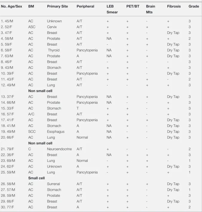
Table 1. Characteristics of the patients
No. Age/Sex BM Primary Site Peripheral LEB PET/BT Brain Fibrosis Grade
Smear Mts
1. 45/M AC Unknown A/T + + - + 3
2. 52/F ASC Cervix A/T + + + + 3
3. 47/F AC Breast A/T + + - Dry Tap 3
4. 58/M AC Prostate A/T NA + + + 2
5. 59/F AC Breast A/T - + + Dry Tap 3
6. 58/F AC Thyroid Pancytopenia NA + - Dry Tap 3
7. 63/M AC Prostate A NA + + Dry Tap 3
8. 46/F AC Breast A/T - + - - 3
9. 43/M AC Stomach A/T + + - + 3
10. 39/F AC Breast Pancytopenia + + - Dry Tap 3
11. 43/F AC Breast A/T + + + - 2
12. 49/M AC Lung A/T - - + + 3
Non small cell
13. 37/F AC Breast Pancytopenia NA + + Dry Tap 3
14. 66/M AC Prostate Pancytopenia NA - - + 3
15. 33/F AC Stomach T + + - + 3
16. 57/F A/C Breast A/T + + - + 3
17. 41/F AC Breast Pancytopenia + + + Dry Tap 3
18. 41/M AC Stomach A NA + - Dry Tap 3
19. 49/M SCC Esophagus A NA - - Dry Tap 3
20. 66/F AC Lung Normal NA + - Dry Tap 3
Non small cell
21. 79/F C Neuroendocrine A/T + - - - 2
22. 36/F AC Breast A NA + + + 3
23. 69/M AC Lung Normal - + + - 1
24. 62/F AC Unknown A + + + Dry Tap 3
25. 59/M AC Lung Pancytopenia - + - + 1
Small cell
26. 58/M AC Surrenal A/T + + + Dry Tap 3
27. 57/M AC Stomach A/T + + - Dry Tap 1
28. 59/M AC Prostate A/T + + - + 3
29. 66/F AC Breast A/T + + - Dry Tap 3
30. 77/F AC Breast A + + - + 2
M= Male, F= Female, AC= Adenocarcinoma, ASC= Adenosquamous carcinoma, SCC= Squamous Cell Carcinoma, C= Carcinoid, A= Anemia, T= Thrombocytopenia, LEB= Leukoerythroblastic blood picture
UHOD
August 2018. 35 patients have been found to have bone marrow metastasis in this population. Two patients who were 15 and 16 years old with rhab-domyosarcoma and one patient who was 16 years old with Ewing’s sarcoma were excluded as they were under 18 years old. Two other patients with prostate and metastatic tumor with unknown pri-mary carcinoma were also excluded because their follow-up data were missing. The data of the re-maining 30 patients were depicted in Table 1. Female to male ratio was 17/13 in the studied population (56.7% vs 43.3%). The mean age at the time of diagnosis was 53.8±12.0 years. The most common malignancy that had metastasis to the bone marrow was breast carcinoma (n= 11, 36.7%). It is followed by prostate (n:4, 13.3%), gastric (n= 4, 13.3%) and lung (n= 4, 13,3%) ad-enocarcinomas. Most of the malignancies were metastatic epithelial adenocarcinomas and there was one adenosquamous carcinoma of the cervix and one squamous cell carcinoma of the esopha-gus. In two cases the primary site could not be de-termined. In 22 patients PET/CT was performed and except two cases (in one of them imaging was 1 year before the bone marrow assessment) bone marrow involvement could be detected in all of the patients (90.9%). In the remaining 8 cases BT was performed and in 6 cases (75%) bone metastasis
was detected. In 2 cases where bone metastasis could not be detected with BT, the images were taken 1 year before the bone marrow assessment. Brain metastasis was present in 12 patients (40%). Presence of brain metastasis was significantly as-sociated with death. 75% of the patients with brain metastasis and 22.2% of the patients without brain metastasis died (p=0.006). 43.3% of the patients died during the study period and the median time from bone marrow assessment to death was 2 months (range= 1 - 36 months).
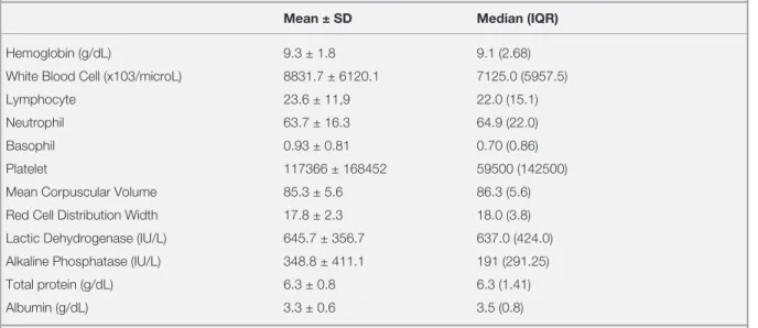
The hematologic results of the patients at the time of bone marrow assessment were shown in table 2. Anemia, thrombocytopenia and leukopenia were present in 90% (n= 27), 73.3% (n= 22) and 20% (n:6) of the patients respectively. Concomitant ane-mia and thrombocytopenia was the most common (50%) presenting hematologic feature followed by pancytopenia (20%). RDW was increased in 24 pa-tients (80%). Lactate dehydrogenase was elevated in 90% (n:27) and alkaline phosphatase was elevat-ed in 80% (n:24) of the patients. Total protein was found to be decreased only in 2 patients (6.7%); however hypoalbuminemia was present in 40% (Table 2). Peripheric smear results of 21 patients were available and in 16 of these (76.2%) a leuko-erythroblastic blood picture was present (Table 1).
Table 2. Hematologic parameters of the patients
Mean ± SD Median (IQR)
Hemoglobin (g/dL) 9.3 ± 1.8 9.1 (2.68)
White Blood Cell (x103/microL) 8831.7 ± 6120.1 7125.0 (5957.5)
Lymphocyte 23.6 ± 11,9 22.0 (15.1)
Neutrophil 63.7 ± 16.3 64.9 (22.0)
Basophil 0.93 ± 0.81 0.70 (0.86)
Platelet 117366 ± 168452 59500 (142500)
Mean Corpuscular Volume 85.3 ± 5.6 86.3 (5.6) Red Cell Distribution Width 17.8 ± 2.3 18.0 (3.8) Lactic Dehydrogenase (IU/L) 645.7 ± 356.7 637.0 (424.0) Alkaline Phosphatase (IU/L) 348.8 ± 411.1 191 (291.25)
Total protein (g/dL) 6.3 ± 0.8 6.3 (1.41)
Albumin (g/dL) 3.3 ± 0.6 3.5 (0.8)
UHOD
All the cases were diagnosed with bone marrow biopsy (n= 30, 100%). Bone marrow aspiration detected infiltration in 12 patients (40%) and in 4 metastatic patients the metastasis could not be detected with aspiration (13.3%). In 14 patients (46.7%) the aspiration resulted with dry tap. Of these 14 dry tap aspiration cases 6 were from bone marrow of breast carcinoma patients (42.9%) (Table 1). In 76.7% of the bone marrow biopsies (23/30), there were grade 3 fibrosis (Table 1, Fig-ure 1A, B). There was a positive correlation be-tween grade of fibrosis and obtaining dry tap with the aspiration (rho= 0.433, p= 0.017).
DISCUSSION
Bone marrow is a common site for solid tumor metastasis as it has a rich blood supply and con-tains various growth factors and therefore any tumor capable of hematologic dissemination can metastasize to the bone marrow. It has been re-ported that bone marrow metastasis is found in 2 to 26% of cases with cancer4 and the most common
tumors are carcinomas arising from breast, pros-tate, lung and gastrointestinal tract with various reported incidences.3,5-8 In this large series of bone
marrow evaluation the most common nonhemato-logic malignancies with bone marrow metastasis were found to be carcinomas of breast, prostate, gastric and lung which is in accordance with the literature. The overall incidence was found to be 1.64% in adults. In 36.7% of these the primary site was breast carcinoma, followed by prostate, lung and gastric carcinomas all having an incidence of 13.3%.
In clinical practice trephine biopsy and aspiration are the two main procedures for the definitive di-agnosis of bone marrow infiltration. These two techniques are considered to be complementary be-cause detection of tumor cells in a biopsy and not in aspirate or vice versa can be seen.9-11 However
in many reports biopsy is considered to be more sensitive9,12 and the gold standard technique when
performed with aspiration simultaneously.2 It is
suggested that in cases in which infiltration cannot be detected with biopsy but positive in aspirates, an adequate bone depth could not be achieved during biopsy.3 In the present study all of the 30 patients
had tumor cells in the biopsy specimens, however, the sensitivity of aspiration was 40% and the false negativity was 13.3%. Syed et al reported that with aspirate they could detect only 35% of the tumors that were diagnosed on bone marrow biopsy13 and
Toi et al reported 50% correlation between aspi-ration and biopsy sections in cases of metastasis from solid tumors.14 Tyagi et al also reported a
100% sensitivity with biopsy and a false negative rate of 14.3% with aspiration.15 Similarly Mishra
et al reported a 100% sensitivity for the biopsy.5
Although in the present study all the cases could be detected with biopsy, there are studies in which tumor cells could be detected with aspiration but not with biopsy16 and therefore aspiration and
bi-opsy must be performed concomitantly as these two techniques complement each other.
Dry tap was seen in 46.7% of the aspirates. It refers to inability to obtain bone marrow on attempted marrow aspirations and is suggested to be due to reactive myelofibrosis which is diagnosed with
re-Figure 1A and B. Bone marrow fibrosis (Reticulin staining, x400, A. Grade 2 myelofibrosis; B. Grade 3 myelofibrosis)
UHOD
ticulin staining of trephine biopsy and represents greater degrees of marrow infiltration.17,18 Marked
myelofibrosis has been reported to be most com-mon in carcinomas of the breast, stomach and lung.19-21 In the present study similarly 42.9% of
the dry tap cases were from aspirates of breast cinoma cases, followed by cases with gastric car-cinoma (14.3%). In 92.9% of the dry tap cases, the fibrosis was very significant. Myelofibrosis with collagen and reticulin was present in all cases, but in 76.7% fibrosis was grade 3 representing an advanced stage. Kaur et al reported myelofibrosis in 6 of the 9 cases (66.7%) however they did not grade the extent of fibrosis 2. Mishra et al reported grade 3 fibrosis in 22.2% of the patients; however, when osteomyosclerotic and necrotic bone marrow biopsies are encountered fibrosis rate increases to 77.8% which is similar to our findings.5
Infiltration of bone marrow by malignant cells is associated with normocytic normochromic ane-mia and thrombocytopenia and there may be myelo-suppression induced leukopenia. In our study anemia was present in 90% of the patients. Thrombocytopenia and leukopenia were detected in 73.3% and 20% of the patients respectively. Anemia and thrombocytopenia were concomitant in 50% and cytopenia was present in 20% of the patients. Ozkalemkas et al in their study reported anemia and thrombocytopenia in all the patients, whereas Kaur et al and Sar et al detected anemia in 77.7% and 66.6% respectively.2,22,23 Various other
studies reveal an incidence of anemia in neoplastic diseases from 55-90%.24-26 Again the reported rates
of thrombocytopenia and leukopenia differ among studied patients, ranging from 33.3% to 100% and 30.3% to 50% respectively.1,2,22,23 In 6.7% of
the studied patients all the hematologic param-eters were within normal ranges which is not an uncommon finding. In these two patients red cell distribution width was found to be increased. RDW was found to be increased in 80% (n= 24) of the patients. Similarly in another study from Turkey, Ozkalemkas et al reported an increased RDW in all of the patients with non-hematologic tumors that had bone marrow metastasis.22 They proposed a
tetrad consisting of anemia, thrombocytopenia, el-evated RDW and hypoproteinemia in patients with disseminated tumors that were diagnosed via bone
marrow examination.22 In the present study
albu-min level was low in 40% of the patients and the other parameters are found to be increased in most of the patients suggesting that this tetrad may be a useful marker for such patients.
A leukoerythroblastic reaction refers to an increase in the peripheral blood of immature red blood cells (normoblasts, immature granulocytes, metamye-locytes, bands), and is suggested to be associated with metastatic cancer, hematopoietic malignancy, hemolytic anemia, Gaucher’s disease, polytrauma and BM infiltration by various processes like infec-tion. Peripheric smear was available in 21 patients. Leukoerythroblastic blood picture was present in 76.2% (n= 16) of these patients. Leukoerythro-blastic blood picture is thought to be resulted from the early release of some cytokines even before the marrow is completely invaded and is associ-ated with the degree of the bone marrow fibrosis rather than the extent of malignant cell invasion.19
In the present study advanced fibrosis was present in 76.6% of the patients.
Alkaline phosphatase was elevated in 80% and lactate dehydrogenase was found to be elevated in 90% of the patients. Similarly Ozkalemkas et al reported an increased level of LDH in 95% of the cases and Wong et al reported an increased ALP in 95% of the cases.22,27 These findings together with
the leukoerythroblastic blood picture indicate an infiltrative process in the marrow.
PET/CT has gained wide acceptance for evalua-tion of the primary site or the metastasis in malig-nancies. In the present study 22 patients had been evaluated with PET/CT and in 20 of the patients (90.9%) FGD entrapment was detected in the bones. And in one PET/CT negative, but infiltra-tion positive patient the imaging was done 1 year before the bone marrow assessment and it may be suggested that rather than missing out the present metastasis, there was no metastasis at the time of the imaging. CT had also a significant sensitivity to detect bone marrow involvement in the studied patients (75%). Again in one of the CT negative patients the imaging was 1 year before the bone marrow assessment. From this point imaging mo-dalities, especially PET/CT, have very high sensi-tivities for detection of bone marrow metastasis.
UHOD
At the time of diagnosis of bone marrow metas-tasis, brain metastasis was also present in 40% of the patients showing the progressive stage of the malignancy. Concomitant presence of brain and bone marrow involvements were associated with poor prognosis. During the study period 43.3% of the patients died after the bone marrow aspiration and biopsy in a median time of 2 months. Survival was similar in the study by Ozkalemkas et al22 in
which it was only a few weeks or months except few patients. The survival of many patients is very short and therefore the therapeutic significance of detection of bone marrow metastasis may be questioned. One may question if it is necessary to confirm bone marrow metastasis in patients whose tumor stage is already known as PET/CT imaging has such a high sensitivity (much more than aspira-tion) in detection of these metastasis. We believe that the only indication of bone marrow assessment in such patients may be the tumors with unknown primary site.
In conclusion bone marrow may be invaded by various non-hematologic tumors; breast, prostate, gastic and lung adenocarcinomas are being the most common ones. Bone marrow involvement is associated with advanced stage and poor progno-sis. Biopsy and aspiration are the two main com-plementary procedures for the definitive diagnosis. There are many laboratory and peripheral blood findings. Bone marrow assessment is a minimally invasive technique and provides very beneficial clinical data, however, because the survival is very short after BM assessment and the PET/CT has a considerable sensitivity it is not necessary to con-firm BM metastasis in patients whose tumor stage is already known.
REFERENCES
1. Meenai AF, Ojha S, Ali MA, et al. Bone marrow involvementin non-hematological malignancy: a clinicopathological study from atertiary hospital. APALM 5: 440-446, 2018.
2. Kaur G, Basu S, Kaur P, Sood T. Metastatic bone marrow tumors: Study of nine cases and review of the literature. J Blood Disord Transfus 2: 110, 2011.
3. Chauhan K, Jain M, Grover S, et al. Bone marrow metastasis in nonhematologic malignancies: Data from a cancer hospital. Clin Cancer Invest J 5: 103-109, 2016.
4. Casciato, DA. Metastatic cancer, in: Cancer Treatment, Haskell, C. H. (Ed.)Boston, W.B. Saunders, 1995, p: 1128. 5. Mishra P, Das S, Kar R, et al. Non-hematopoietic
malignan-cies metastasising to the bone marrow: A 5 year record based descriptive study from a tertiary care centre in South India. Indian J Cancer 51: 30-34, 2014.
6. Kucukzeybek BB, Calli AO, Kucukzeybek Y, et al. The prog-nostic significance of bone marrow metastases: Evaluation of 58 cases. Indian J Pathol Microbiol 57: 3969, 2014. 7. Brahmbhatt B, Parikh B, Shah M. Bone marrow involvement
by metastatic solid tumors. Gujarat Medical Journal 69: 54-57, 2014.
8. Mohanty SK, Dash S. Bone marrow metastasis in solid tu-mors. Indian J Pathol Microbiol 46: 613-616, 2003. 9. Singh G, Krause JR, Breitfeld V. Bone marrow examination
for metastatic tumor aspirate and biopsy. Cancer 40: 2317-2321, 1977.
10. Savage RA, Hoffman GC, Shaker K. Diagnostic problems in-volved in detection of metastatic neoplasms by bone marrow aspirate compared with needle biopsy. Am J Clin Pathol 70: 623-627, 1978.
11. Moid F, DePalma L. Comparison of relative value of bone marrow aspirates and bone marrow trephine biopsies in the diagnosis of solid tumor metastasis and Hodgkin’s lympho-ma. Institutional experience and literature review. Arch Pathol Lab Med 129: 497-501, 2005.
12. Ingle JN, Tormey DC, Tan HK. The bone marrow examina-tion in breast cancer; diagnostic consideraexamina-tions and clinical usefulness. Cancer 41: 670-674, 1978.
13. Syed NN, Moiz B, Adil SN, Khurshid M. Diagnostic impor-tance of bone marrow examination in non-hematological dis-orders. J Pak Med Assoc 57: 123-125, 2007.
14. Toi PC, Varghese RG, Rai R. Comparative evaluation of si-multaneous bone marrow aspiration and bone marrow bi-opsy : an institutional experience. Indian J Hematol Blood Transfus 26: 41-44, 2010.
15. Tyagi R, Singh A, Garg B, Sood N. Beware of bone marrow: Identical detection and primary diagnosis of solid tumours in bone marrow aspiration and biopsies; a study of 22 cases. Iran J Pathol 13: 78-84, 2017.
16. Sharma S, Murari M. Bone marrow involvement by metastatic solid tumors. Indian J Pathol Microbiol 46: 382-384, 2003. 17. Humpries JE. Dry tap bone marrow aspiration: Clinical
signifi-cance. Am J Hematol 35: 247-250, 1990.
18. Hyun BH. Bone marrow examination: Adventures in diagnos-tic hematology. Yonsei Med J 27: 100-105, 1986.
19. Rubins JR. The role of myelofibrosis in malignant myeloscle-rosis. Cancer 51: 308-311, 1983.
20. Kiely JM, Silverstein MN. Metastatic carcinoma simulating ag-nogenic myeloid metaplasia. Cancer 24: 1041-1044, 1969.
UHOD
21. Spector JI, Levine PH. Carcinomatous bone marrow invasion simulating acute myelofibrosis. Am J Med Sci 266: 145-148, 1973.
22. Ozkalemkas F, Ali R, Ozkocaman V, et al. The bone marrow aspirate and biopsy in the diagnosis of unsuspected non-hematologic malignancy: a clinical study of 19 cases. BMC Cancer 5: 144, 2005.
23. Sari R, Aydogdu I, Ozen S, et al. Metastatic bone marrow tumours: a report of six cases and review of the literature. Haematologia 31: 215-223, 2001.
24. Contreras E, Ellis LD, Lee RE. Value of the bone marrow bi-opsy in the diagnosis of metastatic carcinoma. Cancer 29: 778-783, 1972.
25. Hyman GA, Harvey JE. The pathogenesis of anemia in pa-tients with carcinoma. Am J Med 19: 350-356, 1955. 26. Samuels AJ, Bierman HR. Anemia in patients with neoplastic
disease. Calif Med 84: 180, 1956.
27. Wong KF, Chan JK, Ma SK. Solid tumour with initial presenta-tion in the bone marrow--a clinicopathologic study of 25 adult cases. Hematol Oncol 11: 35-42, 1993.
Correspondence:
Dr. Pelin AYTAN
Adana Baskent Universitesi
Dr. Turgut Noyan Uygulama ve Arastirma Hastanesi Hematoloji Bolumu, Eriskin Kemik Iligi Nakli Unitesi Dadaloglu Mah. 2591 Sok. No: 4/A
01250 Yüregir ADANA / TURKEY Tel: (+90-532) 780 35 72 e-mail: [email protected]