The occurrence of epizoic ciliates (Protozoa: Ciliophora) of the
juvenile flounder,
Platichthys flesus L., 1758, from
Sarıkum Lagoon Lake (Sinop, Turkey)
Türkay ÖZTÜRK*, Ahmet ÖZER
Fisheries Faculty, Sinop University, 57000 Sinop - TURKEY
Received: 27.08.2008
Abstract:The occurrence of epizoic ciliates on the juvenile flounder, Platichthys flesus L., 1758, collected monthly by fishing net in Sinop’s Sarıkum Lagoon Lake during the period from May 2003 to April 2004, was investigated. Five epizoic ciliate species, Trichodina jadranica (Raabe, 1958) Haider, 1964; Trichodina domerguei Wallengren, 1897; Riboscyphidia sp.; Ambiphrya sp.; and Vorticella sp., were found after examination of 296 juvenile flounder. Among these, T. jadranica was the dominant species. The prevalence and mean intensity values of the 5 epizoic ciliates were given as pooled data for 2 groups, namely mobiline ciliates and sessiline ciliates. Mobiline ciliates were commonly found on the gills of juvenile flounders, whereas sessiline ciliates were found only on fins. The highest prevalence and mean intensity levels of mobiline ciliates were recorded in the winter and spring seasons. Sessiline ciliates were absent in winter. The largest-sized juvenile flounder among the established 6 length classes had a higher intensity of mobiline ciliates. This study is the first on the epizoic ciliate fauna present on the juvenile flounder in Turkey. While T. jadranica is a new parasite record, the juvenile flounder is a new host record for T. domerguei in Turkey.
Key words:Flounder, Platichthys flesus, epizoic, trichodinids, sessiline ciliates
Sarıkum Lagün Gölü’ndeki (Sinop, Türkiye) yavru dere pisisi
Platichthys flesus L.,
1758 balığında görülen ektoparazitik siliatlar (Protozoa: Ciliophora)
Özet:Sinop Sarıkum Lagün’ünden Mayıs 2003 – Nisan 2004 tarihleri arasında her ay ığrıp çekilerek yakalanan yavru dere pisisi balığının, Platichthyes flesus L., 1758, epizoik siliatları belirlendi. Trichodina jadranica (Raabe, 1958) Haider, 1964;
Trichodina domerguei Wallengren, 1897; Riboscyphidia sp.; Ambiphrya sp.; ve Vorticella sp. olmak üzere 5 epizoik siliat
türü 296 adet yavru dere pisisi balığında tanımlandı. Bu türler arasında Trichodina jadranica dominat türdü. Beş epizoik siliatın enfeste balık başına ortalama yoğunluğu ve enfestasyon oranı mobil ve sesil siliatlar olmak üzere iki grup adı altında toplu olarak verildi. Mobil siliatlar yavru pisi balıkların solungaçlarında yaygın olarak bulunurken, sesil siliatlar yüzgeçlerinde bulundu. Mobil siliatlara ait en yüksek enfestasyon oranı ve yoğunluğu kış ve baharda kaydedildi. Diğer taraftan sesil siliatlar kışın gözlenmedi. Oluşturulan altı boy sınıf içinden en büyük boy sınıfında mobil siliatların yoğunluğu daha yüksek oranda bulundu. Bu çalışma Türkiye’deki yavru pisi balıklarının ektoparazitik siliatları üzerine ilktir. T. jadranica, Türkiye parazit faunası için yeni parazit kaydı iken, T. domerguei için yavru pisi balıkları yeni bir konak kaydıdır.
Anahtar sözcükler:Pisi balığı, Platichthys flesus, epizoik, trichodinidler, sesil siliatlar
Research Article
doi:10.3906/zoo-0808-18Introduction
Among epizoic protozoa, there are important pathogens of wild and hatchery-reared fish. The most frequently observed ectoparasitic protozoans are motile (mobiline) and nonmotile (sessiline) ciliates affecting the gills and skin of aquatic animals. One of the largest and most widely distributed mobiline ciliate genera is Trichodina Ehrenberg, 1838, present on aquatic invertebrate and vertebrate hosts (Van As and Basson, 1989). Sessiline ciliates live as symbionts, commensals, and parasites on various hosts, such as mollusks (Botes et al., 2001), copepods, mysids (Jayasree et al., 2001; Fernandez-Leborans, 2004), and fish (Kuperman et al., 2001). At high densities, these ciliates (mobiline and sessiline) have been reported as causing mortality in juvenile and adult cultured fish populations, leading to severe economic losses in various parts of the world (Van As and Basson, 1987). There are many studies surveying epizoic ciliates of various aquatic species (Xu et al., 1999; Green and Shiel, 2000; Madsen et al., 2000; Basson and Van As, 2002; Dove and O’Donoghue, 2005). In the studies surveying the epizoic ciliates of flounder, P. flesus, more mobiline ciliates than sessiline ciliates were reported to be present. So far, Trichodina borealis, T.
claviformis, T. raabei, T. jadranica, and T. domerguei
have been described by various authors as the epizootic ciliates of flounder (Calenius, 1980; Lüthen, 1989; Palm and Dobberstein, 1999; Dobberstein and Palm, 2000; Chibani et al., 2005). There are few quantitative parasitological studies on flounder in
Turkey (Oğuz, 1991; Aydoğdu and Öztürk, 2003; Oğuz and Öktener, 2007); these have covered larger, commercially caught fish and have mainly described the metazoan parasites. Despite several studies on ectoparasitic fauna of some juvenile fish species (Tokşen, 2004; Ogut and Akyol, 2007), no study on juvenile flounder has been done in Turkey.
The aim of the present study was to investigate and describe epizoic ciliates found on juvenile specimens of flounder and obtain more information on epizoic parasites depending on host characteristics and environmental factors.
Materials and methods
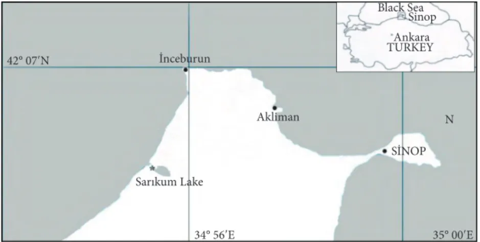
Specimens of juvenile flounder (P. flesus) were collected by fishing net in the estuary of Sarıkum Lagoon Lake (42°00´N; 34°54´E), connected to the Black Sea (Figure 1). It is typically a lagoon, a eutrophic lake with a brackish characteristic and salinity ranging from 1‰ to 5‰.
Sampling was carried out monthly from May 2003 to April 2004. For parasitological examination, fish were transported alive in local water to the Sinop Fisheries Faculty Laboratory and examined in the same water; the following day, they were collected. A total of 296 fish specimens were observed. The total length was measured postmortem. Skin, fins, and gills were examined under a light microscope and scrapings of whole mucus from these parts of the fish were taken on several slides. The total number of epizoic ciliates was determined by screening and
34° 56ʹE 35° 00ʹE 42° 07ʹN SİNOP Sarıkum Lake İnceburun Ankara TURKEY Black Sea Sinop Akliman N
counting of the entire mucus material of each slide. For trichodinids, air-dried smears were stained according to Klein’s silver nitrate (AgNO3) method (Lom and Dykova, 1992) in order to study details of the adhesive disk. All morphological measurements were carried out by an oil-immersion light microscope (Nikon SE), using 20 parasite specimens for each species. All measurements followed the uniform specific characteristics proposed by Lom (1958) and Arthur and Lom (1984). In the case of denticles and radial pins, the mode is given instead of the arithmetic mean. The span of the denticle was measured from the tip of the blade to the tip of the ray. In the description of denticle elements, the format recommended by Van As and Basson (1989) was followed.
Specifically, several references were used in the description of mobilines T. jadranica and T.
domerguei (Lom and Stein, 1966; Lom, 1970; Arthur
and Lom, 1984; Lom and Dykova, 1992) and sessilines
Riboscyphidia sp., Ambiphrya sp., and Vorticella sp.
(Viljoen and Van As, 1983; Lom and Dykova, 1992; Jayasree et al., 2001; Fernandez-Leborans, 2004).
Infestation prevalence (%) and mean intensity levels of the ciliates were determined according to the methods of Bush et al. (1997). The prevalence and mean intensity values of the 5 epizoic ciliates were given as pooled data for 2 groups, namely mobiline ciliates and sessiline ciliates, rather than by individual species.
Water temperature (°C), salinity (‰), and pH levels were recorded using a U-10 Horiba digital water analyzer at the sampling sites.
Normality of the data was checked using the Kolmogorov-Smirnov test. The Kruskal-Wallis test (nonparametric ANOVA) was performed to compare the mean intensity values of mobiline and sessiline ciliates for infestation sites, length classes of fish, and the months in which the study was conducted. The analyses were carried out using the computer programs GraphPad Instat 3.0 and SPSS 9.0 (Software Inc., USA).
Results
Epizoic protozoan ciliates belonging to 2 groups, 2 mobiline and 3 sessiline species, were found on juvenile flounder.
Mobiline ciliates
Two species of trichodinids, T. jadranica and T.
domerguei, the former being the more common, were
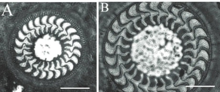
identified (Figures 2 and 3). The preferred infestation sites of these 2 trichodinid species on the juvenile flounder were different. T. jadranica was commonly found on the gills, whereas T. domerguei was commonly present on the skin and fins of the juvenile flounder. In addition, T. jadranica and T. domerguei were observed on stained slides in a ratio of 100:7, respectively.
T. jadranica (Figures 2A and 3A, Table 1) is a
small-sized trichodinid with a disk-shaped body. The center of the adhesive disk of the specimens impregnated with silver nitrate appeared as a clear space. The blade of the denticle was sickle-shaped. The distal margin of the blade was rounded and away from the border membrane. The tangent point was
Figure 2. Parasitic mobiline ciliates of the juvenile flounder, Platichthys flesus: A,
Trichodina jadranica (Raabe, 1958) Haider, 1964; B, Trichodina domerguei
rounded. The anterior margin of the blade sharply curved down. The apex of the blade was round. Anterior apophysis and posterior projection were not present. The central part of the denticle was roundly pointed and extended slightly more than halfway toward the y-1 axis. Rays were slightly curved in a posterior direction with tips extending beyond the y axes, and the point of the ray was rounded. The morphometrical data of the parasites’ body parts are given in Table 1.
T. domerguei (Figures 2B and 3B, Table 1) is a large
trichodinid with a disk-shaped body. The center of the adhesive disk of the specimens impregnated with silver nitrate was clear with numerous dark granules. The blade of the denticle was broad and sickle-shaped, filling a large area between the y axes. The distal margin of the blade was close to the border membrane. The apex of the blade was round. The tangent point was rounded. The posterior margin
y+1
A B
y y-1
Figure 3. Diagrammatic drawings of the denticles of trichodinids: A, Trichodina jadranica (Raabe, 1958) Haider, 1964; B, Trichodina domerguei Wallengren, 1897.
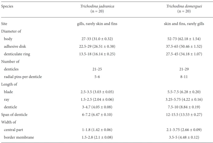
Table 1. Morphometrical data and infestation sites of Trichodina jadranica and T. domerguei in the juvenile flounder, Platichthys flesus (n: number of measured specimens) (range with arithmetic mean and standard error in parentheses) (all measurements in mm).
Species Trichodina jadranica Trichodina domerguei
(n = 20) (n = 20)
Site gills, rarely skin and fins skin and fins, rarely gills
Diameter of body 27-33 (31.0 ± 0.32) 52-73 (62.18 ± 1.54) adhesive disk 22.5-29 (26.51 ± 0.38) 37.5-65 (50.46 ± 1.52) denticulate ring 13.5-18 (16.14 ± 0.25) 27.5-45 (34.18 ± 1.07) Number of denticles 21-25 21-29
radial pins per denticle 5-6 8-11
Length of blade 2.5-3.5 (3.03 ± 0.05) 5.5-7.5 (6.28 ± 0.20) ray 1.5-2.5 (2.04 ± 0.06) 3.25-5.75 (4.22 ± 0.16) denticle 3-4.7 (4.05 ± 0.08) 7.5-10 (8.84 ± 0.19) Span of denticle 6-7.2 (6.47 ± 0.10) 12-15.5 (13.53 ± 0.27) Width of central part 1-1.8 (1.42 ± 0.06) 2.1-3.75 (2.66 ± 0.09) border membrane 1.5-2.8 (2.1 ± 0.08) 3.5-5 (4.48 ± 0.12)
fairly curved down. Blade apophysis was present, but not clearly visible. The blade connection was thin. The central part was well developed, but thin and long, tapering to a rounded point and fitting tightly into the preceding denticle. The ray connection was short and thin. The base of the ray was thin, with the ray being bulbous toward broad, with a rounded point. Rays were short and curved in the posterior direction with tips extending beyond the y axes. The section of the denticle above the x axis was similar to the portion below, with a ratio of one. The morphometrical data are presented in Table 1.
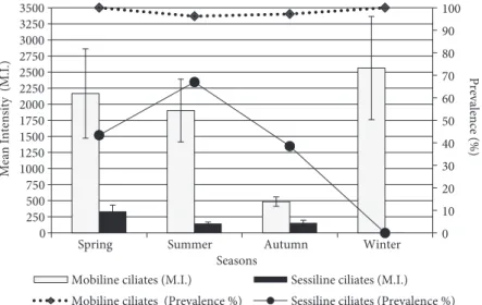
The monthly seasonal prevalence and mean intensities of mobiline ciliates infesting the juvenile flounder are reported in Table 2 and Figure 4. A statistically significant difference among the seasonal mean intensities was detected (P = 0.0074, Table 2). Mean intensities were higher in the spring and winter than in autumn. The overall infestation prevalence (%) and mean intensity levels estimated from a total of 296 fish specimens were 97.97% and 1584.37 ± 242.91 trichodinids/infested fish, respectively (Table 2). Statistically significant differences in the mean intensity values were found in relation to the length classes, months, seasons, and infestation sites (Table 2, Figure 5). However, it must be noted that there were no statistically significant differences among the mean intensities of each of the 6 length classes of fish in any month (P > 0.05). Thus, the data were grouped and
analyzed for the 6 length classes with no respect to sampling months. Larger fish had higher intensities than smaller fish (Table 2).
Sessiline ciliates
A total of 3 sessiline ciliate species, Riboscyphidia sp., Ambiphrya sp., and Vorticella sp., the first one being the most common, were identified. The sites of infestation of these sessiline ciliates on the juvenile flounder were different. Riboscyphidia sp. was commonly found on skin and fins, rarely on gills; for
Ambiphrya sp. and Vorticella sp., it was the reverse. In
addition, Riboscyphidia sp., Ambiphrya sp., and
Vorticella sp. were observed in a respective ratio of
80:15:5.
Riboscyphidia sp. (Figure 6A) has a cylindrical or
conical body, with a length of 60-64 mm (55.50 ± 0.80). The body was divided into nearly equal distinct regions by a circular groove, 30-43 mm in diameter (37.16 ± 0.79). The scopula was extremely large with a thin flat border, 30-43 mm in diameter (37.16 ± 0.79). The peristomial lip opened wider than the body, 35-42 mm in diameter (38.48 ± 0.34), and the infundibulum extended from the ciliary zone to the circular groove.
Ambiphrya sp. (Figure 6B) was similar to Riboscyphidia sp. and its body is also cylindrical. The
macronucleus was ribbon-like. The diameter of the
0 10 20 30 40 50 60 70 80 90 100 0 250 500 750 1000 1250 1500 1750 2000 2250 2500 2750 3000 3250 3500
Spring Summer Autumn Winter
Prevalence (%)
Mean Intensity
(M.I.)
Seasons
Mobiline ciliates (M.I.) Sessiline ciliates (M.I.)
Mobiline ciliates (Prevalence %) Sessiline ciliates (Prevalence %)
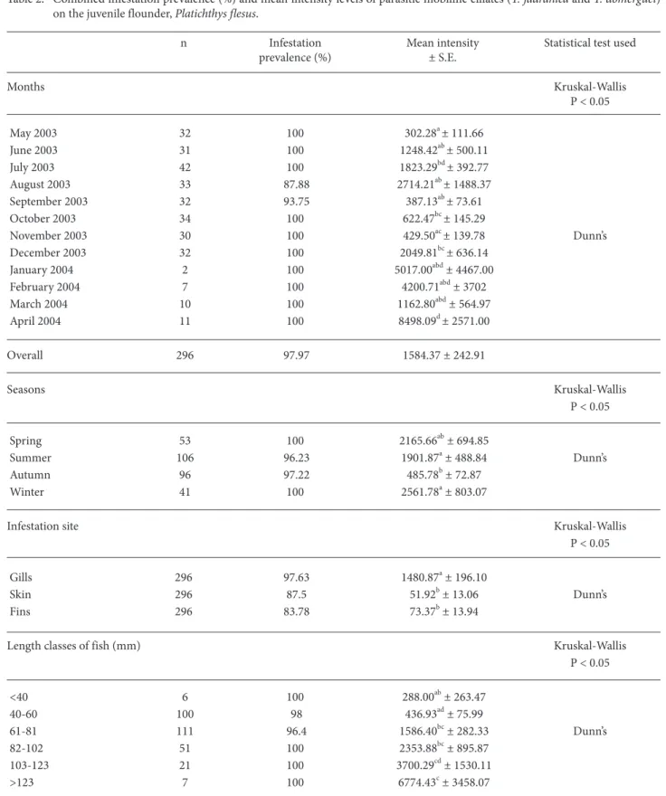
Table 2. Combined infestation prevalence (%) and mean intensity levels of parasitic mobiline ciliates (T. jadranica and T. domerguei) on the juvenile flounder, Platichthys flesus.
n Infestation Mean intensity Statistical test used prevalence (%) ± S.E. Months Kruskal-Wallis P < 0.05 May 2003 32 100 302.28a± 111.66 June 2003 31 100 1248.42ab± 500.11 July 2003 42 100 1823.29bd± 392.77 August 2003 33 87.88 2714.21ab± 1488.37 September 2003 32 93.75 387.13ab± 73.61 October 2003 34 100 622.47bc± 145.29 November 2003 30 100 429.50ac± 139.78 Dunn’s December 2003 32 100 2049.81bc± 636.14 January 2004 2 100 5017.00abd± 4467.00 February 2004 7 100 4200.71abd± 3702 March 2004 10 100 1162.80abd± 564.97 April 2004 11 100 8498.09d± 2571.00 Overall 296 97.97 1584.37 ± 242.91 Seasons Kruskal-Wallis P < 0.05 Spring 53 100 2165.66ab± 694.85 Summer 106 96.23 1901.87a± 488.84 Dunn’s Autumn 96 97.22 485.78b± 72.87 Winter 41 100 2561.78a± 803.07
Infestation site Kruskal-Wallis
P < 0.05
Gills 296 97.63 1480.87a± 196.10
Skin 296 87.5 51.92b± 13.06 Dunn’s
Fins 296 83.78 73.37b± 13.94
Length classes of fish (mm) Kruskal-Wallis
P < 0.05 <40 6 100 288.00ab± 263.47 40-60 100 98 436.93ad± 75.99 61-81 111 96.4 1586.40bc± 282.33 Dunn’s 82-102 51 100 2353.88bc± 895.87 103-123 21 100 3700.29cd± 1530.11 >123 7 100 6774.43c± 3458.07
scopula was wider than the diameter of the circular groove near the scopula.
Vorticella sp. (Figure 6C) has a conical and an
inverted bell-shaped body. The stalk was contractile and longer when compared with the size of the body. The macronucleus was J-shaped. A spherical micronucleus was located close to the macronucleus.
Monthly and seasonal prevalence and mean intensity levels of sessiline ciliates infesting the
juvenile flounder are presented in Table 3 and Figure 4. No sessiline ciliates were observed in November or December 2003, or January, February, March, or April 2004. The mean intensity of sessiline ciliates was slightly greater in the spring than in summer or autumn, although the difference among the seasons was statistically insignificant (P > 0.05, Table 3). The overall infestation prevalence (%) and mean intensity levels were 44.26% and 181.80 ± 24.92 sessiline ciliates/infested fish, respectively (Table 3). Both levels
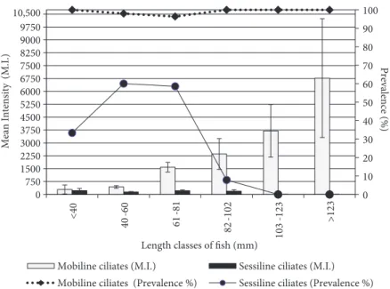
0 10 20 30 40 50 60 70 80 90 100 0 750 1500 2250 3000 3750 4500 5250 6000 6750 7500 8250 9000 9750 10,500 <40 40 -60 61 -81 82 -102 103 -123 >123 Prevalence (%) Mean Intensity (M.I.)
Length classes of fish (mm)
Mobiline ciliates (M.I.) Sessiline ciliates (M.I.)
Mobiline ciliates (Prevalence %) Sessiline ciliates (Prevalence %)
Figure 5. Values of prevalence and mean intensity of epizoic ciliates on juvenile flounder according to fish length classes.
Figure 6. Parasitic sessiline ciliates of the juvenile flounder, Platichthys flesus: A, Riboscyphidia sp.; B, Ambiphrya sp.; C, Vorticella sp. Photomicrographs from fresh material. Scale bar: 10 mm.
Table 3. Combined infestation prevalence (%) and mean intensity levels of parasitic sessiline ciliates (Riboscyphidia sp., Ambiphrya sp., and Vorticella sp.) on the juvenile flounder, Platichthys flesus.
n Infestation Mean intensity Statistical test used prevalence (%) ± S.E. Months Kruskal-Wallis P < 0.05 May 2003 32 71.88 333.57ab± 100.1 June 2003 31 25.81 6.86c± 3.60 July 2003 42 95.24 196.20b± 35.72 Dunn’s August 2003 33 72.73 102.25a± 29.03 September 2003 32 50 195.94ac± 82.40 October 2003 34 61.77 126.76a± 32.82 November 2003 30 0 0 December 2003 32 0 0 January 2004 2 0 0 February 2004 7 0 0 March 2004 10 0 0 April 2004 11 0 0 Overall 296 44.26 181.80 ± 24.92 Seasons Kruskal-Wallis P > 0.05 Spring 53 43.40 333.57 ± 100.04 Summer 106 66.98 145.78 ± 23.48 Autumn 96 38.54 156.68 ± 39.94 Winter 41 0 0
Infestation site Kruskal-Wallis
P < 0.05
Gills 296 23.65 42.61a± 7.80
Skin 296 31.42 44.18a± 8.31 Dunn’s
Fins 296 39.19 144.20b± 23.66
Length classes of fish (mm) Kruskal-Wallis
P > 0.05 <40 6 33.33 223.50 ± 130.5 40-60 100 60 140.95 ± 31.08 61-81 111 58.56 217.83 ± 40.57 82-102 51 7.84 189.00 ± 84.27 103-123 21 0 0 >123 7 0 0
were also recorded for all body parts and the length classes of the juvenile flounder (Table 3). The highest prevalence of sessiline ciliates was observed on the fins (39%), while the skin and gills were found to be less infested (31.42% and 23.65%, respectively). No difference in mean intensities among the various length classes was detected (Table 3).
Water parameters
Monthly water temperature (°C), salinity (‰), and pH values recorded throughout the sampling period are presented in Figure 7. While the highest temperature value was recorded in August (27.8 °C), the lowest temperature was recorded in January (7.1 °C). During the study period, salinity varied from 1.2‰ to 5.1‰ and pH from 5.63 to 7.59. The salinity was at its minimum in February (1.2‰). High salinities were recorded in January (5.1‰) and August (5.0‰)
Discussion
T. jadranica, having a wide geographical
distribution and host range, occurs on the gills and skin of marine and brackish fish in the Adriatic, Baltic, Black, Azov, Yellow, and Bohai Seas; the northeast, northwest, and southeast Atlantic Ocean; and Cuba (Lom, 1970; Arthur and Lom, 1984; Dobberstein and Palm, 2000; Xu et al., 2001; Arthur et al., 2004). In addition, it has been reported from
European eels, Anguilla anguilla, in freshwater (Madsen et al., 2000). This study is the first report of
T. jadranica in Turkish brackish waters of the Black
Sea.
Morphometric data obtained from specimens of
T. jadranica in this study were in agreement with
those reported by Lom (1970), Dobberstein and Palm (2000), Madsen et al., (2000), and Xu et al. (2001), but slightly smaller than those reported by Arthur and Lom (1984) and Arthur et al. (2004). The morphometric data of T. domerguei fall within the size ranges given by several authors (Lom and Stein, 1966; Lom, 1970; Özer, 2003a, 2003b), and this species was found to be among the most variable of the trichodinid species. T. jadranica and T. domerguei are well-known cosmopolitan species. As can be seen in Figure 7, the data collected related to the environmental factors also show fluctuations; thus, the variation in the dimensions of both T. jadranica and T. domerguei throughout the sampling period can be explained by the differences in environmental factors as well as a result of host influences.
The mean intensity levels of the mobiline ciliates (T. jadranica and T. domerguei) fluctuated seasonally (see Table 2). These data were different from the seasonal mean intensity values for T. domerguei, the only species found on the round goby, Neogobius
melanostomus (Özer, 2003a, 2003b). This difference
could be related to the different host species and environmental factors, with the former factor possibly Figure 7. Selected water parameters for Sarıkum Lagoon Lake from May 2003 through April 2004.
0 0.1 0.2 0.3 0.4 0.5 0.6 0 4 8 12 16 20 24 28 37742 June 03 Ju ly 03 A ug. 0 3
Sep. 03 Oct. 03 Nov. 03 Dec. 03 Jan.
04 Feb. 04 March 04 April 0 4 pH -Tem per atur e (°C) Months Temperature pH Salinity Salini ty (%)
having more effect. However, when the environmental factors are considered, it can be seen that the maximum mean intensity values recorded in winter coincided with a decrease in water temperature values in the present study (Figure 7). This result is also supported by the data presented by Kristmundsson et al. (2006), even though the trichodinid species studied were different. Moreover, throughout the investigation period, T. domerguei and T. jadranica were present at the same time. In the present study, T.
jadranica was possibly the main factor causing higher
prevalence and mean intensity levels in winter. Although the data for T. domerguei and T. jadranica were combined and calculated as a total for mobiline ciliates rather than species by species, T. jadranica was found more commonly than T. domerguei on the stained slides (100:7). Palm and Dobberstein (1999) observed a similar increase in the Trichodina population density on flounder in winter, attributing this increase to the bacterial biomass in the environment. Similarly, Ogut and Palm (2005) and Ogut and Akyol (2007) also found an increase in the abundance of trichodinids in winter months. The observed high level of prevalence and mean intensities of infestation in winter and spring was probably due to, as the above authors suggested, seasonal eutrophication or organic pollution in the sampling areas.
The size of the juvenile flounder was a factor affecting the intensity of mobiline ciliates (T. jadranica and T. domerguei) in this study, and the differences in the mean parasite intensities between the different fish length classes were statistically significant. Larger-sized juvenile flounder had higher numbers of parasites. The severity of many ectoparasitic infections increases with host size, possibly as a result of increasing exposure period and a larger space for the feeding and breeding of the parasite. Özer and Erdem (1998) and Özer (2003a, 2003b) noted a tendency of increase in the mean intensity of
Trichodina spp. in relation to the length of the host
fish. Our findings on the intensity levels of mobiline ciliates agree with those reported by the above mentioned authors.
The fins are a preferred attachment site for sessiline ciliates (Riboscyphidia sp., Ambiphrya sp., and Vorticella sp.), probably because the ciliates benefit from nutrients such as bacteria and detritus in their environment. These findings have also been supported by the data of Özer and Erdem (1999) and Kuperman et al. (2001).
The sessiline ciliates on juvenile flounder were observed only from May to October 2003, when water temperatures ranged from 27.5 to 19.1 °C (Figure 7). The present data are comparable to those of Tassaduqe et al. (2003), in which these ciliates were found only in March (14.4 °C), April (29.4 °C), and September (31.7 °C). Similarly, Jayasree et al. (2001) found a tendency of an increase in the abundance of the sessiline ciliates in relation to water temperature. No specimens of sessiline ciliates were found on the larger-sized juvenile flounder (103-123 and >123 mm length classes). The juvenile flounder in these length classes were caught between late autumn and winter months only. The absence of sessiline ciliates in the larger-sized fish length classes could be a result of catching these juvenile flounders only in the colder times of the sampling.
In conclusion, this paper is the first report on the epizoic ciliate fauna of the juvenile flounder in Turkey. While in the present study T. jadranica is a new record for Turkish parasite fauna, the juvenile flounder is a new host record for T. domerguei in Turkey.
Acknowledgement
This study was financed by Ondokuz Mayıs University under Project Number S-082.
Arthur, J.R. and Lom, J. 1984. Trichodinid protozoa (Ciliophora: Peritrichida) from fresh water fishes of Rybinsk Reservoir, USSR. J. Parasitol. 31: 82-91.
Arthur, J.R., Cone, D.K., Cusack, R.R., Barker, D.E. and Burt, M.D.B. 2004. Two species of Trichodina (Ciliophora: Peritrichida) from cultured flatfishes (Pleuronectiformes) in Atlantic Canada. Comp. Parasitol. 71: 247-250.
Aydoğdu, A., and Öztürk, M.O. 2003. Occurrence of Ligula intestinalis and Cucullanellus minutus in flounder, Platichthys flesus L., in Dalyan Lagoon (Karacabey, Bursa, Turkey) from September 1997 to December 1998. Bull. Eur. Ass. Fish Pathol. 23: 287-290. Basson, L. and Van As, J.G. 2002. Trichodinid ectoparasites (Ciliophora: Peritrichia) of freshwater fishes of the family Anabantidae from the Okavango River and Delta (Botswana). Folia Parasitol. 49: 169-181.
Botes, H., Basson, L. and Van As, L.L. 2001. Two new species of
Mantoscyphidia Jankowski, 1980 (Ciliophora: Peritrichia), gill
symbionts of Haliotis Linnaeus, 1758 (Mollusca:
Archaaeogastropoda) from the south coast of South Africa. Acta Protozol. 40: 131-140.
Bush, A.O., Lafferty, K.D., Lotz, J.M. and Shostak, A.W. 1997. Parasitology meets ecology on its own terms: Margolis et al. revisited. J. Parasitol. 83: 575-583.
Calenius, G. 1980. Parasites of fish in Finland. III. Ciliates of the family Urceolariidae (Dujardin, 1851). Acta Acad. Abo. Ser B 40: 1-16.
Chibani, M., Kijewska, A. and Rokicki, J. 2005. Sex and age of flounder Platichthys flesus (L.) and parasitic infection in the Gulf of Gdańsk. Oceanol. and Hydrobiol. Stud. 17: 85-96.
Dobberstein, R. and Palm, H.W. 2000. Trichodinid ciliates (Peritrichia: Trichodinidae) from the Bay of Kiel, with description of Trichodina claviformis sp. n. Folia Parasitol. (Praha) 47: 81-90.
Dove, A.D.M. and O’Donoghue, P. 2005. Trichodinids (Ciliophora: Trichodinidae) from native and exotic Australian freshwater fishes. Acta Protozool. 44: 51-60.
Fernandez-Leborans, G. 2004. Protozoan epibionts on Mysis relicta Loven, 1862 (Crustacea, Mysidacea) from Lake Lüšiai (Lithuania). Acta Zool. (Stockholm) 85: 101-112.
Green, J.D. and Shiel, R.J. 2000. Mobiline peritrich riders on Australian calanoid copepods. Hydrobiologia. 437: 203-212. Jayasree, L., Janakiram, P. and Madhavi, R. 2001. Epibionts and
parasites of Macrobrachium rosenbergii and Metapenaeus
dobsoni from Gosthani estuary. J. Nat. Hist. 35: 157-167.
Kristmundsson, A., Eydal, M. and Helgason, S. 2006. Progress of co-infections of Trichodina cooperi and T. murmanica parasitising farmed Atlantic cod Gadus morhua juveniles in Iceland. Dis. Aquat. Org. 71: 213-223.
Kuperman, B.I., Matey, V.E. and Hurlbert, S.H. 2001. Parasites of fish from the Salton Sea, California, U.S.A. Hydrobiologia. 466: 195-208.
Lom, J. 1958. A contribution to the systematics and morphology of endoparasitic trichodinids from amphibians, with a proposal of uniform specific characteristics. J. Parasitol. 5: 251-263. Lom, J. 1970. Trichodinid ciliates (Peritrichida: Urceolariidae) from
some marine fishes. Folia Parasitol. (Praha) 17: 113-125.
Lom, J. and Stein, G.A. 1966. Trichodinids from sticklebacks and a remark on the taxonomic position of Trichodina domerguei (Wall.). Acta Soc. Zool. Bohemoslov. 1: 39-48.
Lom, J. and Dykova L. 1992. Protozoan Parasites of Fish (Developments in Aquaculture and Fisheries Science, 26). Elsevier, Amsterdam.
Lüthen, K. 1989. Fischkrankheiten und Parasiten von Flunder, Scholle, Kliesche und Steinbutt in den Küstengewässern der DDR. Dissertation, Lieselotte Herrmann Academy, Güstrow. Madsen, H.C.K., Buchmann, K. and Mellergaard, S. 2000. Trichodina
sp. (Ciliophora: Peritrichida) in eel Anguilla anguilla in recirculation systems in Denmark: host-parasite relations. Dis. Aquat. Org. 42: 149-157.
Ogut, H. and Akyol, A. 2007. Prevalence and intensity of ectoparasites in rainbow trout (Oncorhynchus mykiss) from larvae stage to market size in Turkey. Isr. J. Aquac. - Bamidgeh 59: 23-31. Ogut, H. and Palm, H.W. 2005. Seasonal dynamics of Trichodina spp.
on whiting (Merlangius merlangus) in relation to organic pollution on the eastern Black Sea coast of Turkey. Parasitol. Res. 96: 149-153.
Oğuz, M.C. 1991. A parasitological investigation on flounders (Pleuronectes flesus luscus L. 1758) which were caught in Ekinli Lagoon. Turk J. Zool. 15: 150-163 (in Turkish).
Oğuz, M.C. and Öktener, A. 2007. Four parasitic Crustacean species from marine fishes of Turkey. Türkiye Parasitol. Derg. 31: 79-83. Özer, A. 2003a. The occurrence of Trichodina domerguei Wallengren, 1897 and Trichodina tenuidens Fauré-Fremiet, 1944 (Peritrichia) on three-spined stickleback, Gasterosteus aculeatus L., 1857 found in a brackish and freshwater environment. Acta Protozool. 42: 41-46.
Özer, A. 2003b. Trichodina domerguei Wallengren, 1897 (Ciliophora: Peritrichia) infestations on the round goby, Neogobius
melanostomus Pallas, 1811 in relation to seasonality and host
factors. Comp. Parasitol. 70: 132-135.
Özer, A. and Erdem O. 1998. Ectoparasitic protozoa fauna of the common carp (Cyprinus carpio L., 1758) caught in the Sinop region of Turkey. J. Nat. Hist. 32: 441-454.
Özer, A. and Erdem O. 1999. The relationship between occurrence of ectoparasites, temperature and culture conditions: a comparison of farmed and wild common carp (Cyprinus carpio L., 1758) in the Sinop region of northern Turkey. J. Nat. Hist. 33: 483-491. Palm, H.W. and Dobberstein R. 1999. Occurrence of trichodinid
ciliates (Peritricha: Urceolariidae) in the Kiel Fjord, Baltic Sea, and its possible use as a biological indicator. Parasitol. Res. 85: 726-732.
Tassaduqe, K., Ali, M., Salam, A., Latif, M. and Zahra T. 2003. Study of the seasonal variations in the physico chemical and biological aspects of Indus River Pakistan. Pak. J. Biol. Sci. 6: 1795-1801. Tokşen, E. 2004. The effect of formaldehyde baths on Trichodiniasis of
juvenile sea bream (Sparus aurata L., 1758). E.U. J. Fish. Aquat. Sci. 21: 31-33 (in Turkish).
Van As, J.G. and Basson, L. 1987. Host specificity of trichodinid ectoparasites of freshwater fish. Parasitol. Today 3: 88-90. Van As, J.G. and Basson, L. 1989. A further contribution to the
taxonomy of the Trichodinidae (Ciliophora: Peritrichia) and a review of the taxonomic status of some fish ectoparasitic trichodinids. Syst. Parasit. 5: 245-247.
Viljoen, S. and Van As, J.G. 1983. A taxonomic study of sessile peritrichians of a small impoundment with notes on their substrate preferences. J. Limnol. Soc. Sth. Afr. 9: 33-42.
Xu, K., Song, W. and Warren, A. 1999. Trichodinid ectoparasites (Ciliophora: Peritrichida) from the gills of mariculture molluscs in China, with the descriptions of four new species of
Trichodina Ehrenberg, 1838. Syst. Parasitol. 42: 229-237.
Xu, K., Song, W., Warren, A. and Choi, J.K. 2001. Trichodinid ectoparasites (Ciliophora: Peritrichida) of some marine fishes from coastal regions of the Yellow Sea and Bohai Sea. Syst. Parasitol. 50: 69-79.