R E S E A R C H A R T I C L E
Open Access
Epidemiological features of primary
glomerular disease in Turkey: a multicenter
study by the Turkish Society of Nephrology
Glomerular Diseases Working Group
Aydin Turkmen
1, Abdullah Sumnu
2*, Egemen Cebeci
3, Halil Yazici
1, Necmi Eren
4, Nurhan Seyahi
5, Kamil Dilek
6,
Fatih Dede
7, Ulver Derici
8, Abdulkadir Unsal
9, Garip Sahin
10, Murat Sipahioglu
11, Mahmut Gok
12, Erhan Tatar
13,
Belda Dursun
14, Savas Sipahi
15, Murvet Yilmaz
16, Gultekin Suleymanlar
17, Sena Ulu
18, Ozkan Gungor
19, Sim Kutlay
20,
Zerrin Bicik Bahcebasi
21, Idris Sahin
22, Ilhan Kurultak
23, Kultigin Turkmen
24, Zulfikar Yilmaz
25,
Rumeyza Turan Kazancioglu
26, Caner Cavdar
27, Ferhan Candan
28, Zeki Aydin
29, Duriye Deren Oygar
30,
Cuma Bulent Gul
31, Mustafa Arici
32, Saime Paydas
33, Dilek Guven Taymez
34, Mehmet Kucuk
35, Sinan Trablus
36,
Kenan Turgutalp
37, Leyla Koc
38, Siren Sezer
39, Murat Duranay
40, Simge Bardak
41, Lutfullah Altintepe
42,
Izzet Hakki Arikan
43, Alper Azak
44, Ali Riza Odabas
45, Gulizar Manga Sahin
12and Savas Ozturk
3Abstract
Background: The largest data on the epidemiology of primary glomerular diseases (PGDs) are obtained from the databases of countries or centers. Here, we present the extended results of the Primary Glomerular Diseases Study of the Turkish Society of Nephrology Glomerular Diseases (TSN-GOLD) Working Group.
Methods: Data of patients who underwent renal biopsy and received the diagnosis of PGD were recorded in the database prepared for the study. A total of 4399 patients from 47 centers were evaluated between May 2009 and May 2019. The data obtained at the time of kidney biopsy were analyzed. After the exclusion of patients without light microscopy and immunofluorescence microscopy findings, a total of 3875 patients were included in the study. Results: The mean age was 41.5 ± 14.9 years. 1690 patients were female (43.6%) and 2185 (56.3%) were male. Nephrotic syndrome was the most common biopsy indication (51.7%). This was followed by asymptomatic urinary abnormalities (18.3%) and nephritic syndrome (17.8%). The most common PGD was IgA nephropathy (25.7%) followed by membranous nephropathy (25.6%) and focal segmental glomerulosclerosis (21.9%). The mean total number of glomeruli per biopsy was 17 ± 10. The mean baseline systolic blood pressure was 130 ± 20 mmHg and diastolic blood pressure was 81 ± 12 mmHg. The median proteinuria, serum creatinine, estimated GFR, and mean albumin values were 3300 (IQR: 1467–6307) mg/day, 1.0 (IQR: 0.7–1.6) mg/dL, 82.9 (IQR: 47.0–113.0) mL/min and (Continued on next page)
© The Author(s). 2020 Open Access This article is licensed under a Creative Commons Attribution 4.0 International License, which permits use, sharing, adaptation, distribution and reproduction in any medium or format, as long as you give appropriate credit to the original author(s) and the source, provide a link to the Creative Commons licence, and indicate if changes were made. The images or other third party material in this article are included in the article's Creative Commons licence, unless indicated otherwise in a credit line to the material. If material is not included in the article's Creative Commons licence and your intended use is not permitted by statutory regulation or exceeds the permitted use, you will need to obtain permission directly from the copyright holder. To view a copy of this licence, visithttp://creativecommons.org/licenses/by/4.0/. The Creative Commons Public Domain Dedication waiver (http://creativecommons.org/publicdomain/zero/1.0/) applies to the data made available in this article, unless otherwise stated in a credit line to the data.
* Correspondence:[email protected]
2Medical Faculty, Nephrology, Istanbul Medipol University, Istanbul, Turkey Full list of author information is available at the end of the article
(Continued from previous page) 3.2 ± 0.9 g/dL, respectively.
Conclusions: The distribution of PGDs in Turkey has become similar to that in other European countries. IgA nephropathy diagnosed via renal biopsy has become more prevalent compared to membranous nephropathy. Keywords: Epidemiology, Glomerulonephritis, Kidney biopsy, Primary glomerular diseases; the Turkish Society of Nephrology glomerular diseases (TSN-GOLD) working group, Turkish Society of Nephrology
Background
Primary glomerular diseases (PGDs) are among the lead-ing causes of chronic kidney disease (CKD) and end-stage renal disease (ESRD) both in Turkey and in the world. According to the registry data of Turkey, Europe, and the United States, PGDs are the third most common cause of ESRD after diabetes mellitus and hypertension among patients starting dialysis. According to the Turkish Society of Nephrology (TSN) 2017 annual regis-try report, the incidence of PGDs in patients starting hemodialysis and peritoneal dialysis was 6.01 and 12.27%, respectively [1]. In the European Dialysis and Transplant Association (ERA-EDTA) 2017 annual re-port, the incidence of PGDs in patients receiving renal replacement therapy was 6.3–20.0% [2]. Similarly, in the United States Renal Data System (USRDS) 2016 annual report, the prevalence of PGDs among patients starting renal replacement therapy was 8.6%, and this rate did not change significantly in recent years [3].
On the other hand, since PGDs are potentially pre-ventable or treatable diseases, several research studies are being done to understand and ultimately treat these diseases. Additionally, PGDs are heterogeneous diseases that may show different clinical pictures at different ages, the course of the diseases usually lasts for many years and, their relative rarity makes it difficult to con-duct studies on the epidemiology of these diseases at a single center. Although the epidemiological studies examining the prevalence, geographic distribution, and disease trends of PDGs are an important part of these researches, such studies are still scarce. Therefore, national glomerulonephritis (GN) registry systems, in-cluding epidemiological data, have been established.
Epidemiological data on PGDs vary between countries due to geographical, genetic and environmental differ-ences as well as changes in medical approach, indica-tions for biopsy, the prevalence of GNs, etc. For example, it has been reported that the incidence of membranous nephropathy (MN) has increased in China in recent years due to increased air pollution [4]. There-fore, data-based studies from different regions of the world can contribute to the assessment and management of PGDs.
We published the result of a study, including 1274 biopsy-proven PGD patients, performed by the TSN
Glomerular Diseases (TSN-GOLD) Working Group in 2014 [5]. The most frequent PGD was MN (28.8%) followed by focal segmental glomerulosclerosis (FSGS) (19.3%) and IgA nephropathy (IgAN) (17.2%). This study investigated the changes in the presentation and fre-quency of PGDs and indications for biopsy in our coun-try in the last 10 years.
Methods
This multicenter cross-sectional study was conducted using data from a web-based database formed by the TSN-GOLD Working Group. At the time of the study, there were 60 authorized nephrology clinics in our coun-try. Among these, 47 centers were included in the TSN-GOLD Group. Only data on patients with PGDs older than 16 years of age are entered into this database. Secondary glomerular diseases are not recorded in this system. IgAN, MN, FSGS, minimal change disease (MCD), membranoproliferative glomerulonephritis (MPGN), and crescentic glomerulonephritis (CGN) were accepted as the main PGD. The remaining less common PGDs were classified as“others”.
The database was prepared mostly using multiple-choice questions for standardized data. The demo-graphic and clinical characteristics of the patients, in addition to the biochemical and histopathological find-ings, were asked. Ethical approval was obtained from The Ethics Committee of Istanbul University, Istanbul Medical Faculty.
In this study, only data collected during renal biopsy were included. Data from the follow-up period after biopsy were not included in the study. Non-diagnostic biopsy samples were excluded. Biopsy specimens were examined in the pathology laboratory of each center. Demographic parameters, history of chronic diseases such as diabetes mellitus, hypertension, and cardiovascu-lar disease, indication for renal biopsy, pathological diag-nosis, the department at which the biopsy was carried out, and comprehensive description of pathological findings were recorded in the database. The biopsy indications were classified as asymptomatic urinary abnormalities (AUA), nephrotic syndrome, nephritic syndrome, including rapidly progressive glomeruloneph-ritis (RPGN), mixed nephrotic syndrome, and others. Persistent non-nephrotic proteinuria and/or isolated
microscopic hematuria were defined as AUA. Nephrotic syndrome was defined by the presence of proteinuria (protein excretion > 3.5 g/24 h) along with hypoalbu-minemia, edema, and hyperlipidemia. Nephritic syn-drome was defined by hematuria, proteinuria (< 3.5 g/ day), hypertension and decreased glomerular filtration rate (GFR). Mixed nephrotic syndrome was defined by nephrotic syndrome comprising findings of neph-ritic syndrome. RPGN was defined by a rapid de-crease in GFR within days or weeks associated with any GN. Estimated GFR (eGFR) was calculated using the Chronic Kidney Disease Epidemiology Collabor-ation (CKD-EPI) equCollabor-ation [6].
The Statistical Package for the Social Sciences version 17.0 for Windows (SPSS Inc., Chicago, IL, USA) was used for statistical analyses. Normally-distributed vari-ables were presented as percent or mean ± standard de-viation and non-normally distributed variables as median and interquartile range.
Results
A total of 4399 patients from 47 centers recorded be-tween May 2009 and May 2019 were evaluated. Data from a total of 3875 patients were analyzed after the ex-clusion of patients without light microscopy and im-munofluorescence microscopy (IF) findings. 1690 patients were female (43.6%) and 2185 (56.4%) were
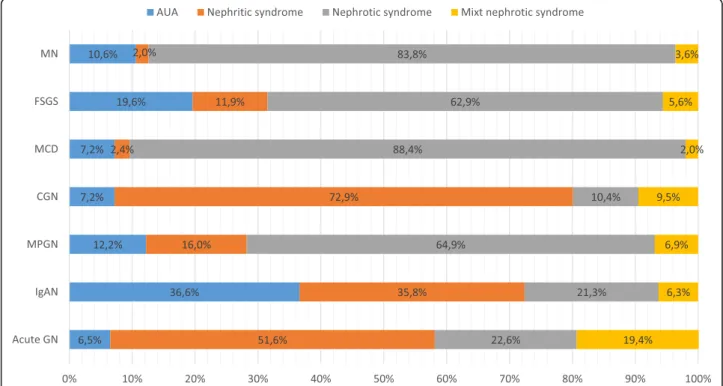
male. Nephrotic syndrome was the most common indi-cation for renal biopsy (52.7%) followed by AUA (18.7%) and nephritic syndrome (18.2%). Figure 1shows the dis-tribution of renal biopsy indications according to the PGDs. The most common PGD was IgAN (25.7%) followed by MN (25.6%) and FSGS (21.9%). Figure 2
shows the distribution of PGDs in our current data and in the previous data published in 2014. While the inci-dence of IgAN and FSGS increased, MN, MPGN and mesangioproliferative GN were found to be decreased. There was no significant change in the frequency of MCD. When the patients were divided into two age groups (Fig.3), the leading etiology was MN in patients aged > 40 years and IgAN in those aged < 40 years. There was no difference in FSGS between these age groups. However, MCD and MPGN were almost twice frequent in patients under 40 years of age compared to those over 40. On the other hand, CGN was almost twice frequent in patients over 40 years of age than in those under 40.
Patients’ data obtained during kidney biopsy are pre-sented in Table1. The data of patients, according to the PGD type, are presented in Table 2. The mean baseline systolic and diastolic blood pressure were 130 ± 20 mmHg and 81 ± 12 mmHg, respectively. About half of the patients (1722 of 3456 patients, 49.8%) had edema at presentation. The median proteinuria was 3300 (IQR: 1467–6307) mg/day, mean serum creatinine, eGFR and
Fig. 1 Indications for renal biopsy according to the PGDs. (Abbreviations: Acute GN: acute glomerulonephritis, AUA: Asymptomatic urinary abnormalities, CGN: crescentic glomerulonephritis, FSGS: Focal segmental glomerulosclerosis, IgAN: IgA nephropathy, MCD: minimal change disease, MN: membranous nephropathy, MPGN: membranoproliferative glomerulonephritis)
albumin values were 1.4 ± 1.5 mg/dL, 80.7 ± 39.1 mL/ min/m2 and 3.2 ± 0.9 g/dL, respectively. Medical history revealed type 1 diabetes mellitus in 0.26% (10 of 3730 patients), type 2 diabetes mellitus 8.7% (327 of 3730 patients), and hypertension in 32.4% (1213 of 3735 patients) of the patients. About 33.7% of the patients
(1308 of 3875 patients) were using a renin-angiotensin-aldosterone-system blocker before the renal biopsy.
Nephrotic syndrome was the most frequent indication for renal biopsy (2002 patients, 51.7%) followed by AUA (709 patients, 18.3%) and nephritic syndrome, including RPGN (691 patients, 17.8%). Biopsy indications for
Fig. 2 Diagnosis of patients according to the renal biopsy results. The data are presented separately for each PDG based on the current study and the data of our group published in 2014 [5]. (Abbreviations: Acute PGN: acute proliferative glomerulonephritis CGN: crescentic
glomerulonephritis, FSGS: Focal segmental glomerulosclerosis, IgAN: IgA nephropathy, MCD: minimal change disease, Mes.PGN: Mesangioproliferative glomerulonephritis (non-IgA), MN: membranous nephropathy, MPGN: membranoproliferative glomerulonephritis)
Fig. 3 Distribution of PDG in age groups < 40 and > 40 years. (Abbreviations: Acute PGN: acute proliferative glomerulonephritis CGN: crescentic glomerulonephritis, FSGS: Focal segmental glomerulosclerosis, IgAN: IgA nephropathy, MCD: minimal change disease, Mes.PGN:
different pathological diagnoses are presented in Table
1. In more than 80% of patients with MN and MCD, the renal biopsy was performed due to nephrotic syndrome. In addition, nephrotic syndrome was the indication for renal biopsy in more than 60% of those with FSGS and type 1 MPGN. In more than half of the patients with IgAN, AUA and nephritic syndrome, including RPGN, was the indication for renal biopsy.
Renal biopsies were performed in departments of nephrology and radiology in 75.4 and 24.5% of patients, respectively. The mean total number of glomeruli per biopsy was 17 ± 10, and the number of globally scler-otic glomeruli per biopsy was 3 ± 4. The biopsies per-formed between 1994 and 2019 were included in the study. However, about half of the data were recorded from patients over the period 2013–2019. The patho-logical diagnoses in patients who had renal biopsy due to nephrotic syndrome were MN, FSGS and MCD, respectively. The most frequent diagnosis was IgAN in patients with AUA.
Examination of urinary sediment revealed microscopic hematuria (> 5 erythrocytes per high power field) in 50.1% (1699 of 3391 and pyuria (> 5 leukocytes per high power field) in 19.2% (645 of 3230) of patients.
Comparison of the data with 2014 data
We compared our patient data in our article published in 2014 [5] with the new patient data added to our data-set from 2014 to 2019. The changes in the rates of PGDs
are given in Fig.4. The changes in the rates of all PGDs, except CGN, were statistically significant. Although nephrotic syndrome continued to be the most common renal biopsy indication, its frequency decreased signifi-cantly (57.8% in 2014, 48.7% in 2019). Moreover, the median age of the patients increased significantly [me-dian (interquartile range-IQR): 39 (29–51) years in 2014 vs. 42.5 (32–54) years in 2019, p < 0.05]. Figure 5 shows the age distribution curve of most frequently diagnosed PGDs (IgAN, MN, and FSGS) according to biopsy indi-cations. The mean age of all these PGDs increased in 2019 compared to the 2014 database (ages according to the databases 2014 and 2019: IgAN: 35.2 ± 12.2 vs. 39.5 ± 13.1; MN: 43.4 ± 14.5 vs. 48.6 ± 14.1; FSGS: 36.0 ± 13.3 42.1 ± 14.2, respectively). When the percentage of the biopsy indication for each PGD between the time pe-riods was analysed (Table 4), biopsy indication in IgAN due to AUA significantly increased from 25.5% in 2014 to 39.8% in 2019, but NS indication in MN was not con-siderably changed (88.2% in 2014 and 81.3% in 2019). The average number of total glomeruli and the number of globally sclerotic glomeruli per biopsy were statisti-cally significantly higher in 2019 data when compared to 2014 data [median (IQR): 16 [7–20] vs. 13 [7–17,21,22] and 1 (0–4) vs. 1 (0–3), respectively].
Discussion
In this study, we presented comprehensive, up-to-date data of the multicentre glomerular diseases study per-formed by the TSN-GOLD Working Group. There were 1274 patients in the first data published in 2014 [5]. The number of cases has increased by approximately three-fold (3875 patients) and has reached one of the highest published PGD data. The most significant change be-tween these data is that the frequency of IgAN increased from 17.2 to 25.7%, and become the most common PGD diagnosed via renal biopsy in Turkey. Another important point is that the frequency of biopsy indication due to AUA and nephritic syndrome significantly increased (from 10.8 to 17.8% and from 16.6 to 18.2%, respect-ively). IgAN was found to be the most common PGD in our patients who underwent kidney biopsy due to AUA (Fig. 1). When these data are combined, it can be inter-preted that the increase in the frequency of IgAN in our patients is mainly related to the change in biopsy indica-tions rather than the increase in the frequency of IgAN. As is known, there are significant regional differences in the prevalence of PGDs.
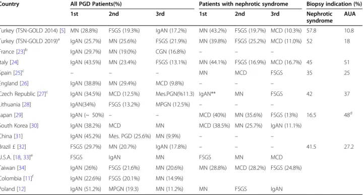
For the comparison of similar international data, we created a table (Table 3) from similar articles reporting the frequency of GN published in different parts of the world. We tried to obtain a more formal table by elimin-ating the secondary GN causes from the data and create an easily understandable and comparable data. However,
Table 1 Patients’ data at the time when kidney biopsy was performed
Mean ± SD
Systolic blood pressure (mmHg) 130 ± 20
Diastolic blood pressure (mmHg) 81 ± 12
BMI (kg/m2) 27.3 ± 9.7
Glucose (mg/dL) 97 ± 31
BUN (mg/dL) 23 ± 19
Creatinine (mg/dL)a 1.0 (0.7–1.6)
eGFR (CKD-EPI, mL/min/1.73m2)a 82.9 (47.0–113.0) The total number of glomeruli (per biopsy) 17 ± 10
Uric acid (mg/dL) 6.2 ± 1.8 Triglyceride (mg/dL)a 175.5 (121.0–256.0) LDL-cholesterol (mg/dL) 163 ± 85 ALT (IU/L)a 17 (13.0–24.0) Calcium (mg/dL) 8.8 ± 0.8 Hemoglobin (g/dL) 12.9 ± 2.1 Serum albumin (g/dL) 3.2 ± 0.9 Proteinuria (mg/day)a 3300 (1467–6307)
BMI Body mass index, eGFR Estimated glomerular filtration rate.a
Median (interquartile range)
Table 2 The data of patients according to the type of PGD MCD (n = 259) FSG S (n = 848) MN (n = 992) IgAN (n = 995) M es. PGN (n = 97) MP GN (n = 349) Acute PGN (n = 31) CGN (n = 244) Age Mean ± SD 36.1 ± 15.4 40 .4 ± 14.2 46.7 ± 14.5 38.4 ± 13 36 .8 ± 13.8 37.3 ± 15 .6 43.1 ± 17.2 48 ± 16.6 Gen der n (%) Wom an 118 (45. 9) 41 5 (48.9 ) 409 (41.3 ) 371 (37.3) 56 (58.9 ) 168 (48.3) 13 (40. 6) 112 (45.5) Creat inine (mg /dL ) Mean ± SD 0.89 ± 0.65 1. 29 ± 1.17 1 ± 0.8 1.55 ± 1.26 0. 97 ± 0.69 1.51 ± 1.41 2.23 ± 2.22 4.1 ± 3.34 eGFR (CKD-EP I) mL/min Mean ± SD 105 ± 33 82 ± 36 94 ± 33 72 ± 36 99 ± 33 77 ± 40 62 ± 43 30 ± 30 Serum alb umin (g /dL) Mean ± SD 2.61 ± 1.03 3. 39 ± 0.93 2.77 ± 0.86 3.85 ± 0.69 3. 52 ± 0.9 3.15 ± 0.89 3.19 ± 0.85 3.24 ± 0. 73 Protei nuri a (mg/ day) Medi an (IQR) 5835 (2400 –9000) 34 00 (177 0– 5740) 6000 (3400 –9280 ) 1822 (1000 –3400) 16 00 (540 –39 00) 3500 (1400 –6120) 2310 (900 –74 35) 2100 (1230 –3690) Hem aturia n n (%) 47 (18.2 ) 27 7 (32.6 ) 288 (29.1 ) 633 (63.7) 42 (43.8 ) 170 (48.7) 22 (68. 8) 191 (77.6) Pyuri a n (%) 31 (13.5 ) 11 3 (15.2 ) 115 (13.2 ) 178 (20.0) 15 (18.8 ) 83 (31.4 ) 8 (28.6 ) 92 (41.1 ) SBP (mmHg) Me an ± SD 122 ± 17 13 1 ± 19 129 ± 19 130 ± 21 12 9 ± 25 132 ± 23 142 ± 23 136 ± 20 DBP (mm Hg) Mean ± SD 76 ± 10 81 ± 11 81 ± 11 82 ± 12 81 ± 15 82 ± 13 84 ± 13 82 ± 11 Abbreviations : Acute PGN acute proliferative glomerulonephritis, CGN crescentic glomerulonephritis, DPB diastolic blood pressure, eGFR estimated glomerular filtration rate, FSGS Focal segmental glomerulosclerosis, IgAN IgA nephropathy, MCD minimal change disease, Mes.PGN Mesangioproliferative glomerulonephritis (non-IgA), MN membranous nephropathy, MPGN membranoproli ferative glomerulonephritis, SPB systolic blood pressure, PGD primary glomerular disease
as can be seen in this table, both the first publication and present data present biopsy results in patients with nephrotic syndrome in our country; biopsy results are more consistent with European data and MN is detected more frequently. However, IgAN is common in Asian countries and FSGS in the United States.
When all PGD patients were included, IgAN became the most common PGD in our country and the rate be-came similar to that in other European and Asian coun-tries. Although IgA deposits may be seen in biopsies of individuals without renal disease, these cases generally do not have clinical features of GN [21, 35], and it is known that differences in the indications for biopsy in management practices across countries can affect the in-cidence and prevalence of diagnosed PGDs [22]. Routine urine screening is performed in some countries such as Japan and Korea. Such applications may allow the detec-tion and biopsy of AUA cases and thus high AUA rates in GN registries. On the other hand, although our data show that the frequency of biopsies due to AUA seems to be increasing (from 10.8 to 17.8%) in our country, it is still relatively low compared to European and Asian countries. For example, the rate of patients undergoing biopsy for AUA is 51% in Italy, 25% in Spain, 37% in the Czech Republic, and 48% in Japan (Table3).
The prevalence of IgAN is higher in Asian cohorts and Caucasians, whereas FSGS is more common in North
America [7–9]. Another finding consistent with these data is that the percentage of patients diagnosed with MN decreased in parallel with the decrease in the pro-portion of patients undergoing biopsy for nephrotic syn-drome indication. There were significant changes in both biopsy indications and age groups between the 2014 and 2019 data, as presented in Figs. 4 and 5. Out data clearly showed that the peak of PGD in our country moved to older age groups. This might suggests aging may cause an increase of IgAN and the decrease of MN. But biopsy indication in IgAN due to AUA significantly increased from 25.5% in 2014 to 39.8% in 2019, and neph-rotic syndrome indication in MN was not considerably changed (88.2% in 2014 and 81.3% in 2019) (Table4). In the last decade, many studies have shown that even pro-teinuria at the level of 0.5-1 g/day has a significant effect on the prognosis of IgAN [10–12] and steroid treatment may have a positive effect on prognosis in such IgAN pa-tients [13–16]. This may be the most important reason for the recent increase in the frequency of renal biopsy for AUA and, hence IgAN, in our country. In addition, the in-crease in the number of nephrologists and the number of centres that can perform a kidney biopsy in our country is likely to play an important role in performing biopsy earl-ier in patients with AUA.
Another important point in our study was the in-creased prevalence of FSGS in all patient groups, more
Fig. 4 The biopsy diagnoses of the patients compared between the participants involved in the 2014 article [5] and newly diagnosed patients after then. The changes in the rates of all PGDs, except CGN, were statistically significant. *Includes only the patients diagnosed after 2014 manuscript. (Abbreviations: MN: membranous nephropathy, FSGS: Focal and segmental glomerulosclerosis, IgAN: IgA nephropathy, MCD: minimal change disease, MPGN: membranoproliferative glomerulonephritis, CGN: crescentic glomerulonephritis)
prominent in patients with nephrotic syndrome (Table
3, Fig. 2). FSGS is the most common cause of nephrotic syndrome in the United States [17–19]. However, there are publications indicating that the incidence of FSGS decreases relatively in biopsy patients due to the in-creased prevalence of diabetic nephropathy [20]. There are similar publications from Brazil (Table 3). In other regions, FSGS is the second most common cause of nephrotic syndrome after MN. In order to explain the increase in the frequency of FSGS in our data, we need more detailed data. However, compared to our previous report, we found that our rate of patients with diabetes (possibly also with high body mass index) and hyperten-sion has increased (from 6 to 10% and from 26 to 32.4%, respectively) and this might have eventually increased the frequency of secondary FSGS. Therefore, it cannot be ignored that some of these secondary FSGS patients might have been registered as having primary FSGS in our database.
MN was more common in patients over 40 years of age (Fig.3). This finding is compatible with many Euro-pean data [24–26]. On the other hand, IgAN was the most common PGD in the group under 40 years of age.
This finding is consistent with European and Asian data, as well as the United States and Brazilian data (where FSGS was the most common PGD when all patients were included) [17,18,32].
There are some limitations of this study. We included only the data obtained at the time of renal biopsy. As we are currently collecting data on our long-term results, we have not included these data (renal outcomes, treat-ments, etc.) in this study. Misclassification due to differ-ent definition or measuremdiffer-ent variables could be a problem in all centres. Since we excluded patients with-out light microscopy and IF findings, errors in the diag-nosis of glomerular diseases such as FSGS, which are likely to have such diagnostic confusion, were mini-mized. For this purpose, we excluded more than 500 pa-tients. In addition, the identity information and centre information of each patient registered in the database were recorded anonymously in order to minimize the patient selection bias. Another point is, as in every retro-spective study, there were patients who lacked some data. Also, although we asked the centers to include all their patients’ data complying with the inclusion criteria, there might have been some patients that were recorded
Fig. 5 Age distribution curves of major PGDs according to the biopsy period and biopsy indication. 2019 dataset includes only the data of the patients diagnosed after 2014 manuscript. The mean age of all these PGDs increased in 2019 compared to the 2014 database (ages according to the databases 2014 and 2019: IgAN: 35.2 ± 12.2 vs. 39.5 ± 13.1; MN: 43.4 ± 14.5 vs. 48.6 ± 14.1; FSGS: 36.0 ± 13.3 42.1 ± 14.2,
respectively).(Abbreviations: MN: membranous nephropathy, FSGS: Focal segmental glomerulosclerosis, IgAN: IgA nephropathy, NS: Nephrotic syndrome)
or data that some centres failed to input to the database. The biopsy specimens were examined only by local pa-thologists and the diagnosis could not be confirmed by an-other pathologist. However, all these limitations are common problems in most recording systems. The sub-types of FSGS and MPGN were not specified because pathology reports do not routinely include FSGS subtypes, and the new classification of MPGN, according to IF find-ings, have recently begun to be included in renal biopsy reports in our country. Therefore, we did not present these subgroups data. On the other hand, only PGDs were included in our study, and the database has been specially
prepared for it. This is the major advantage of the present study because previous epidemiological studies involved both primary and secondary glomerular diseases. Eventu-ally, we assume that our data has the power to represent PGD patients across the country.
Conclusions
The distribution of PGDs in Turkey has become similar to that in other European countries. IgAN diagnosed with renal biopsy has become more prevalent compared to MN, becoming the most common PGD in Turkey.
Table 3 Similar data from some of the published primary glomerulonephritis studies generated from international data
Country All PGD Patients(%) Patients with nephrotic syndrome Biopsy indication (%)
1st 2nd 3rd 1st 2nd 3rd Nephrotic
syndrome AUA Turkey (TSN-GOLD 2014) [5] MN (28.8%) FSGS (19.3%) IgAN (17.2%) MN (43.2%) FSGS (19.7%) MCD (10.3%) 57.8 10.8 Turkey (TSN-GOLD 2019)a IgAN (25.7%) MN (25.6%) FSGS (21.9%) MN (39.8%) FSGS (25.2%) MCD (11.0%) 52 18
France [23]b IgAN (29.7%) MN (19.0%) CGN (16.8%) – – –
Italy [24] IgAN (43.5%) MN (23.4%) FSGS (13.1%) MN (44.1%) FSGS (16.9%) MCD (16.7%) 45 51
Spain [25]c – – – MN MCD FSGS 35 25
England [26] IgAN (38.8%) MN (29.4%) MCD (9.8%) – – –
Czech Republic [27]c IgAN (34.5%) MCD (12.5%) Mes.PGN(%11.3) IgAN** MN FSGS 42 37
Lithuania [28] IgAN(34%) FSGS (13.2%) MPGN (12.5%) – – –
Japan [29] IgAN (~ 50%) – – MCD (40%) MN (35.6%) FSGS (13%) 16.5 48d
South Korea [30] IgAN (38.2%) MCD MN MCD (38.5%) MN (25.7%) IgAN (11.1%)
China [31] IgAN (45.2%) Mes. PGD (25.6%) MN (9.9%) – – –
Brazil £ [32] FSGS (29.7%) MN (20.7%) IgAN (17.8%) – – – 41.5 27.2
U.S.A. [18,33]e FSGS IgAN MN FSGS MN MCD
Taiwan [34] IgAN (26%) FSGS (21.6%) MN (20.6%) MN (28.8%) MCD (28.2%) FSGS (24.8%)
Colombia [11]f IgAN (22.6%) FSGS (20.1%) MN (14.9%)
Poland [12] IgAN (51.2%) MPGN (19.3) MN (11.2%) MN FSGS IgAN
Abbreviations: AUA Asymptomatic urinary abnormalities, CGN crescentic glomerulonephritis, FSGS Focal segmental glomerulosclerosis, IgAN IgA nephropathy, MCD minimal change disease, Mes.PGN Mesangioproliferative glomerulonephritis (non-IgA), MN membranous nephropathy, MPGN membranoproliferative
glomerulonephritis, PGD primary glomerular disease, TSN-GOLD Turkish Society of Nephrology Glomerular Diseases
a
Current study;b
cumulative data of different age and periods,c
including pediatrics cases,d
This percentage was presented as the percentage of nephritic syndrome, but asymptomatic urinary abnormalities seem to be involved also.eThese results are the combination of two different studies [9,10],fincluding secondary cases
Table 4 The percentage of the biopsy indication for each PGD according to the biopsy period
2014 Data (%) 2019 Data (%)
AUA Mixt NS Nephritic syndrome NS AUA Mixt NS Nephritic syndrome NS
IgAN 25.5 9.5 37.0 28.0 39.8 5.3 35.5 19.4 FSGS 14.7 8.0 13.8 63.6 21.5 4.7 11.1 62.7 MN 5.2 3.8 2.9 88.2 13.7 3.5 1.5 81.3 MCD 2.7 2.7 1.4 93.2 9.0 1.7 2.8 86.4 MPGN 7.3 8.7 15.3 68.7 16.6 5.3 16.6 61.5 CGN 5.1 10.2 71.2 13.6 8.0 9.3 73.5 9.3
Abbreviations: AUA asymptomatic urinary abnormalities, NS Nephrotic syndrome, IgAN IgA nephropathy, FSGS Focal segmental glomerulosclerosis, MN membranous nephropathy, MCD minimal change disease, CGN crescentic glomerulonephritis
MN is still the most common cause of nephrotic syn-drome among PGDs.
Abbreviations
Acute PGN:Acute proliferative glomerulonephritis; AUA: Asymptomatic urinary abnormalities; CGN: Crescentic glomerulonephritis; CKD: Chronic kidney disease; CKD-EPI: Chronic kidney disease epidemiology collaboration; ERA-EDTA: European Dialysis and Transplant Association; ESRD: End-stage renal disease; IF: Immunofluorescence microscopy; IgAN: IgA nephropathy; FSGS: focal segmental glomerulosclerosis; GN: Glomerulonephritis; MCD: Minimal change disease; Mes.PGN: Mesangioproliferative glomerulonephritis (non-IgA); MN: Membranous nephropathy;
MPGN: Membranoproliferative glomerulonephritis; PGD: Primary glomerular disease; PGDs: Primary glomerular diseases; TSN: Turkish Society of Nephrology; TSN-GOLD: Turkish Society of Nephrology Glomerular Diseases; USRDS: United States Renal Data System
Acknowledgments Not applicable.
Ethics approval and informed consent
The Ethics Committee of Istanbul University, Istanbul Medical Faculty approved the study. Due to the retrospective nature of the study, informed consent was not received.
Authors’ contributions
Concept - A.T., A.S., E.C., H.Y., N.E., N.S., K.D., F.D., U.D., A.U., G.S., M.S., M.G., E.T., B. D, S.S., M.Y., G.S., S.U., O.G., S.K., Z.B.B, I.S., I.K., K.T., Z.Y., R.T.K., C.C., F.C., Z.A., D.D.O., C.B.G., M.A., S.P., D.D.T., M.K., S.T., K.T., L.K., S.S., M.D., S.B., L.A., I.H.A., A.A., A.R.O., G.M.S., S.O.; Design - A.T., A.S., E.C., H.Y., N.E., N.S., K.D., F.D., U.D., A.U., G.S., M.S., M.G., E.T., B. D, S.S., M.Y., G.S., S.U., O.G., S.K., Z.B.B, I.S., I.K., K.T., Z.Y., R.T.K., S.O.; Supervision -A.T., S.O., M.A.; Resources- A.S., E.C., N.E., H.Y., O.G.; Data Collection and/or Processing:; A.T., A.S., E.C., H.Y., N.E., N.S., K.D., F.D., U.D., A.U., G.S., M.S., M.G., E.T., B. D, S.S., M.Y., G.S., S.U., O.G., S.K., Z.B.B, I.S., I.K., K.T., Z.Y., R.T.K., C.C., F.C., Z.A., D.D.O., C.B.G., M.A., S.P., D.D.T., M.K., S.T., K.T., L.K., S.S., M.D., S.B., L.A., I.H.A., A.A., A.R.O., G.M.S., S.O. Analysis and/or Interpretation -A.T., A.S., C.C., F.C., Z.A., D.D.O., C.B.G., M.A., S.P., D.D.T., M.K., S.T.,S.O.; Literature Search- A.S., E.C., N.E., H.Y., O.G., K.T., L.K., S.S., M.D., S.B., L.A., I.H.A., A.A., A.R.O., G.M.S.; Writing Manuscript -A.S., S.O. All authors read and approved the manuscript.
Funding
Financial supports (establishing the registry database) have been obtained from Turkish Society of Nephrology and logistic support from the authors’ affiliation departments.
Availability of data and materials
We created a database for primary glomerular diseases with the name of ‘Turkish Society of Nephrology Glomerular Diseases Working Group (TSN-GOLD) in 04.04.2008. We published the first results of our study in 2014 and now, we present here the extended results. The data can be found at the following address:
http://pgh.tsn.org.tr/login.php
Consent for publication Not applicable.
Competing interests
The authors declare that they have no competing interests.
Author details
1Istanbul Medical Faculty, Nephrology, Istanbul University, Istanbul, Turkey. 2Medical Faculty, Nephrology, Istanbul Medipol University, Istanbul, Turkey. 3
Haseki Training and Research Hospital, Nephrology, Istanbul, Turkey. 4Medical Faculty, Nephrology, Kocaeli University, Kocaeli, Turkey.5Cerrahpasa Medical Faculty, Nephrology, Istanbul University, Istanbul, Turkey.6Medical Faculty, Nephrology, Uludag University, Bursa, Turkey.7Ankara Numune Training and Research Hospital, Nephrology, Ankara, Turkey.8Medical Faculty, Nephrology, Gazi University, Ankara, Turkey.9Hamidiye Sisli Etfal Training and Research Hospital, Nephrology, Istanbul, Turkey.10Medical Faculty, Nephrology, Eskisehir Osmangazi University, Eskisehir, Turkey.11Medical
Faculty, Nephrology, Erciyes University, Kayseri, Turkey.12Sultan Abdulhamit Han Training and Research Hospital, Nephrology, Istanbul, Turkey.13Izmir Bozyaka Training and Research Hospital, Nephrology, Izmır, Turkey.14Medical Faculty, Nephrology, Pamukkale University, Denizli, Turkey.15Medical Faculty, Nephrology, Sakarya University, Adapazari, Sakarya, Turkey.16Bakirkoy Sadi Konuk Training and Research Hospital, Nephrology, Istanbul, Turkey. 17Medical Faculty, Nephrology, Akdeniz University, Antalya, Turkey.18Medical Faculty, Nephrology, Afyonkarahisar University, Afyon, Turkey.19Medical Faculty, Nephrology, Sutcuİmam University, Kahramanmaras, Turkey. 20Medical Faculty,İbni Sina Hospital, Nephrology, Ankara University, Ankara, Turkey.21Dr. Lutfi Kirdar Kartal Training and Research Hospital, Nephrology, Istanbul, Turkey.22Medical Faculty, Nephrology, Inonu University, Malatya, Turkey.23Medical Faculty, Nephrology, Trakya University, Edirne, Turkey. 24Meram Medical Faculty, Nephrology, Necmettin Erbakan University, Konya, Turkey.25Medical Faculty, Nephrology, Dicle University, Diyarbakir, Turkey. 26
Medical Faculty, Nephrology, Bezmialem Vakif University, Istanbul, Turkey. 27Medical Faculty, Nephrology, Dokuz Eylul University, Izmir, Turkey. 28Medical Faculty, Nephrology, Cumhuriyet University, Sivas, Turkey.29Darica Farabi Training and Research Hospital, Nephrology, Kocaeli, Turkey.30Doktor Burhan Nalbantoglu State Hospital, Lefkosa, Cyprus.31Bursa Yuksek Ihtisas Training and Research Hospital, Nephrology, Bursa, Turkey.32Medical Faculty, Nephrology, Hacettepe University, Ankara, Turkey.33Medical Faculty, Nephrology, Cukurova University, Adana, Turkey.34Kocaeli State Hospital, Nephrology, Kocaeli, Turkey.35Okmeydani Training and Research Hospital, Nephrology, Istanbul, Turkey.36Istanbul Training and Research Hospital, Nephrology, Istanbul, Turkey.37Medical Faculty, Nephrology, Mersin University, Mersin, Turkey.38Taksim Training and Research Hospital, Nephrology, Istanbul, Turkey.39Medical Faculty, Nephrology, Baskent University, Ankara, Turkey.40Ankara Training and Research Hospital, Nephrology, Ankara, Turkey.41Batman State Hospital, Nephrology, Batman, Turkey.42Selcuk Medical Faculty, Nephrology, Selcuk University, Konya, Turkey.43Marmara University, Medical Faculty, Nephrology, Istanbul, Turkey. 44Balikesir Training and Research Hospital, Nephrology, Balikesir, Turkey. 45Goztepe Training and Research Hospital, Nephrology, Istanbul, Turkey.
Received: 15 May 2020 Accepted: 29 October 2020
References
1. The Registry of The Nephrology, Dialysis And Transplantation In Turkey. 2018.http://www.nefroloji.org.tr/folders/file/REGISTRY_2018pdf. Accessed 9 Feb 2020. .
2. ERA-EDTA Registry. ERA-EDTA Registry Annual Report 2017. Amsterdam: Amsterdam UMC, location AMC DoMI; 2019.
3. United States Renal Data System. 2018 USRDS annual data report: epidemiology of kidney disease in the United States. National Institutes of Health NIoDaDaK.
4. Xu X, Wang G, Chen N, Lu T, Nie S, Xu G, et al. Long-term exposure to air pollution and increased risk of membranous nephropathy in China. J Am Soc Nephrol. 2016;27(12):3739–46.
5. Ozturk S, Sumnu A, Seyahi N, Gullulu M, Sipahioglu M, Artan S, et al. Demographic and clinical characteristics of primary glomerular diseases in Turkey. Int Urol Nephrol. 2014;46(12):2347–55.
6. Inker LA, Schmid CH, Tighiouart H, Eckfeldt JH, Feldman HI, Greene T, et al. Estimating glomerular filtration rate from serum creatinine and cystatin C. N Engl J Med. 2012;367(1):20–9.
7. McGrogan A, Franssen CFM, de Vries CS. The incidence of primary glomerulonephritis worldwide: a systematic review of the literature. Nephrol Dialysis Transplantation. 2011;26(2):414–30.
8. Galla JH. IgA nephropathy. Kidney Int. 1995;47(2):377–87. 9. Floege J, Amann K. Primary glomerulonephritides. Lancet (London,
England). 2016;387(10032):2036–48.
10. Le W, Liang S, Hu Y, Deng K, Bao H, Zeng C, et al. Long-term renal survival and related risk factors in patients with IgA nephropathy: results from a cohort of 1155 cases in a Chinese adult population. Nephrol Dialysis Transplantation. 2012;27(4):1479–85.
11. Maixnerova D, Bauerova L, Skibova J, Rysava R, Reiterova J, Merta M, et al. The retrospective analysis of 343 Czech patients with IgA nephropathy--one Centre experience. Nephrol Dialysis Transplantation. 2012;27(4):1492–8.
12. Reich HN, Troyanov S, Scholey JW, Cattran DC, Toronto GR. Remission of proteinuria improves prognosis in IgA nephropathy. J Am Soc Nephrol. 2007;18(12):3177–83.
13. Pozzi C, Bolasco PG, Fogazzi GB, Andrulli S, Altieri P, Ponticelli C, et al. Corticosteroids in IgA nephropathy: a randomised controlled trial. Lancet (London, England). 1999;353(9156):883–7.
14. Manno C, Torres DD, Rossini M, Pesce F, Schena FP. Randomized controlled clinical trial of corticosteroids plus ACE-inhibitors with long-term follow-up in proteinuric IgA nephropathy. Nephrol Dialysis Transplantation. 2009; 24(12):3694–701.
15. Locatelli F, Del Vecchio L, Pozzi C. The patient with IgA glomerulonephritis--what is the role of steroid treatment? Nephrol Dialysis Transplantation. 1999;14(5):1057–60.
16. Lv J, Xu D, Perkovic V, Ma X, Johnson DW, Woodward M, et al. Corticosteroid therapy in IgA nephropathy. J Am Soc Nephrol. 2012;23(6): 1108–16.
17. Kitiyakara C, Eggers P, Kopp JB. Twenty-one-year trend in ESRD due to focal segmental glomerulosclerosis in the United States. Am J Kidney Dis. 2004; 44(5):815–25.
18. Haas M, Meehan SM, Karrison TG, Spargo BH. Changing etiologies of unexplained adult nephrotic syndrome: a comparison of renal biopsy findings from 1976-1979 and 1995-1997. Am J Kidney Dis. 1997;30(5): 621–31.
19. Hommos MS, De Vriese AS, Alexander MP, Sethi S, Vaughan L, Zand L, et al. The incidence of primary vs secondary focal segmental Glomerulosclerosis: a Clinicopathologic study. Mayo Clin Proc. 2017;92(12):1772–81. 20. O'Shaughnessy MM, Hogan SL, Poulton CJ, Falk RJ, Singh HK, Nickeleit V,
et al. Temporal and demographic trends in glomerular disease
epidemiology in the southeastern United States, 1986-2015. Clin J Am Soc Nephrol. 2017;12(4):614–23.
21. Suzuki K, Honda K, Tanabe K, Toma H, Nihei H, Yamaguchi Y. Incidence of latent mesangial IgA deposition in renal allograft donors in Japan. Kidney Int. 2003;63(6):2286–94.
22. Donadio JV, Grande JP. IgA nephropathy. N Engl J Med. 2002;347(10): 738–48.
23. Simon P, Ramee M-P, Boulahrouz R, Stanescu C, Charasse C, Ang KS, et al. Epidemiologic data of primary glomerular diseases in western France. Kidney Int. 2004;66(3):905–8.
24. Gesualdo L, Di Palma AM, Morrone LF, Strippoli GF, Schena FP. Italian immunopathology group ISoN. The Italian experience of the national registry of renal biopsies. Kidney Int. 2004;66(3):890–4.
25. Rivera F, López-Gómez JM, Pérez-García R. Spsnish registry of G. frequency of renal pathology in Spain 1994-1999. Nephrol Dialysis Transplantation. 2002;17(9):1594–602.
26. Davison AM. The United Kingdom Medical Research Council's glomerulonephritis registry. Contrib Nephrol. 1985;48:24–35.
27. Rychlík I, Jancová E, Tesar V, Kolsky A, Lácha J, Stejskal J, et al. The Czech registry of renal biopsies. Occurrence of renal diseases in the years 1994-2000. Nephrol Dialysis Transplantation. 2004;19(12):3040–9.
28. Brazdziute E, Miglinas M, Gruodyte E, Priluckiene J, Tamosaitis A, Bumblyte IA, et al. Nationwide renal biopsy data in Lithuania 1994-2012. Int Urol Nephrol. 2015;47(4):655–62.
29. Sugiyama H, Yokoyama H, Sato H, Saito T, Kohda Y, Nishi S, et al. Japan renal biopsy registry: the first nationwide, web-based, and prospective registry system of renal biopsies in Japan. Clin Exp Nephrol. 2011;15(4): 493–503.
30. Chang JH, Kim DK, Kim HW, Park SY, Yoo T-H, Kim BS, et al. Changing prevalence of glomerular diseases in Korean adults: a review of 20 years of experience. Nephrol Dialysis Transplantation. 2009;24(8):2406–10.
31. Li L-S, Liu Z-H. Epidemiologic data of renal diseases from a single unit in China: analysis based on 13,519 renal biopsies. Kidney Int. 2004;66(3):920–3. 32. Malafronte P, Mastroianni-Kirsztajn G, Betônico GN, Romão JE Jr, Alves MAR, Carvalho MF, et al. Paulista registry of glomerulonephritis: 5-year data report. Nephrol Dialysis Transplantation. 2006;21(11):3098–105.
33. Nair R, Walker PD. Is IgA nephropathy the commonest primary glomerulopathy among young adults in the USA? Kidney Int. 2006; 69(8):1455–8.
34. Chiu H-F, Chen H-C, Lu K-C, Shu K-H. Taiwan Society of N. Distribution of glomerular diseases in Taiwan: preliminary report of National Renal Biopsy
Registry-publication on behalf of Taiwan Society of Nephrology. BMC nephrology. 2018;19(1):6.
35. Waldherr R, Rambausek M, Duncker WD, Ritz E. Frequency of mesangial IgA deposits in a non-selected autopsy series. Nephrol Dialysis Transplantation. 1989;4(11):943–6.
Publisher’s Note
Springer Nature remains neutral with regard to jurisdictional claims in published maps and institutional affiliations.
![Fig. 2 Diagnosis of patients according to the renal biopsy results. The data are presented separately for each PDG based on the current study and the data of our group published in 2014 [5]](https://thumb-eu.123doks.com/thumbv2/9libnet/5456162.105185/4.892.92.807.131.467/diagnosis-patients-according-results-presented-separately-current-published.webp)
![Fig. 4 The biopsy diagnoses of the patients compared between the participants involved in the 2014 article [5] and newly diagnosed patients after then](https://thumb-eu.123doks.com/thumbv2/9libnet/5456162.105185/7.892.86.809.129.527/diagnoses-patients-compared-participants-involved-article-diagnosed-patients.webp)