Journal section: Oral Surgery Publication Types: Research
Posıtıve effect of platelet rich fibrin on osseointegration
Elif Öncü 1, Burak Bayram 2, Alpdoğan Kantarcı 3, Serap Gülsever 2, Emine-Elif Alaaddinoğlu 4
1 Necmettin Erbakan University, Departmant of Periodontolgy, Konya, Turkey
2 Baskent University, Departmant of Oral and Maxillofacial Surgery, Ankara, Turkey
3 Department of Applied Oral Sciences, Center for Periodontology, The Forsyth Institute, Cambridge, MA, USA
4 Baskent University, Departmant of Periodontolgy, Ankara, Turkey
Correspondence:
Karaciğan Mah.Ankara
cad, Necmettin Erbakan Üniversitesi Diş Hekimliği Fakültesi Karatay Konya- Turkey
Received: 25/08/2015 Accepted: 23/03/2016
Abstract
Background: Leukocyte-platelet rich fibrin (L-PRF) is a second generation platelet concentrate clinically used to accelerate tissue healing and bone regeneration. Achieving reduced implant osseointegration time could provide immediate or early loading of implants. The aim of this study was to evaluate the L-PRF-induced osseointegration and bone-implant contact (BIC) in an experimental animal model.
Material and Methods: Twelve 4-month-old New Zealand white rabbits were used. Following general anesthesia, 3-5 mL of blood was obtained from the central artery in rabbit ear and L-PRF was prepared. Two implant cavi-ties (5 mm long and 3 mm in diameter) were created in each tibia with a total of four cavicavi-ties in each animal. Two of these cavities were selected and covered with PRF (test group). The remaining L-PRF was used to soak the implants placed into the L-PRF covered sockets. Other cavities were left as controls. In total, 48 implants were placed. Animals were sacrificed after two, three, or four weeks. Histological samples were obtained and peri-implant tissues were histomorphometrically evaluated for bone-to-peri-implant contact and new bone formation. Results: Histomorphometric analyses of the defects revealed that the L-PRF was detectable up to the second week. Application of L-PRF increased the rate and amount of new bone formation in the experimental group compared to the control group. Bone-to-implant contact was enhanced when the surface was pre-wetted with L-PRF (p<0.01).
Conclusions: The results of this study demonstrated that L-PRF application may increases amount and rate of new bone formation during the early healing period and provides a faster osseointegration around implants.
Key words: Dental implants, platelet rich fibrin, osseointegration, bone regeneration, matrix for growth factors.
Öncü E, Bayram B, Kantarcı A, Gülsever S, Alaaddinoğlu EE. Posıtıve effect of platelet rich fibrin on osseointegration. Med Oral Patol Oral Cir Bucal. 2016 Sep 1;21 (5):e601-7.
http://www.medicinaoral.com/medoralfree01/v21i5/medoralv21i5p601.pdf
Article Number: 21026 http://www.medicinaoral.com/
© Medicina Oral S. L. C.I.F. B 96689336 - pISSN 1698-4447 - eISSN: 1698-6946 eMail: [email protected]
Indexed in:
Science Citation Index Expanded Journal Citation Reports Index Medicus, MEDLINE, PubMed Scopus, Embase and Emcare Indice Médico Español
doi:10.4317/medoral.21026
Introduction
Osseointegration of dental implants is critical for the long-term success and stability. Various strategies have been used to accelerate the time required for osseointe-gration without compromising the mechanical out-comes and tissue integration (1,2). The implant surface topography plays a key role in the early stages of bone-to-implant contact (BIC); peri-implant bone forma-tion depends on the healing capacity of the bone (2,3). Modification of the surface properties of the implants by chemical methods such as incorporation of inorganic phases on or into the titanium oxide layer and physical enhancement of the materials by increasing the level of roughness have successfully increased the BIC (3). An-other strategy to reduce the osseointegration time has been the modulation of the healing response after the implant placement (2,3). This has been accomplished by biologically active molecules during implant place-ment to induce osteoconductivity, increase osteoblas-tic differentiation and enhance healing of peri-implant bone (3,4). Growth factors, bone-specific proteins, bone morphogenetic proteins induce better osseointegration through replication and differentiation of osteoprogeni-tor cells and interfacial tissue maturation (3-8).
Platelet-based preparations from patient’s own blood provide an inexpensive alternative to commercially available bioactive materials. Activated platelets secrete a wide range of proteins and growth factors including, Bone Morphogenetic Protein (BMP), Platelet-Derived Growth factor (PDGF), Insulin - like Growth Factor (IGF), Vascular Endothelial Growth Factor (VEGF), Transforming Growth Factor-β1 (TGF-β1) and Trans-forming Growth Factor-β2 (TGF-β2), which play key roles in bone healing (5-9). They attract undifferentiated mesenchymal cells to the injured site and facilitate an-giogenesis, chemotaxis and cell proliferation. Growth factors also control the synthesis and degradation of extracellular matrix proteins, enhance osteogenesis and potentially accelerate peri-implant wound healing and osteointegration (8-11).
Leukocyte-Platelet rich fibrin (L-PRF) is a second gen-eration of autologous platelet concentration and a fibrin mesh consisting of leukocytes, growth factors, proteins and cytokines (9). L-PRF, platelet rich plasma (PRP) and platelet rich growth factor (PRGF) are structurally different materials. L-PRF has advantages over PRP and PRGF by having a strong fibrin structure and not requiring any biochemical modification through bovine thrombin or anticoagulants (12,13). L-PRF has a very significant slow sustained release of key growth fac-tors for at least 1 week and up to 28 days, which stimu-lates its environment for a significant time during early phases of wound healing (10-14). Because of its natural fibrin framework properties, growth factors can keep their activity for a relatively longer period and promote
tissue regeneration (15). When L-PRF was applied to the titanium implant surfaces, growth factors covered the implants forming a fibrin layer for platelets to ad-here (5-8,15). Recently evaluated the effect of PRP and PRF on proliferation and differentiation of rat osteo-blasts. This study showed that PRF released autologous growth factors gradually and expressed stronger and more durable effect on proliferation and differentiation of rat osteoblasts than PRP in vitro (14,15).
Yet, in vivo justification for L-PRF application during implant placement and its impact on osseointegration is limited. Therefore, we have hypothesized that imple-mentation L-PRF around dental implants may lead to faster healing rates in peri-implant bone and decrease osseointegration time. In order to test this hypothesis, we have used a rabbit model and studied the effects of L-PRF on new bone formation and BIC around dental implants.
Material and Methods
- Animal Model, Preparation of the L-PRF and Surgical Procedure
The research protocol was submitted to and approved by the Institutional Animal Care and Use Committee and ethical board of animal investigations. Twelve 4-month old New Zealand white rabbits with an average weight of 3.0-3.5 kg were used. Each rabbit was individually caged and fed. General anesthesia was induced by intra-muscular injection of a combination of 0.5 mL ketamine (20 mg/kg by body weight; Ketalar, Eczacıbası, Istanbul, Turkey) and 0.5 mL xylazine (10 mg/kg by body weight; Rompun, Bayer, Leverkusen, Germany). After general anesthesia, 3 to 5 mL of blood was obtained from the central artery of the ear. Samples were collected in 9 mL glass-coated plastic tubes without anti-clotting agent (Becton Dickinson Vacutainer, Franklin Lakes, NJ, USA) and immediately centrifuged at 2700 rpm for 12 min-utes with a table centrifuge (PC-02, Process Ltd., Nice, France; Fig. 1A). The leukocyte-rich fibrin clot formed in the middle part of the tube was taken and remnants of red blood cells were scraped off with gauze. The clot was then transferred to the PRF box (Process Ltd., Nice, France), compressed and PRF membranes were obtained (Fig. 1B). Serum obtained during compression of the fi-brin clot was transferred to a syringe.
Distal surfaces of right and left tibias were shaved and disinfected with a povidine-iodine solution. Local anesthesia was accomplished (Ultracaine D-S®, Hoechst A.G, Turkey). Surgical procedures were performed by the same surgeon (E.Ö.). After a crestal incision, the muscles were dissected and a sharp subperiosteal dissection was used to reflect the periosteum to expose the tibia bone. Two implant site preparation, which were 5-mm apart from each other, were prepared. Mini implants specially produced for application in rabbit
tibia were used (SLA surface; Nucleoss, Izmir, Turkey). Two implants in left tibia were used as “test” and other two implants in the right tibia of the same animal were assigned as “control”. A total of 48 implants (3 mm width and 5 mm length) were used. L-PRF membranes were applied to test sockets (Fig. 1C). Test implants were thoroughly soaked with L-PRF before implant insertion (Fig. 1D) and placed (Fig. 1E). Control implants were placed without the PRF application (Fig. 1F). The periosteum and skin were closed in layers using 5-0 resorbable sutures (Fig. 1G).
All animals were postoperatively medicated with in-tramuscular amoxicillin (1.5 mg/kg body weight) for infection control and buprenorphine (10 mg) for pain relief. Animals were sacrificed on 2nd, 3rd and 4th weeks post-surgery and histological samples were obtained (Fig. 2).
- Histological Analyses
Samples were fixed in neutral buffered formalin, dehy-drated in 70%, 90%, 95% and 100% alcohol and embed-ded in a blue light-curing resin (Technovit 7200 VLC; Kulzer, Wehrheim, Germany) for 48 hours. After the dehydration, each specimen was embedded in methyl-methacrylate. Samples were then sectioned along the long axis of the implant using high-speed rotational
microtome (Micromet, Remet, Italy). Finally, the sec-tions were ground to a thickness of 40-µm and then were stained with 1% toluidine blue. Histological ex-amination was made with Zeiss Axiovert 200 micro-scope (Carl Zeiss inverted micromicro-scope for transmitted light and epifluorescence, Germany), which was con-nected to a computer and photomicrographs were taken with a under UV light with a same microscope. Before performing image analysis, a specific region of inter-est (ROI) had been determined. The proportion of new bone formation related areas of bone detected inside the ROI divided by the total surface area and analyzed the effect of L-PRF on bone formation, the newly formed bone on the surface of the implant and BIC were evalu-ated. The total amount of new bone was calculated ac-cording to percentage of the total region on the surface of the implant. The bone-to-implant contact was calcu-lated by the percentage of the bony contact to the im-plant surface.
- Statistical Analysis
Statistical computations were carried out using IBM PASW/SPSS software (v.18.0.0 2009, IBM Corpora-tion, Somers, NY, USA). Implants were included in the statistical analysis as independent values. Mean values and standard deviations were calculated for each vari-able and group. The difference between groups was an-alyzed with ANOVA and the difference within groups was analyzed with Student t-test.
Results
All implants successfully healed without any postop-erative complications. No mortality, infection or wound exposure or change in body weight was noted.
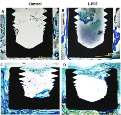
Histomorphometric analyses of the defects revealed that the L-PRF was detectable up to the second week (Fig. 2D,3B). In the test group, L-PRF around implants was obvious on the third and fourth weeks with a substantial new bone formation. At the end of 4 weeks, new bone formation as stained with toluidine blue around implant was limited with almost no bone contact on the implant surface in the control group (Fig. 3C). Widely stained bone formation and almost complete healing around implants were observed in the L-PRF group (Fig. 3D). Application of L-PRF increased the rate and amount of new bone formation in the experimental group com-pared to the control group (31.7% ± 6.8% vs 12.1% ± 7.7% at third week and 39.9% ± 3.9% vs 26.4% ± 9.0% at fourth week, p<0.01).
There was a rapid increase in the percentage of new bone formation and BIC in the test group (Fig. 4). Bone-to-implant contact was enhanced when the surface was pre-wetted with L-PRF (52.6% ± 21.7% vs 36.0% ± 23.2% at third week and 54.6% ± 5.2% vs 39.0% ± 8.9% at fourth week p<0.01).
There was no evidence for fibrotic tissue layer formation Fig. 1.Study design and sequence of events. Panel A. L-PRF clot
in the middle of the tube, Panel B. Fibrin clots transferred to L-PRF Box, Panel C. The implants were washed completely, Panel D. L-PRF membrane placed in implant socket, Panel E. Implants placed in sockets which coated with L-PRF, Panel F. Implants placed in sockets in control group, Panel G. The periosteum and skin were closed in a layer using 5-0 vicryl resorbable sutures.
in any groups. The percentage of new bone formation around the implants in the test group was significantly higher than the control group (P < 0.01). Likewise, the BIC was significantly higher in the test group than the control group on the third and fourth weeks (P < 0.01). Discussion
We have investigated the potential effects of L-PRF on stimulating bone regeneration and accelerating the os-seointegration of dental implants. The histomorphomet-ric results showed the rate of bone formation and BIC were enhanced in the L-PRF treated sockets compared to the empty sockets after the implant placement. In this study mean bone-to-implant contact in the 4th week was 54.61% in the experimental group and 26.44% in the control group. The findings of this study were in agreement with the results reported by Anitua et al. Us-ing a similar approach, these authors reported a 51% BIC when implants were coated with PRGF compared to the 22% BIC of the control group after 8 weeks of healing
time (5-8). Our results also confirm the data presented by Lee et al. who have reported that the mean new bone formation was 29% in the experimental group and 11% in the control group with a BIC of 39% vs 17% (16). Fur-thermore, our results support the findings by Fontana et
al. and those of another study where PRP improved BIC
for roughened implants inserted in rat tibias (11,15). Us-ing a similar approach, Fuerst et al. reported a 55% BIC when implants were coated with PRGF versus 39% of the control group after 4 weeks of healing time (17). In another rabbit study, histomorphometric results showed that during the first 2-3 weeks, integration of tita-nium mini-screws and bones was weak but after 4 weeks of healing integration of titanium mini-screws and bones was significantly stronger. They stated that four weeks is a critical time point during the progress of integration of titanium mini-screws and bones (18). In this study there is virtually no difference in BIC in the control group be-tween 2, 3 and 4 weeks, these results may have been due to the lack of sufficient time for osseointegration in the Fig. 2. Second, third and fourth weeks macroscopic view of the test and
control groupsafter sacrifice. Panel A. The image of the control group at 2nd week, Panel B. The image of the control group at 3rdweek, Panel C.The im-age of the control group at 4th week, Panel D.The imim-age of the test group at 2nd week, L-PRF was not resolved in the second week of the test groups was shown by the red arrow, Panel E. The image of the test group at 3rd week, implants uppermost coated with bone tissue, Panel F. The image of the test group at 4th week, implants cannot be observed due to new bone formation at the peak of the implants.
Fig. 3. Photographs of the histological sections seen by light microscopy at sec-ond and fourth week view of the image in test and control groups. Sections were stained with toluidine blue. Original magnification, 20×. Panel A.Photographs of the histological sections seen by light microscopy at 2nd weeks in control group, percentage of new bone formation and bone to implant contact was shown. Panel B Photographs of the histological sections seen by light microscopy at 2nd weeks in test group, L-PRF was not resolved in the second week of the test groups was shown. Panel C. Photographs of the histological sections seen by light micros-copy at 4th weeks in control group, percentage of new bone formation and bone to implant contact was seen, there was almost no bone contact in the uppermost on the implant surface . Panel D. Photographs of the histological sections seen by light microscopy at 4th weeks in test group, the regenerated bone covered nearly all the surface in this group.
!
! Fig. 4.Percentage of new bone formation in the test and control groups.
control groups. Histomorphometric results showed BIC in theexperimental group than in the control in the 3rd and 4th weeks. Differences between groups may be from the fact that either follow-up period is short for osseointe-gration, or PRF speeds up the healing.
Some researchers argue that the growth factors re-leased by platelets play an important role in enhancing the bone response and in the transformation of mar-row bone cells into osteoblasts (7,10-15). Other studies have also demonstrated the potential effects of different platelet rich products in accelerating the regeneration of bone tissues. It is thought that the implant surfaces activate the platelets leading to a rapid osteointegration (5,8,19). Platelet Rich Fibrin is a recognized support ma-trix for BMP transplants. Therefore, the fibrin mama-trix associated with BMPs has angiotrophic, hemostatic, and osseous conductive properties (10,20). L-PRF may improve and accelerate osteogenesis (10). Previously, PDGF and PRP were applied around implants in order to provide the regeneration of bone and increase BIC in many studies (7-10). We preferred L-PRF, because L-PRF, PRP and PRGF are structurally different mate-rials. The advantages of PRF technique over PRP and PRGF include shorter time of preparation, lack of re-quiring anticoagulant and bovine thrombin. In course of the platelet and fibrinogen activation of PRGF and PRP growth factors and some other proteins are not en-meshed in the fibrin network, because the fibrin polym-erization is incomplete. These molecules are therefore released quickly during the first hours after preparation. Furthermore these products do not contain leukocytes and cannot sustain the production of new growth factors after the initial release. On the other hand, the strong fi-brin architecture of the L-PRF allows an intense slow release throughout the entire span of the experiment, and the release is further supported by the production of new growth factors by the leukocytes living in the L-PRF membrane (4,11,17,18,21). When L-PRF applied to the implant surface, a protein layer involving mas-sive growth factors are constituted. This regeneration potential may stimulate the healing of implant on the surrounding bone with the platelet rich layer (4). To our knowledge, the present study is the first experi-mental study, which evaluated the effect of L-PRF on osseointegration. Within the limits of our study, we concluded that L-PRF application during implant place-ment may increases the rate and enhances the osseointe-gration providing a convenient and affordable choice for implant placement especially in sites where earlier loading may be required. Clinical studies are needed to demonstrate efficacy in humans.
References
1. Brånemark R, Ohrnell LO, Nilsson P, Thomsen P. Biomechanical characterization of osseointegration during healing: an experimen-tal in vivo study in the rat. Biomaterials. 1997;18:969-78.
2. Germanier Y, Tosatti S, Broggini N, Textor M, Buser D. Enhan-ced bone apposition around biofunctionalized sandblasted and acid-etched titanium implant surfaces. A histomorphometric study in mi-niature pigs. Clin Oral Implants Res. 2006;17:251-7.
3. Wennerberg A, Albrektsson T. Effects of titanium surface topog-raphy on bone integration: a systematic review. Clin Oral Implants Res. 2009;20 Suppl 4:172-84.
4. Sanchez-Ilarduya MB, Trouche E, Tejero R, Orive G, Reviakine I, Anitua E. Time-dependent release of growth factors from implant surfaces treated with plasma rich in growth factors. J Biomed Mater Res A. 2013;101:1478-88.
5. Anitua E. Plasma rich in growth factors: preliminary results of use in the preparation of future sites for implants. Int J Oral Maxillofac Implants. 1999;14:529-35.
6. Anitua E, Andia I, Ardanza B, Nurden P, Nurden AT. Autologous platelets as a source of proteins for healing and tissue regeneration. Thromb Haemost. 2004;91:4-15.
7. Anitua E, Orive G, Pla R, Roman P, Serrano V, Andia I. The ef-fects of PRGF on bone regeneration and on titanium implant osse-ointegration in goats: a histologic and histomorphometric study. J Biomed Mater Res A. 2009;91:158-65.
8. Anitua EA. Enhancement of osseointegration by generating a dynamic implant surface. J Oral Implantol. 2006;32:72-6.
9. Choukroun J, Diss A, Simonpieri A, Girard MO, Schoeffler C, Dohan SL, et al. Platelet-rich fibrin (PRF): a second-generation plate-let concentrate. Part IV: clinical effects on tissue healing. Oral Surg Oral Med Oral Pathol Oral Radiol Endod. 2006;101:e56-60. 10. Dohan Ehrenfest DM, Bielecki T, Jimbo R, Barbé G, Del Cor-so M, Inchingolo F, et al. Do the fibrin architecture and leukocyte content influence the growth factor release of platelet concentrates? An evidence-based answer comparing a pure platelet-rich plasma (P-PRP) gel and a leukocyte- and platelet-rich fibrin (L-PRF). Curr Pharm Biotechnol. 2012;13:1145-52.
11. Fontana S, Olmedo DG, Linares JA, Guglielmotti MB, Crosa ME. Effect of platelet-rich plasma on the peri-implant bone response: an experimental study. Implant Dent. 2004;13:73-8.
12. Dohan Ehrenfest DM, Del Corso M, Diss A, Mouhyi J, Char-rier JB. Three-dimensional architecture and cell composition of a Choukroun’s platelet-rich fibrin clot and membrane. J Periodontol 2010;81:546-55.
13. Dohan Ehrenfest DM, Diss A, Odin G, Doglioli P, Hippolyte MP, Charrier JB. In vitro effects of Choukroun’s PRF (platelet-rich fibrin) on human gingival fibroblasts, dermal prekeratinocytes, preadipocy-tes, and maxillofacial osteoblasts in primary cultures. Oral Surg Oral Med Oral Pathol Oral Radiol Endod. 2009;108:341-52.
14. He L, Lin Y, Hu X, Zhang Y, Wu H. A comparative study of platelet-rich fibrin (PRF) and platelet-rich plasma (PRP) on the effect of proliferation and differentiation of rat osteoblasts in vitro. Oral Surg Oral Med Oral Pathol Oral Radiol Endod. 2009;108:707-13. 15. Simonpieri A, Del Corso M, Vervelle A, Jimbo R, Inchingolo F, Sammartino G, et al. Current knowledge and perspectives for the use of platelet-rich plasma (PRP) and platelet-rich fibrin (PRF) in oral and maxillofacial surgery part 2: Bone graft, implant and reconstruc-tive surgery. Curr Pharm Biotechnol. 2012;13:1231-56.
16. Lee JW, Kim SG, Kim JY, Lee YC, Choi JY, Dragos R, et al. Restoration of a peri-implant defect by platelet-rich fibrin. Oral Surg Oral Med Oral Pathol Oral Radiol. 2012;113:459-63.
17. Fuerst G, Gruber R, Tangl S, Sanroman F, Watzek G. Enhanced bone-to-implant contact by platelet-released growth factors in man-dibular cortical bone: a histomorphometric study in minipigs. Int J Oral Maxillofac Implants. 2003;18:685-90.
18. Wu J, Bai YX, Wang BK. Biomechanical and histomorphometric characterizations of osseointegration during mini-screw healing in rabbit tibiae. Angle Orthod. 2009;79:558-63.
19. Öncü E, Alaaddinoğlu EE. The effect of platelet-rich fibrin on implant stability. Int J Oral Maxillofac Implants. 2015;30:578-82. 20. Bagno A, Piovan A, Dettin M, Chiarion A, Brun P, Gambaret-to R, et al. Human osteoblast-like cell adhesion on titanium subs-trates covalently functionalized with synthetic peptides. Bone. 2007;40:693-9.
21. Chang IC, Tsai CH, Chang YC. Platelet-rich fibrin modula-tes the expression of extracellular signal-regulated protein kinase and osteoprotegerin in human osteoblasts. J Biomed Mater Res A. 2010;95:327-32.
Conflict of Interest