Original Paper
Comparison of Carvedilol and Metoprolol
for Preventing Contrast-Induced Nephropathy
after Coronary Angiography
Mustafa Yılmaz a Alp Aydınalp a Kaan Okyay a Abdullah Tekin a
Uğur Abbas Bal a Nilüfer Bayraktar b Aylin Yıldırır a
Haldun Müderrisoğlu a
Departments of a Cardiology and b Biochemistry, Baskent University, Ankara , Turkey
Key Words
Contrast-induced nephropathy · Metoprolol · Carvedilol · Cystatin C · Malondialdehyde
Abstract
Aims: Contrast-induced nephropathy (CIN) is one of the most common causes of hospital-acquired acute renal failure. Oxidative stress and vasoconstriction might play key roles in its pathogenesis. In a few experimental models, antioxidant properties of carvedilol have been documented. The aim of this study was to analyze and compare the effects of carvedilol and metoprolol on the development of CIN in patients undergoing coronary angiography.
Meth-ods: One hundred patients currently taking metoprolol and 100 patients currently taking
carvedilol were enrolled into the study. Venous blood samples were obtained before and 48 h after contrast administration. Cystatin C and malondialdehyde values were examined and compared. CIN was defined as a creatinine increase of at least 25% or 0.5 mg/dl from the baseline value. Results: Seven patients in the carvedilol group (7%) and 22 patients in the metoprolol group (22%) developed CIN (p = 0.003). In the metoprolol group, the median cystatin C concentration increased significantly from 978 to 1,086 ng/ml (p = 0.001) 48 h af-ter radiocontrast administration. In the carvedilol group, the median cystatin C concentration did not change significantly (1,143 vs. 1,068 ng/ml; p = 0.94). In the metoprolol group, the mean malondialdehyde concentration increased significantly from 7.09 ± 1.48 to 8.38 ± 2.6 nmol/l (p < 0.001). In the carvedilol group, the mean serum malondialdehyde concentration did not change significantly (7.44 ± 1.21 vs. 7.56 ± 1.11 nmol/l; p = 0.59). Conclusion: When compared to metoprolol, carvedilol might decrease oxidative stress and subsequent devel-opment of CIN. © 2015 S. Karger AG, Basel
Mustafa Yılmaz, MD
Department of Cardiology, Baskent University
Adana Research Center, Dadaloğlu Mah. 2591 Sok. No. 4/A Yuregir, Adana (Turkey)
Introduction
Hypoxic injury to the renal medulla plays a major part in the pathogenesis of contrast-induced nephropathy (CIN) [1] . However, factors other than hypoxic medullary injury may be involved in CIN pathogenesis. They include tubular obstruction by radiocontrast media, precipitated crystals of oxalate or uric acid [2] and direct tubular toxicity possibly involving the generation of oxygen free radicals and lipid peroxidation [2–4] . These processes may coexist and act in concert with hypoxic medullary injury [1] . Several interventions and drugs have been advocated to reduce contrast-associated morbidity and mortality, but very few have consistently shown benefit [5] . Periprocedural hydration and the use of small amounts of low-osmolality contrast agents have been considered to be the first-line measures for CIN prevention.
Carvedilol is a third-generation nonselective β-blocker with the features of a β- and α-receptor blocker. It also has antioxidant and vasodilator effects [6–8] . In this study, we sought to examine the efficacy of carvedilol when compared to metoprolol, an old-generation β-blocker, without any proven antioxidant effect, for the prevention of CIN in patients under-going coronary angiography.
Methods
All consecutive patients admitted to Baskent University Ankara Hospital on an outpatient basis between December 2009 and August 2010 were evaluated for possible enrollment into the study. The study was conducted in accordance with the guidelines proposed in the Declaration of Helsinki and was approved by the local ethics committee. Each patient provided a signed informed consent form.
The following patients were excluded from the study: patients with known malignancy, liver disease, active infection or allergy to contrast media, patients using vitamins or other known antioxidant agents, patients who had used any nephrotoxic agent in the previous 48 h or who had had exposure to a contrast agent in the previous 7 days, patients with unstable angina, hemodynamically unstable patients or patients who required immediate percutaneous coronary intervention.
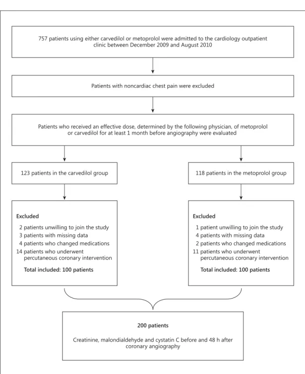
All patients had been taking a sustained dose, determined by the following physician, of carvedilol or metoprolol for at least 1 month before possible enrollment into the study. The doses of the drugs were not changed during the study period. The study design is summarized in figure 1 .
In order to evaluate each patient’s kidney functions, serum creatinine and cystatin C levels were evaluated using venous blood samples before and 48 h after contrast administration. Similarly, to evaluate oxidative status, malondialdehyde levels were analyzed from venous blood samples before and 48 h after coronary angiography. Glomerular filtration rate was estimated using the Cockcroft-Gault formula. CIN was defined as a creatinine increase of at least 25% or 0.5 mg/dl at 48 h of contrast exposure. A nonionic low-osmolar contrast agent (iohexol) was used in all patients. The amount of contrast sub-stance used was recorded for each patient. Saline infusion with a rate of 1 ml/kg/h was given for 12 h before contrast exposure; after contrast exposure, saline infusion with a rate of 1 ml/kg/h was continued for 24 h.
Biochemical Analysis
Serum creatinine was assayed using the kinetic Jaffe method, rate-blanked and compensated using a Hitachi Modular PP Analyzer (Roche Diagnostics GmbH, Mannheim, Germany) according to the manufac-turer’s specifications. Serum cystatin C was measured by enzyme-linked immunosorbent assay (ELISA) using 96-well microplates (BioVendor Laboratory Medicine, Inc., CTPark Modrice, Modrice, Czech Republic) according to the manufacturer’s instructions of the DYNEX Technologies DSX Modal Microelisa Analyzer. The detection limit was 0.2 ng/ml. The intra- and interassay variabilities were 9.6 and 6.2%, respectively. Serum malondialdehyde levels were determined using the spectrophotometric method at 532 nm after boiling the sample and condensing it with thiobarbituric acid [9] .
Statistical Analysis
Continuous variables are presented as means ± SD if normally distributed or as medians with ranges. Normality was tested using the Shapiro-Wilk test. Categorical variables are presented as numbers and percentages and were compared using the Fisher exact test. For comparison of the continuous variables, the t tests, the Mann-Whitney U test or the Wilcoxon signed-rank test were used, whichever was appropriate. A p value of <0.05 was considered statistically significant. The SPSS for Windows version 11.5 statistical software (SPSS Inc., Chicago, Ill., USA) was used for all the statistical calculations.
Patients who received an effective dose, determined by the following physician, of metoprolol or carvedilol for at least 1 month before angiography were evaluated
757 patients using either carvedilol or metoprolol were admitted to the cardiology outpatient clinic between December 2009 and August 2010
Patients with noncardiac chest pain were excluded
123 patients in the carvedilol group 118 patients in the metoprolol group
Excluded
2 patients unwilling to join the study 3 patients with missing data 4 patients who changed medications
Total included: 100 patients
14 patients who underwent
percutaneous coronary intervention
Excluded
1 patient unwilling to join the study 4 patients with missing data 2 patients who changed medications
Total included: 100 patients
11 patients who underwent
percutaneous coronary intervention
200 patients
Creatinine, malondialdehyde and cystatin C before and 48 h after coronary angiography
Results
Two hundred patients were evaluated prospectively. There were no significant differ-ences in baseline clinical characteristics between the carvedilol and metoprolol groups ( table 1 ). The average doses of carvedilol and metoprolol were 25 (range 12.5–50) and 67.5 (range 50–200) mg, respectively.
The mean serum creatinine concentration for all patients was 0.87 ± 0.24 (range 0.4–1.9) mg/dl. In the metoprolol group, the mean serum creatinine concentration in-creased significantly from 0.89 ± 0.23 to 0.96 ± 0.28 mg/dl (p = 0.007) 48 h after radiocon-trast administration. In the carvedilol group, the mean serum creatinine concentration did not change significantly (0.86 ± 0.23 vs. 0.86 ± 0.25 mg/dl; p = 0.76). In the metoprolol group, the median cystatin C concentration increased significantly from 978 (range 145–4,004) to 1,086 (range 303–3,840) ng/ml (p = 0.001). In the carvedilol group, the median cystatin C concentration did not change [1,143 (range 240–3,011) vs. 1,068 (range 262–3,640) ng/ml; p = 0.94]. In the metoprolol group, the mean malondialdehyde concen-tration increased significantly from 7.09 ± 1.48 to 8.38 ± 2.6 nmol/l (p < 0.001). In the carvedilol group, the mean serum malondialdehyde concentration did not change signifi-cantly (7.44 ± 1.21 vs. 7.56 ± 1.11 nmol/l; p = 0.4). There were positive correlations between
Characteristics Carvedilol (n = 100) Metoprolol (n = 100) p Age, years 63±9 65±9 0.1 Male gender 57 62 0.4 BMI 29.13±3.58 28.16±3.87 0.06 Hypertension 98 98 1 Diabetes 43 44 0.8 Hyperlipidemia 75 76 0.8 Smoking 26 35 0.6 History of MI 6 3 0.49 History of PCI 30 19 0.10 History of CABG 10 14 0.52 LVEF, % 54.3±10.3 54.6±8.9 0.84 Medication use ACE inhibitor 47 51 0.57 ARB 36 28 0.22 Ca channel blocker 17 17 1 α-blocker 6 9 0.4 Statins 73 80 0.24 ASA 97 95 0.7 Clopidogrel 20 32 0.053 Spironolactone 9 4 0.152 Furosemide 6 8 0.57 Insulin 24 18 0.29 Metformin 20 25 0.39
Volume of contrast media, ml 97±51 105±49 0.2 Hydration before angiography, ml 1,223±162 1,210±155 0.4 Hydration after angiography, ml 2,518±383 2,466±398 0.3
Values are means ± SD or percentages. BMI = Body mass index; MI = myocardial infarction; PCI = percutaneous coronary intervention; CABG = coronary artery bypass grafting; LVEF = left-ventricular ejection fraction; ACE = angiotensin-converting enzyme; ARB = angiotensin receptor blocker; Ca = calcium; ASA = acetylsalicylic acid.
Table 1. Baseline characteristics
change in creatinine concentration and change in malondialdehyde levels (r = 0.156; p = 0.027; fig. 2 ) and between change in creatinine concentration and change in cystatin C (r = 0.183; p = 0.009; fig. 3 ). The glomerular filtration rate was unchanged in both groups after contrast exposure. The laboratory parameters at baseline and 48 h after contrast exposure are shown in table 2 .
CIN occurred in 22 patients in the metoprolol group and in 7 patients in the carvedilol group (p = 0.003). Baseline median creatinine concentrations were not different between patients with CIN and patients without CIN [0.8 (0.5–1.2) vs. 0.8 (0.4–1.9) mg/dl; p = 0.29]. In a separate analysis, when CIN was defined only as an increase in creatinine concentration of at least 0.5 mg/dl, there were 9 patients with CIN. All of these 9 patients were in the meto-prolol group (p = 0.003). The dose of metometo-prolol was not significantly different between patients who developed CIN and those without CIN (71.4 ± 25.3 vs. 66.6 ± 28.6 mg; p = 0.49). Similarly, the dose of carvedilol was not significantly different between patients who developed CIN and those without CIN (25 vs. 23.9 ± 4.7 mg; p = 0.55).
Change in creatinine (mg/dl)
Change in malondialdehyde (nmol/l)
20 10 0 –0.6 –0.4 –0.2 0 0.2 0.4 0.6 0.8 1.0 –10 Change in creatinine (mg/dl) Change in cystatin C (ng/ml) 2,000 1,000 0 –1,000 –2,000 –0.6 –0.4 –0.2 0 0.2 0.4 0.6 0.8 1.0
Fig. 2. Correlations between
change in creatinine and change in malondialdehyde (r = 0156; p = 0.027).
Fig. 3. Correlations between
change in creatinine and change in cystatin C (r = 0.183; p = 0.009).
Discussion
In this study, we observed that carvedilol had a protective effect against CIN when compared to metoprolol after coronary angiography. In addition, cystatin C, a marker of renal function, and malondialdehyde, an oxidative end-product, were found to be increased in patients using metoprolol, while they were unchanged in patients taking carvedilol.
CIN is the third most common cause of hospital-acquired acute renal failure, and its diag-nosis is associated with numerous diagnostic and interventional procedures in daily practice [10] . Serum creatinine is the most commonly used parameter for evaluating renal function, but serum creatinine is affected by sex, age and muscle mass. Cystatin C is a nonglycosylated low-molecular-weight cysteine protease inhibitor belonging to the cystatin superfamily. Its structure is not affected by inflammation, dietary factors, sex, age or muscle mass [11–13] . Several studies examined the role of cystatin C in predicting CIN [14–18] . Ebru et al. [14] showed that only cystatin C increase after contrast media exposure is in concordance with the Mehran risk scoring system for predicting the risk of CIN. Another study found that baseline serum cystatin C levels significantly predicted the occurrence of CIN in patients with moderate renal insufficiency [15] . A study from Germany demonstrated that CIN was predicted by baseline cystatin C, whereas creatinine, creatinine clearance and blood urea nitrogen were not predictive [16] . The best predictive capabilities were provided by the cystatin C/creatinine ratio. In a recent study by Cicek et al. [17] , serum cystatin C levels were found to be elevated in diabetic patients who developed CIN compared to those who did not. In contrast, Ribichini at al. [18] observed that variations from the serum creatinine baseline offered a better diagnostic accuracy for predicting CIN at an earlier stage than similar varia-tions in cystatin C. In the present study, serum cystatin C levels were significantly increased in the metoprolol group compared to the carvedilol group, suggesting that less deterioration in renal function occurred in the carvedilol group after contrast exposure.
Oxidative stress also results in lipid peroxidation, producing malondialdehyde as an advanced lipoxidation end-product [19, 20] . Malondialdehyde levels were found to be increased in plasma samples and kidney tissues of patients and rats with diabetic nephropathy [21] . In an experimental study by Devrim et al. [22] , serum malondialdehyde levels increased and nitric oxide (NO) levels decreased in rat kidney tissues after contrast exposure. As a
Table 2. Serum creatinine, cystatin C and malondialdehyde levels at baseline and 48 h after contrast exposure
and prevalence of acute reduction in renal function in the carvedilol and metoprolol groups
Variable Carvedilol group
(n = 100) Metoprolol group (n = 100) p Baseline creatinine, mg/dl 0.86±0.23 0.89±0.23 0.25 Post-contrast creatinine, mg/dl 0.86±0.25a 0.96±0.28b 0.05 Baseline cystatin C, ng/ml 1,143 (240–3,011) 978 (145–4,004) 0.1 Post-contrast cystatin C, ng/ml 1,068 (262–3,640)a 1,086 (303–3,840)b 0.26
Baseline malondialdehyde, nmol/l 7.44±1.21 7.09±1.48 0.07
Post-contrast malondialdehyde, nmol/l 7.56±1.11a 8.38±2.6b 0.001
Baseline creatinine clearance 98±26 94±27 0.29
Post-contrast creatinine clearance 98±24a 91±31a 0.32
Contrast-induced nephropathy, n 7 22 0.003
Values are means ± SD or medians with ranges in parentheses. a The p value is nonsignificant when compared to baseline. b The p value is <0.05 when compared to baseline.
result, they hypothesized that an increase in oxidative substances and a decrease in vasodi-latation were responsible for the development of renal damage. Following their study, Colbay et al. [23] similarly demonstrated an increase in malondialdehyde levels in the renal tissues of rats exposed to contrast media. The observation in our study was that malondialdehyde levels were found to be increased in patients using metoprolol, while they were unchanged in patients taking carvedilol. This finding suggests that the antioxidant properties of carvedilol might be the underlying mechanism in preventing CIN. In addition, carvedilol has a vasodi-latory effect which might also be beneficial in reducing the risk of CIN.
Patients with coronary artery disease are frequently prescribed antiischemic medica-tions before the catheterization procedure. Because β-blockers are the first-line agents for patients suspected of having stable coronary artery disease [24] , it is important to delineate the effects of this group of drugs on the development of CIN. Metoprolol is still one of the most frequent β-blocker agents prescribed by clinicians. It does not have a vasodilatory effect like carvedilol and nebivolol [25] . This β-blocker was found to be ineffective in protecting the kidneys from ischemic episodes in a study performed on dogs [26] . Yet, metoprolol prevented lipid peroxidation in experimental adult rat hearts, although it was the least effective drug among the lipophilic β-blockers [27] . Tissue studies have demonstrated that nebivolol increases renal blood flow and glomerular filtration rate by causing dilatation in the afferent and efferent arterioles and that it converts reactive oxygen products formed by the nicotin-amide adenine dinucleotide phosphate oxidase to NO by way of endothelial constitutive NO synthase and increases the total antioxidant action [28, 29] . It was previously reported that nebivolol was effective in preventing CIN [30–32] . The incidence of CIN was shown to be statistically significantly lower in the nebivolol group when compared to the metoprolol group in patients undergoing coronary angiography [30] . Akgüllü et al. [33] support our results by reporting that the histopathological findings after contrast exposure were re-markably lower in rats administered carvedilol or nebivolol when compared to a control group. Carvedilol and nebivolol both were able to prevent the development of nephropathy related to contrast media by decreasing oxidative stress [33] . In an experimental model, carvedilol provided protective effects against cisplatin-induced oxidative stress [34] .
The present study has several limitations. The subjects had already been using meto-prolol and carvedilol at the time of recruitment. There were minor differences in individual dosing, since we selected patients who had already been using carvedilol or metoprolol depending on their own physician’s preferences. This might be a drawback of our study. The retrospective aspect of the study needs to be verified prospectively. The relatively well-preserved renal function of the study population should also be considered when interpret-ing the results. Renal function should be tested in patients with an elevated creatinine con-centration who are particularly at risk of CIN and its complications. We excluded patients who required percutaneous interventions; thus, a relatively low dose of radiocontrast agents was used. There were more patients using clopidogrel in the metoprolol group when compared to the carvedilol group. There is no clinical study that has examined the effect of clopidogrel on the development of CIN. In addition to its antiplatelet effect, clopidogrel also has anti-inflammatory properties and improves endothelial function [35] . High-maintenance doses (150 mg) of clopidogrel were also associated with a stronger improvement in endothelial function and a reduction of systemic inflammation compared with the standard dose (75 mg daily) [35] . Thus, clopidogrel is probably not associated with susceptibility to CIN. Serum creatinine levels were measured immediately before and 48 h after contrast exposure. Evalu-ating serum creatinine beyond the first 48 h would have revealed more patients with CIN.
In conclusion, compared to metoprolol, carvedilol might protect against CIN develop-ment by decreasing oxidative stress in patients undergoing coronary angiography. When a β-blocker therapy is indicated, carvedilol might be preferred to metoprolol in these patients.
Disclosure Statement
The authors declare that they have no conflicts of interest.
References
1 Heyman SN, Reichman J, Brezis M: Pathophysiology of radiocontrast nephropathy: a role for medullary hypoxia. Invest Radiol 1999; 34: 685–691.
2 Berkseth RO, Kjellstrand CM: Radiologic contrast-induced nephropathy. Med Clin North Am 1984; 68: 351– 370.
3 Messana JM, Cieslinski DA, Nguyen VD, Humes HD: Comparison of the toxicity of the radiocontrast agents, iopamidol and diatrizoate, to rabbit renal proximal tubule cells in vitro. J Pharmacol Exp Ther 1988; 244: 1139–1144.
4 Yoshioka T, Fogo A, Beckman JK: Reduced activity of antioxidant enzymes underlies contrast media-induced renal injury in volume depletion. Kidney Int 1992; 41: 1008–1015.
5 Cox CD, Tsikouris JP: Preventing contrast nephropathy: what is the best strategy? A review of the literature. J Clin Pharmacol 2004; 44: 327–337.
6 Freuerstein GZ, Sponer G: The adrenergic pharmacology of carvedilol. Heart Fail Rev 1999; 4: 21–27. 7 Keith M, Geranmayegan A, Sole MJ, Kurian R, Robinson A, Omran AS, Jeejeebhoy KN: Increased oxidative stress
in patients with heart failure. J Am Coll Cardiol 1998; 31: 1352–1356.
8 Flesch M, Maack C, Cremers B, Baumer AT, Südkamp M, Böhm M: Effect of β-blockers on free radical-induced cardiac contractile dysfunction. Circulation 1999; 100: 346–353.
9 Satoh K: Serum lipid peroxide in cerebrovascular disorders determined by new colorimetric method. Clin Chim Acta 1978; 90: 37–43.
10 Rihal CS, Textor SC, Grill DE, Berger PB, Ting HH, Best PJ, Singh M, Bell MR, Barsness GW, Mathew V, Garratt KN, Holmes DR Jr: Incidence and prognostic importance of acute renal failure after percutaneous coronary intervention. Circulation 2002; 105: 2259–2264.
11 Woo KS, Choi JL, Kim BR, Kim JE, Han JY: Clinical usefulness of serum cystatin C as a marker of renal function. Diabetes Metab J 2014; 38: 278–284.
12 Harmoinen A, Lehtimaki T, Korpela M, Turjanmaa V, Saha H: Diagnostic accuracies of plasma creatinine, cystatin C and glomerular filtration calculated by the Cockcroft-Gault and Levey (MDRD) formulas. Clin Chem 2003; 49: 1223–1225.
13 Rickli H, Benou K, Ammann P, Fehr T, Brunner-La Rocca HP, Petridis H, Riesen W, Wuthrich RP: Time course of cystatin C levels in comparison with serum creatinine after application of radiocontrast media. Clin Nephrol 2004; 61: 98–102.
14 Ebru AE, Kilic A, Korkmaz FS, Seker R, Sasmaz H, Demirtas S, Biyikli Z: Is cystatin-C superior to creatinine in the early diagnosis of contrast-induced nephropathy? A potential new biomarker for an old complication. J Postgrad Med 2014; 60: 135–140.
15 Ishibashi Y, Yamauchi M, Musha H, Mikami T, Kawasaki K, Miyake F: Impact of contrast-induced nephropathy and cardiovascular events by serum cystatin C in renal insufficiency patients undergoing cardiac catheter-ization. Angiology 2010; 61: 724–730.
16 Wacker-Gussmann A, Bühren K, Schultheiss C, Braun SL, Page S, Saugel B, Schmid S, Mair S, Schoemig A, Schmid RM, Huber W: Prediction of contrast-induced nephropathy in patients with serum creatinine levels in the upper normal range by cystatin C: a prospective study in 374 patients. AJR Am J Roentgenol 2014; 202: 452–458.
17 Cicek M, Yıldırır A, Okyay K, Yazici AC, Aydinalp A, Kanyilmaz S, Muderrisoglu H: Use of α-lipoic acid in prevention of contrast-induced nephropathy in diabetic patients. Ren Fail 2013; 35: 748–753.
18 Ribichini F, Gambaro G, Graziani MS, Pighi M, Pesarini G, Pasoli P, Anselmi M, Ferrero V, Yabarek T, Sorio A, Rizzotti P, Lupo A, Vassanelli C: Comparison of serum creatinine and cystatin C for early diagnosis of contrast-induced nephropathy after coronary angiography and interventions. Clin Chem 2012; 58: 458–464.
19 Halliwel B: Free radicals, antioxidants, and human disease: curiosity, cause, or consequence? Lancet 1994; 344: 721–724.
20 Gutteridge JM, Halliwel B: Free radicals and antioxidants in the year 2000. A historical look to the future. Ann NY Acad Sci 2000; 899: 136–147.
21 Chang JM, Kuo MC, Kuo HT, Chıu YW, Chen HC: Increased glomerular and extracellular malondialdehyde levels in patients and rats with diabetic nephropathy. J Lab Clin Med 2005; 146: 210–215.
22 Devrim E, Cetin M, Namuslu M, Erguder IB, Cetin R, Durak I: Oxidant stress due to nonionic low osmolar contrast medium in rat kidney. Indian J Med Res 2009; 130: 433–436.
23 Colbay M, Yuksel S, Uslan I, Acarturk G, Karaman O, Bas O, Mollaoglu H, Yagmurca M, Ozen OA: Novel approach for the prevention of contrast nephropathy. Exp Toxicol Pathol 2010; 62: 81–89.
24 Task Force Members, Montalescot G, Sechtem U, Achenbach S, et al; ESC Committee for Practice Guidelines, Zamorano JL, Achenbach S, Baumgartner H, et al; Document Reviewers, Knuuti J, Valgimigli M, Bueno H, et al: 2013 ESC guidelines on the management of stable coronary artery disease: the Task Force on the management of stable coronary artery disease of the European Society of Cardiology. Eur Heart J 2013; 34: 2949–3003. 25 Fares H, Lavie CJ, Ventura HO: Vasodilating versus first-generation β-blockers for cardiovascular protection.
Postgrad Med 2012; 124: 7–15.
26 Módolo NS, Castiglia YM, Ganem EM, Braz JR, Vianna PT, Vane LA: Acute renal ischemia model in dogs: effects of metoprolol. Ren Fail 2001; 23: 1–10.
27 Jenkins RR, Del Signore CM, Sauer P, Skelly C: The effect of β blocking drugs on lipid peroxidation in rat heart in vitro. Lipids 1992; 27: 539–542.
28 Maffei A, Lembo G: Nitric oxide mechanisms of nebivolol. Ther Adv Cardiovasc Dis 2009; 3: 317–327. 29 Oelze M, Daiber A, Brandes RP, Hortmann M, Wenzel P, Hink U, Schulz E, Mollnau H, von Sandersleben A,
Kleschyov AL, Mülsch A, Li H, Förstermann U, Münzel T: Nebivolol inhibits superoxide formation by NADPH oxidase and endothelial dysfunction in angiotensin II-treated rats. Hypertension 2006; 48: 677–684. 30 Avcı E, Yeşil M, Bayata S, Postacı N, Arıkan E, Cirit M: The role of nebivolol in the prevention of
contrast-induced nephropathy in patients with renal dysfunction. Anadolu Kardiyol Derg 2011; 11: 613–617. 31 Günebakmaz Ö, Kaya MG, Koç F, Akpek M, Kasapkara A, İnanç MT, Yarlioglues M, Calapkorur B, Karadag Z,
Oguzhan A: Does nebivolol prevent contrast-induced nephropathy in humans. Clin Cardiol 2012; 35: 250–254. 32 Akgüllü C, Eryılmaz U, Güngör H, Huyut A, Zencir C, Hekim T: A clinical study about contrast nephropathy: risk
factors and the role of β blockers. Anatol J Cardiol 2015; 15: 232–240.
33 Akgüllü Ç, Hekim T, Eryılmaz U, Boyacıoğlu M, Güngör H, Meteoğlu İ, Karul A: The usefulness of carvedilol and nebivolol in preventing contrast nephropathy in rats. Ren Fail 2015; 22: 1–7.
34 Rodrigues MA, Rodrigues JL, Martins NM, Barbosa F, Curti C, Santos NA, Santos AC: Carvedilol protects against cisplatin-induced oxidative stress, redox state unbalance and apoptosis in rat kidney mitochondria. Chem Biol Interact 2011; 189: 45–51.
35 Patti G, Grieco D, Dicuonzo G, Pasceri V, Nusca A, Di Sciascio G: High versus standard clopidogrel maintenance dose after percutaneous coronary intervention and effects on platelet inhibition, endothelial function, and inflammation results of the ARMYDA-150 mg (antiplatelet therapy for reduction of myocardial damage during angioplasty) randomized study. J Am Coll Cardiol 2011; 57: 771–778.