Ankara Üniv Vet Fak Derg, 58, 285-288, 2011
Short Communication / Kısa Bilimsel Çalışma
Anatomical and pathological findings of renal aplasia and
compensatory hypertrophy in a cat
Okan EKİM1, Mehmet Fatih BOZKURT2, Çağdaş OTO1
1Ankara University Faculty of Veterinary Medicine Department of Anatomy, Ankara; 2Afyon Kocatepe University Faculty of
Veterinary Medicine Department of Pathology, Afyonkarahisar, Turkey.
Summary: In this study incidentally detected renal aplasia in right kidney and compensatory hypertrophy in left kidney have been defined with the anatomical and pathological findings in a 2 years old male crossbred cat which had suffered from infirmity, vomiting, anorexia and depression. In macroscopic examination, it was observed that the left kidney was located on the left side of the median line, at the ventral part of the transverse processes of the 1st – 4th lumbar vertebrae. The right kidney was at the right side
of median line, ventral part of the transverse process of the 2nd lumbar vertebra. Left kidney had a yellow color and a firm
consistency and also the cut sections were smooth. Besides, right kidney had a grayish-yellow color and the distinction between renal cortex and medulla in cut sections couldn’t be distinguished. As of anatomical location and morphology, no abnormality was observed in the other organs of the cat. In right kidney, tubular structures were recognized histopathologically in between intense collagen tissue. Numerous of vascular formations were found near to the renal capsule. In hypertrophic kidney, in many of the tubules, degenerative alterations were observed significantly. Microscopically the most of the glomeruli were enlarged with mesangial cell hyperplasia and filled completely the Bowman’s space. In the light of the detailed anatomical and pathological findings, the aim of this study was to make a contribution to such unilateral aplasia and compensatory hypertrophy cases that were rarely detected both in human and veterinary medicine.
Key words: Agenesis, aplasia, cat, hypertrophy, kidney.
Bir kedide renal aplazi ve kompenzatorik hipertrofi olgusunda anatomik ve patolojik bulgular
Özet: Bu çalışmada, kusma, depresyon, anoreksi şikayetiyle gelen, 2 yaşlı, erkek, tekir bir kedide rastlantısal olarak karşılaşılan sağ böbrekte renal aplazi ile sol böbrekte hipertrofi olguları anatomopatolojik bulgularıyla tanımlanmıştır. Makroskobik incelemede; sol böbreğin median hattın sol tarafında, 1.-4. vertebrae lumbales’lerin, processus transversus’larının iz düşümü hizasına, sağ böbreğin ise median hattın sağında, 2. lumbal vertebra’nın processus transversus’unun ventral’ine yerleştiği görüldü. Sağ böbreğin grimsi-sarı renkte olup kesit yüzüne bakıldığında korteks-medulla ayrımının yapılamadığı göze çarparken, sol böbrek sarı renkte, sert kıvamda ve kesit yüzü düz olarak gözlendi. Hayvanın diğer organlarında konum ve anatomik yapı itibarı ile herhangi bir anormallik tespit edilmedi. Histopatolojik olarak aplazik böbrekte yoğun kollagen doku arasında tubulus taslakları dikkati çekti. Kapsüle yakın yerlerde çok miktarda damarsal oluşumlar tespit edildi. Hipertrofik böbrekte ise çok sayıda tubulusta dejeneratif değişiklikler ön plandaydı. İncelenen sahaların çoğunda, glomerulusların mezangiyal hücre hiperplazisi ile genişlediği ve Bowman boşluğunu doldurduğu dikkati çekti. Bu çalışmanın amacı; detaylı anatomik ve patolojik bulgular ışığında, gerek beşeri gerekse veteriner hekimlikte nadiren tespit edilen, bu tür unilateral aplazi ve kompenzatorik hipertrofi olgularına katkı sağlamaktır.
Anahtar Sözcükler: Agenezi, aplazi, böbrek, hipertrofi, kedi.
Renal aplasia or agenesis is mostly congenital disorder which has frequently being encountered in humans (6, 7) and also in domestic mammals (9). In addition to the high prevalence of the aplasia in even organs like testicles or ovaries (8), especially in renal aplasia, malformations can be observed in the other urogenital organs which are functionally related with kidneys (5). Renal agenesis in dogs has been detected by many researchers and is often related with the congenital anomalies of the urinary organs such as the ureters or the
urinary bladder (2, 10, 11). It has been reported that the renal agenesis in cats was detected together with the bilateral or unilateral uterine aplasias (4). In most of the cases unilateral renal aplasia can be incidentally detected in postmortem examinations because of the compensation of opposite kidney (12). Therefore patients can maintain the vital functions with the opposite organ and the statistical percentage of the cases are limited only with the detected ones (1). The aim of this study was to make a contribution to such unilateral aplasia and compensatory
Okan Ekim - Mehmet Fatih Bozkurt - Çağdaş Oto 286
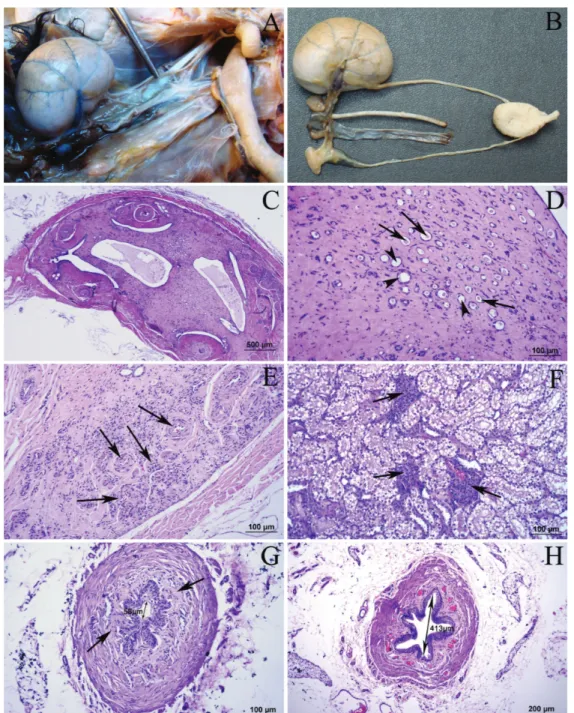
Figure 1. Anatomic and histopathologic images of the kidneys and the ureters. A; Ventral view of the left and the right kidneys in the abdominal cavity.
B; Hypertrophic and aplastic kidneys dissected out together with the ureters, the urinary bladder, the renal artery and vein. C; Microscopic view of the aplastic kidney. Renal cortex and the medulla can’t be distinguished from each other.
D; Some of the tubular formations had pinkish-red proteinous liquid in their lumen (arrows) and the rest of those were dilatated or cystic (arrowheads) in between the hyalinized collagen tissue in aplastic kidney.
E; Large number of vascular formations (arrows) were detected near to the renal capsule in aplastic kidney.
F; In most of the microscopic areas observed in hypertrophic kidney, glomeruli were enlarged with mesangial cell hyperplasia (arrows) and filled completely the Bowman’s space.
G; The diameter of the ureter of the aplastic kidney was shorter. Hyalinized areas were observed in the submucosa (arrows). H; The transversal section of the ureter of the hypertrophic kidney.
Şekil 1. Böbrekler ve üreterlerin anatomik ve histopatolojik görüntüleri A; Sağ ve sol böbreğin karın boşluğu içinde ventral’den görünümü.
B; Üreterler, sidik kesesi, a. ve v. renalis ile birlikte diseke edilmiş hipertrofik ve aplazik böbrek. C; Aplazik böbreğin mikroskobik görünümü. Renal korteks ile medulla sınırı ayırdedilememektedir.
D; Aplazik böbrekte hiyalinize kollagen doku arasında bazıları kistik ya da genişlemiş (okbaşları), bazılarının lümeninde pembemsi kırmızı renkte proteinöz sıvı bulunan (oklar) tubuler yapılar.
E; Aplazik böbrekte, kapsüle yakın olarak çok miktarda damarsal oluşumlar (oklar) tespit edildi.
F; Hipertrofik böbrekte incelenen sahaların çoğunda, glomerulusların mezangiyal hücre hiperplazisi ile genişlemiş ve Bowman kapsülünü doldurmuş durumdaydı (oklar).
G; Aplazik böbreğe ait üreter çapı, hipertrofik böbreğe göre daha dardı. Submukozada hyalinize alanlar (oklar) gözlendi. H; Hipertrofik böbreğe ait üreterin transversal kesidi.
Ankara Üniv Vet Fak Derg, 58, 2011 287
hypertrophy cases that were rarely detected both in human and veterinary medicine. A 2 years old, 4.2 kg weighted male tabby cat suffering from infirmity, vomiting, anorexia and depression had been euthanized in a private clinic after taking the consent of the owner and the body was donated to the Ankara University Faculty of Veterinary Medicine Department of Anatomy for inspection. Anatomic features and the positions of the organs in the abdominal and pelvic cavities were inspected macroscopically. After the inspection visceral organs with pathological alterations were exenterated out and fixed in a 10% formalin solution and embedded in paraffin. Tissue sections taken from paraffin blocks were stained with hematoxylin-eosin. The macroscopic photographs were taken by Sony DSC-HX20 digital camera and the microscopic ones were taken by Olympus DP-71 real time digital camera attached to Olympus BX-51 light microscope. The dimensions of the kidneys were determined by Mitutoyo digital caliper and the microscopic measurements were performed by Olympus DP Controller software.
In macroscopic examination, it has been observed that the left kidney was located on the left side of the median line, at the ventral part of the transverse processes of the 1st – 4th lumbar vertebrae. The right
kidney was at the right side of median line, ventral part of the transverse process of the 2nd lumbar vertebra
(Figure1.A). The craniocaudal lengths of the left and the right kidney were 4.9 cm and 1.4 cm. At the level of renal hilus the mediolateral diameters were 2.6 cm and 0.4 cm, the dorsoventral diameters were 1.9 cm and 0.3 cm, respectively. Left kidney had a yellow color and a firm consistency (Figure 1.A, B) and also the cut sections were smooth. Besides, right kidney had a grayish-yellow color (Figure 1.A, B) and the distinction between renal cortex and medulla in cut sections couldn’t be distinguished in macroscopic and also in microscopic inspections (Figure 1.C).
The ureters of both left and right kidneys run caudally on the lateral walls of the pelvis and entered dorsally to the urinary bladder in accordance with the other anatomical features (Figure 1.B). As of anatomical location and morphology, no abnormality was observed in the other abdominal or pelvic organs of the cat. The morphological and the histopathological findings of the testicles, the seminal ducts and the bladder were normal.
Histopathologically in aplastic kidney, on a wide area beginning from renal hilus outwards, as some of the tubular formations had pinkish-red proteinous liquid in their lumen (Figure 1.D/arrows), the rest of those were dilatated or cystic (Figure 1.D/arrowheads) in between the hyalinized collagen tissue. A large number of vascular formations (Figure 1.E/arrows) were detected near to the renal capsule. Extensive hemorrhage was also determined in those areas (Figure 1.E). The diameter of
the ureter of the mentioned kidney was shorter (Figure 1.G) and also the epithelium was found more degenerated as compared to the hypertrophic kidney (Figure 1.H). Extensive hyalinized areas (Figure 1.G/arrows) were observed in the submucosa.
In hypertrophic kidney, in many of the tubules, degenerative alterations were observed significantly (Figure 1.F). In most of the microscopic areas observed, glomeruli were enlarged with mesangial cell hyperplasia and filled completely the Bowman’s space (Figure 1.F/arrows).
In certain references (3, 9) the presence of metanephric structures in the histopathological examination of the kidney is indicated as a renal dysplasia. However in this case only primitive tubular structures were recognized histopathologically in between intense collagen tissue in right kidney and none of the glomerular formations could be determined. On account of that, this case was considered as a renal aplasia or agenesis.
Depending on the dysfunction of agenesic kidney, it was detected that the left kidney became hypertrophic to compensate the right one. Nevertheless, it is still a question mark if the compensation is adequate to maintain the vital activities in this case. It was indicated that renal aplasia or agenesis might be unilateral or bilateral and caused by developmental failure of pronephros, mesonephros or ureteral bud, by absence or totaly degeneration of metanephric blastema. Renal agenesis might be accompanied with genital organ deformities. Also the ureters may be absent or terminate in a connective tissue with blind end (9). In this case on contrary with those above, a spesific malformation has not been determined in the genital organs. Altough the right ureter was quite narrower than the left one, both of them were present. Besides, a satisfactory study could not be found for the renal aplasia observed together with the malformations in urogenital organs in male cats. Therefore, the histopathologic and anatomic findings of the present study are remarkable. Genetic factors that take role in the ethiology of the renal agenesis might be quite effective in cats according to their breeds but current researchs are inadequate to say something about genetic effects.
In conclusion, the renal aplasia cases like mentioned in this study are mostly detected incidentally and should be investigated in detailed manner to understand the interactions of different malformations or abnormalities in cats and to make a contribution to such unilateral aplasia and compensatory hypertrophy cases that were encountered in both human and veterinary medicine.
References
1. Acién P, Acién M, Sánchez-Ferrer M (2004): Complex malformations of the female genital tract. New types and revision of classification. Hum Reprod, 19, 2377–2384.
Okan Ekim - Mehmet Fatih Bozkurt - Çağdaş Oto 288
2. Agut A, Fernandez del Palacio MJ, Laredo FG, Murciano J, Bayon A, Soler M (2002): Unilateral renal
agenesis associated with additional congenital abnormalities of the urinary tract in a Pekingese bitch. J Small Anim Pract, 43, 32–35.
3. Azizi S, Kheirandish R, Yazdanpour H (2010):
Histopathologic features of a unilateral renal dysplasia in a cat (Felis domestica). Comp Clin Pathol, 19, 445–447
4. Chang J, Jung JH, Yoon J, Choi MC, Park JH, Seo KM, Jeong SM (2008): Segmental aplasia of the uterine
horn with ipsilateral renal agenesis in a cat. J Vet Med Sci, 70, 641–643.
5. Hazıroğlu R, Milli ÜH (2001): Veteriner Patoloji. Cilt II. 2. Baskı. Medipres, Ankara.
6. Hiraoka M, Tsukahara H, Ohshima Y, Kasuga K, Ishihara Y. Mayumi M (2002): Renal aplasia is the
predominant cause of congenital solitary kidneys. Kidney Int, 61, 1840–1844.
7. Kissane JM (1974): Development of the kidney and congenital malformations. 69-109. In: Hepstinstall RH (Ed), Pathology of the Kidney. Vol 1. Little, Brown and Co, Boston.
8. Köküuslu C (1996): Genel Patoloji. 1. Baskı. Medisan Yayınevi, Ankara.
9. Maxie MG, Newman SJ (2007): The urinary system. 425-522. In: MG Maxie (Ed), Jubb, Kennedy & Palmer's Pathology of Domestic Animals. Vol 2. Saunders Elsevier, Philadelphia.
10. Morita T, Michimae Y, Sawada M, Uemura T, Araki Y, Haruna A, Shimada A (2005): Renal dysplasia with
unilateral renal agenesis in a dog. J Comp Pathol, 133, 64-67.
11. Taney KG, Moore KW, Carro T, Spencer C (2003): Bilateral ectopic ureters in a male dog with unilateral renal agenesis. J Am Vet Med Assoc, 223, 817–820. 12. Wadsworth PF, Squires PF (1980): Renal aplasia and
hypoplasia in the rhesus monkey (Macaca mulatta). Lab Anim, 14, 1-2.
Geliş tarihi: 11.10.2010 / Kabul tarihi: 24.03.2011
Adress for correspondence
Lect.Off.Dr. Okan Ekim Department of Anatomy, Faculty of Veterinary Medicine, Ankara University,
06110, Dışkapı-Ankara/ TURKEY.
Tel: +90312 3170315 Fax: +90312 3175559 e-mail: [email protected]