Case Report / Olgu Sunumu
The right displacement of the abomasum complicated with abomasitis
in a calf
İbrahim YURDAKUL
1, Uğur AYDOĞDU
21Department of Surgery, Faculty of Veterinary Medicine, Cumhuriyet University, 58140 Sivas, Turkey, 2Department of Internal
Medicine, Faculty of Veterinary Medicine, Balıkesir University, 58140 Balıkesir, Turkey.
Summary: In this case, it was intended to define the right displacement of the abomasum complicated with abomasitis in 45 days old unweaned Brown Swiss male calf with clinical and ultrasonographic examination and further to report the operative treatment results. Metallic ping and splashing sounds were heard on the right side in the percussion of the 10-13th intercostal space. Right
abomasal displacement was confirmed by ultrasonography. After the diagnosis of the illness, its treatment was done operatively through the right abdominal wall. During the operation, approximately 6 L content was emptied from the abomasum. Edema, ulceration and inflammation were seen on the abomasum wall. As a result, it was determined that abomasitis could develop together with the displacement of the abomasum to the right, which is rarely seen in calves. Successful results can be obtained by bringing the abomasum to its normal position during the treatment made through the right abdominal wall.
Keywords: Abomasitis, abomasum, calf, right displacement, ultrasonography.
Bir buzağıda abomasitis ile komplike sağa abomasum deplasmanı
Özet: Bu olguda 45 günlük sütten kesilmemiş montofon ırkı erkek bir buzağıda karşılaşılan abomazitis ile komplike abomasumun sağa deplasmanı olgusunun klinik ve ultrasonografik olarak tanımlanması ve operatif sağaltım sonuçlarının bildirilmesi amaçlandı. Hastalığın teşhisi sağ karın duvarının aynı anda oskültasyon ve perküsyonunda ping ve çalkantı seslerinin duyulması ve ultrasonografi aracılığıyla konulduktan sonra tedavisi sağ karın duvarından operatif olarak yapıldı. Operasyon sırasında abomasumdan yaklaşık 6 L içerik boşaltıldı. Abomazum duvarında ödem ve kalınlaşma ile ülseratif ve yangısal alanlar görüldü. Sonuç olarak, buzağılarda ender görülen abomasumun sağa deplasmanı ile birlikte abomazitisinde gelişebileceği belirlendi. Sağ karın duvarından yapılan tedavide abomasumun normal konumuna getirilmesi ile gayet başarılı sonuçlar elde edilebilir.
Anahtar sözcükler: Abomasitis, abomazum, buzağı, sağa deplasman, ultrasonografi.
Abomasum displacement, which is one of the most important metabolic diseases of the digestive system and which is seen in dairy cows, is commonly related to stress, nutrition and metabolic disorders. Abomasal atony, which leads to meteorism in the abomasum, is thought to cause abomasum displacements. Abomasum displacements are seen more frequently in dairy cows which are fed with concentrated food. Consumption of grain feed at a high-level cause extreme fatty acid and gas production and they lead to abomasum displacements (8, 12). Abomasum dilatation and its displacement to the right which are usually seen in adult cattle are rarely seen in calves (3). While displacements of the abomasum to the right are observed in unweaned calves, displacements of the abomasum to the left are more common in weaned calves (6). Earlier findings show that left and right abomasum displacements are seen in calves which are lack of appetite, poor weight gain, repeated tympani, abdominal
distension, colic, depressive behavior and diarrhea (1, 3, 12). Diagnosis of the right displacement of the abomasum in cattle is based on the fundamental of hearing ping and splashing sounds over 10-13 ribs during the simultaneous auscultation and percussion on the right abdominal wall (1). Moreover, ultrasonography is used as a very valuable diagnosis method in the diagnosis of the right displacement of the abomasum (9).
The purpose of this case presentation is to define the abomasitis case together with the right displacement of the abomasum in a 45 days old unweaned Brown Swiss male calf considering clinical, ultrasonographical and operative findings. A 45 days-old male Brown Swiss calf was referred to the Animal Hospital of Sivas Cumhuriyet University Faculty of Veterinary Medicine with complaints of loss appetite, abdominal distension, colic and absence of defecation.
Mild hypothermia (Temperature=37,5oC), tachycardia
(>120), dehydration (10%), symptoms of colic (kicking the abdomen, lying down and getting up), depression and anorexia were determined in the routine clinical examination of the calf. The mucous membranes were hyperemic and the scleral vessels were filled. Characteristic metallic ping and splashing sounds were heard during the simultaneous auscultation and percussion of the right abdominal wall at the 10-13th intercostal space
for the diagnosis of right displacement in the calf. Five ml blood was taken for blood gas (Epoc, Ottawa, Canada) and hematological analyses (Mindray BC-2800Vet, PRC) from jugular vein anaerobically and the analyses were made just after taking blood. Hypokalemia, hyperbasemia (increase in HCO3 and BE), hypercapnia, hyperlactatemia,
leukocytosis, and increase in PCV were determined (Table 1).
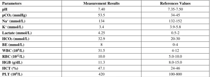
Table 1. Pre-operation blood gases and hematological analysis results in a calf with right displacement of the abomasum. Tablo 1. Sağa abomasum deplasmanlı buzağının operasyon öncesi kan gazları ve hematolojik analiz sonuçları.
Parameters Measurement Results References Values
pH 7.40 7.35-7.50 pCO2 (mmHg) 53.5 34-45 Na+ (mmol/L) 134 132-152 K+ (mmol/L) 3.4 3.9-5.8 Lactate (mmol/L) 4.25 0.5-2 HCO3 (mmol/L) 32.9 20-30 BE (mmol/L) 8 0-4 WBC (109/L) 31.5 4-12 RBC (1012/L) 10.0 5.0-10.0 HGB (g/dL) 11.3 8.0-15.0 HCT (%) 47.1 24-46 PLT (109/L) 420 100-800
pH: concentration of hydrogen ions, pCO2: partial pressure of carbon dioxide, Na+; sodium, K+; potassium HCO3-:bicarbonate, BE:
base excess, WBC; white blood cell, RBC; red blood cell, HGB; haemoglobin, HCT; haematocrit, PLT; platelet.
Fig 1. Ultrasonographic image of the abomasum. Ultrasonographic examination of the abomasum of a 45-day-old calf with the right displacement of the abomasum complicated with abomasitis. The abomasal content was hypoechogenic. 1: Abomasum, 2: Abomasal wall, 3: Abdominal wall.
Şekil 1. Abomasumun ultrasonografik görüntüsü. Abomasitis ile komplike sağa abomazum deplasmanlı 45 günlük bir buzağının abomazumunun ultrasonografik incelemesi. Abomasal içerik hipoekojenik görünümdeydi. 1: Abomasum with hypoechogenic ingesta, 2: Abomasal duvar, 3: Abdominal duvar.
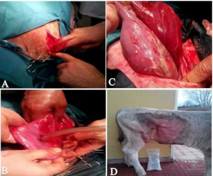
Fig 2. A. Abomasal tension, Fig 2. B. Emptying of abomasal content, Fig 2. C. Abomasitis and fibrin, Fig 2. D. Appearance of the calf after the operation.
Şekil 2. A. Abomasal gerginlik, Şekil 2. B. Abomasal içeriğin alınması, Şekil 2. C. Abomasitis ve fibrin, Şekil 2. D. Operasyon sonrası buzağının görünümü.
Ultrasonographic analysis of the abomasum was made in the right abdominal area through the 10-13 intercostal space in the dorso-ventral way with real time 3.5 MHz convex transducer (Mindray DP-20 Vet, China). In ultrasonography, it was observed that abomasum became larger towards the right and upper parts of the abdomen and there was hypoechogenic liquid in it (Fig 1) (Fig 2. A). Moreover, it was found that the liver was not in its normal position. In consequence of the clinical, hematological and ultrasonographic examination, right displacement of the abomasum was diagnosed and the calf was operated. A catheter was placed in vena auricularis and totally 4 L 0.9% NaCl (Polifarma, Turkey) was applied before and after the operation. In addition, before the operation, intravenous 6 ml Scoban (500 mg metamisol+ 4 mg scopolamin N-butil bromid/ml, Provet, Istanbul, Turkey) was applied.
The right paralumbar approach was used for right abomasal displacement. The right paralumbar area was clipped and prepared for aseptic surgery. Local anesthesia was administered by local infiltration. A right flank laparotomy was performed on the standing animal and the abomasum was right upper abdominal wall and it was dilated with gas and content. The abomasum was also twist on itself (abomasal volvulus) and peritonitis was also noted in the calf.
The abomasum was punctured with a 2 mm diameter needle attached to a sterile rubber tubing and the gas of abomasum emptied. Then the abomasum was cut of 3 cm long and approximately 6 L abomasal content was emptied. (Fig. 2. B). Severe edema, thickening and extensive ulcerative areas and inflammation were found (Fig. 2. C). Incision line in the abomasum was inserted inside with two sero-muskuler suture. Then, a
seromuscular Ford-interlocking suture -using non-absorbable material was performed in the abomasal body, parallel to and at a distance of about 3 cm from the greater curvature omentum. Both ends of the suture was kept long. Both sutures perforated the ventral abdominal wall at a distance of 6 cm from each other from inside and was tied outside the abdominal wall by an assistant. The abomasum was brought to the normal position. Surgical incision line was closed with three layers. Peritoneum, fascia and transversus muscle were sutured together with simple continuous pattern with chromic catgut. M. obliqus internus and m. obliqus externus were sutured with simple continuous pattern, both with chromic catgut. Skin was closed with supramid (Fig. 2. D).
Reptopen-S (penicillin-streptomicine, CEVA, Istanbul, Turkey) for 2 ml/25 kg body weight was applied to the calf in an intramuscular way after the operation. Ulcuran (Ranitidine 25 mg/ml, Abfar, Istanbul, Turkey) and diet were recommended for abomasitis. Medical treatment was applied for 10 days. Skin sutures were removed on the 10th day after the operation. The calf was
followed up for 1 month and no complications were encountered.
It is reported that the dilatation and the right displacement of the abomasum are rarely seen in calves and usually 6 and 14 weeks old calves are very sensitive to abomasal illnesses. Displacement of the abomasum is seen in male calves more than in females (1, 3, 14). Although the calf was male, it was not overweight. The etiology of the right displacement of the abomasum has not been exactly understood but probably it is similar to the left displacement of the abomasum. Abomasal atony is thought to lead displacement and volvulus. In calves, during the right displacement of the abomasum and volvulus, clinical findings such as sudden onset of anorexia, acute abdominal pain, depression, bellowing and straining are observed. Tachycardia and abdominal distention are seen and ping sound is heard on the upper part of right abdomen during the auscultation and percussion. There is pain in palpation of the abdomen. In this case, anorexia, depression, colic symptoms such as kicking of the abdomen, lying down and the anamnesis findings such as absence of defecation were compatible with the findings reported before. The clinical findings (dehydration, hypothermia, tachycardia, pain in the palpation of the abdomen, ping and splashing sound in the auscultation and percussion of the right part of the abdomen) were also similar to the previous studies (1, 12). It is reported that the volatile fatty acids coming out in the rumen as a result of nutrition in animals which are fed with highly concentrated food cause hypomotility in the abomasum. They also increase the osmotic pressure of the abomasum and cause the extracellular liquid to pass
into the rumen, which cause dilatation (2, 4). In the anamnesis, it was observed that the calf fed with high concentrate feed. This condition, demonstrates the importance of concentrated feeds in the formation of abomasum displacement.
Hypokalemia, hyperbasemia (increase in HCO3 and
BE), hypercapnia, hyperlactatemia, leukocytosis and increase in PCV were determined in this case with the right displacement of the abomasum (Table 1). Hemoconcentration (increase in PCV), alkalosis, hypochloremia and hypokalemia are seen at various levels in the laboratory findings of the abomasum displacements (5, 11, 13). Excessive base is also important in the evaluation of the prognosis. It is expressed that the chance of recovery is higher in cattle with right displacement whose base excess is high (12). As for this case, the pH was at its normal levels although the bicarbonate and base excess values were high. This situation is probably related to pCO2 being high. Hypokalemia is probably related to
K+’s passing into the cell in consequence of alkalosis and
anorexia (13). The high level in the hematocrit value confirms severe dehydration (10%). High level of leukocyte in cattle with displacement of the abomasum to the right can be seen as a result of an immunological response related to endotoxemia, peritonitis and abomasitis (7, 15). In this case, the serious increase in the level of leucocyte is probably related to abomasitis and peritonitis. Hyperlactatemia related to decrease in the perfusion of the abomasal tissue can be observed in cattle with abomasum displacement (10). In this case, increase in blood lactate level was determined and this situation is compatible with the determination of the severe ulcerative focuses in the abomasum at the moment of the operation. The abomasum can easily be seen from the right side in cattle and calves with RDA (1, 9). The right displaced abomasum can easily be separated from other organs due to its content. The right displaced abomasum is usually observed as dilated. In addition, the displaced abomasum changes the position of the liver (9). Our ultrasound findings were consistent with this information.
In conclusion, it was determined that abomasitis could develop together with the displacement of the abomasum to the right, which is rarely seen in calves. In addition, right displacement of the abomasum should be considered in the definitive diagnosis of the calves with abdominal distension. Quite successful results can be obtained by bringing the abomasum to its normal position during the treatment made through the right abdominal wall. Moreover, treatment for abomasitis can increase the chance to get well. However, ultrasonographical examination can be a reliable tool for definitive diagnosis of the cases in which the abomasum is displaced to the right side. It is thought that the presentation of the case
with right displacement of the abomasum in a 45 day’s old unweaned calf will contribute to clinic veterinary surgery and literature.
References
1. Altan S, Alkan F, Koç Y (2012): The right displacement of
abomasum with ulceration in a calf. Kafkas Univ Vet Fak
Derg. 18, (2), 343-346.
2. Aslan V, Ok M, Boydak M, et al. (1997): Süt ineklerinde
abomasum deplasmanlarının yağlı karaciğer sendromu ile ilişkisi. Vet Bil Derg, 13, (2), 77-82.
3. Cruz MM, Roblesgil AP, Escamilla MRG, et al. (1990):
Description of abomasal displacements in dairy calves. Bov
Pract, 25, 95-98.
4. Çeçen G (2012): Mide ve bağırsak hastalıkları. 269- 282. In: Veteriner Özel Cerrahi. 1nd ed, Medipres, Malatya, Türkiye.
5. Dezfouli MM, Alidadi N, Heidari SM, et al. (2016): Case
report: a rare right abomasal displacement in a feedlot bull calf. Comp Clin Pathol, 25, 667-670.
6. Fubini S, Divers TJ (2008): Noninfectious diseases of the
gastrointestinal tract. 130-199. In: Rebhun’s diseases of
dairy cattle 2nd ed. (Rebhun, WC, ed): Saunders, USA. 7. Maden M, Sagkan OA, Bulbul A, et al. (2012):
Acute-phase proteins, oxidative stress, and enzyme activities of blood serum and peritoneal fluid in cattle with abomasal displacement. J Vet Intern Med, 26, 1470-1475.
8. Niehaus A (2009): Displaced abomasum in cattle. 40-43. In: Food Animal Practice 5th ed. (Anderson DE, Rings DM, eds): Saunders, Missouri, USA.
9. Ok M, Arican M, Turgut K (2002): Ultrasonographic
findings in cows with left and right displacement of abomasum. Revue Méd Vét, 15, 15-18.
10. Ok M, Yıldız R, Naseri A (2014): Ultrasonographic
finding in anterior displacement of abomasum in a cow.
Kafkas Univ Vet Fak Derg, 20, (2): 317-319.
11. Oman RE, Streeter RN, Reppert EJ, et al. (2016): Left
displacement of the abomasum in 4 beef calves. J Vet Intern
Med, 30, 1376-1380.
12. Radostits OM, Gay CC, Hinchcliff KW, et al. (2007):
Veterinary Medicine. 293-382. 10 nd Ed, W.B. Saunders,
London, UK.
13. Sahinduran S, Albay MK (2006): Haematological and
biochemical profiles in right displacement of abomasum in cattle. Revue Méd Vét, 157, 352-356.
14. Trent AM (2004): Abomasal disease. Surgery of the calf
gastrointestinal system. 461-466. In: Farm Animal Surgery
(Fubini SL, Ducharme NG, eds): Elsevier, USA.
15. Zadnik TA (2003): Comparative study of the
hemato-biochemical parameters between clinically healthy cows and cows with displacement of the abomasum. Acta Vet
Beograd, 53, 297-209.
Geliş tarihi: 09.03.2017 / Kabul tarihi: 10.08.2017
Correspondence Author:
Assist. Prof. Dr. İbrahim YURDAKUL
Department of Surgery, Faculty of Veterinary Medicine, Sivas Cumhuriyet University, TR- 58140 Sivas, Turkey e-mail: [email protected]