Acta virologica 44: 29 - 33, 2000
29
A HIGHLY SENSITIVE AND SPECIFIC ENZYME-LINKED
IMMUNOSORBENT ASSAY OF ANTIBODIES TO HEPATITIS C VIRUS
C. EROGLU1, E. YILDIZ1, M. OZTURK1, E. PINARBASl1·!"'Department of Molecular Biology and Genetics, Science faculty, Bilkent University, Bilkcnt Ankara, Turkey; !Department of Medical Biology and (icnetics, Medicine Faculty, Cumhuriyct University, 58140 Sivas, Turkey
Receil'ed Octohcr 22, /1)1)9; accepted DecemhC'/" 8, /1)1)1)
Summary. - In this study, a 178 amino acids long portion of the hepatitis C virus (HCV) core gene was cl once~ sequenced, expressed in F:.w:IIC'richia coli, and purified. The resulting antigen (C 178) w',as tested with human scra enzyme-linked immunosorbent assay (ELISA) in order to assess its ability to diagnose HCV. It was shown by ELISA that 92%, of the patients scra, diagnosed previously by a 3"1 generation enzyme immunoassay (EIA) as HCV-positivc, had antibodies against the C 178 antigen. This antigen gave no false positive results when tested with anti-HCY-ncgative sera.
Key words: hepatitis C virus; core protein gene; cloning; nucleotide sequencing; expression; E. coli; purification; ELISA; Rl:J>cR
Introduction
HCV is the major causative agent of the non-A and non-B hepatitis. It has been first identified using molecular cloning techniques (Kao
et al.,
1989; Chooet al.,
1989). IICV is a single-stranded positive-sense RNA virus classified within the familyFlaviviridae.
A hundred million individuals are estimated to be chronically infected with I ICY and this number increases every year (Murphyet al.,
1996). Over 80% of those exposed to llCV become chronically infected, and 20% of them develop cirrhosis, possibly leading to hepatoeellular carcinoma. There is no vaccine available tu prevent HCV (Urdeaet al.,
1997). As HCV infection can have such serious consequences and its treatment is rarely'Corresponding author. E-mail: epinar(1iJ,cu111huriyet.cclu.tr; fax: +90346-22615 13.
Abbreviations: anti-HCV
= antibodies to HCV; bDNJ\
=
bran-ched DNA; El/\ = enzyme immunoassay; ELISA=
enzyme-linked immunosorbent assay; HCV = hepatitis C virus; NS = non-structural; PBS = phosphate-buffered saline; PI3ST = PBS with 0.05% Tween 20; PCR = polymerase chain reaction; RT-PCR = reverse transcription-PCRefficacious, the tests that can identity HCV-infected patients are crucial for addressing this potentially life-threatening viral disease. The main screening assays for detecting and confirming antibodies to HCV (anti-HCV) are EIA and radio-immunublot assay. Tests for I-ICY RNA including PCR-hased and branched DNA (bDNA) ones, are used for therapeutic monitoring and prognostics (Younossi and Mcllutchison. 1996; Urdea
et al.,
1997). A I" generation anti-I·TCV test (ElA-1) uses a single HCV recombinant antigen derived from the non-structural (NS) 4 gene designated CI 00-3. Only 80% of the patients with clinical und molecular evidence of HCV infection were positive for anti-HCV by this method (Kaoet al.,
1989; Nagayamaet
al.,
1993). A 21111 generation anti-HCV EIA (EIA-2), developed in 1992, employes the HCV antigens from the core (C22) and NS3 (C33) genes in addition to the NS4-derived (CI 00) antigen (Mimmset al.,
1990). Introduction of new antigens led to a substantial improvement in sensitivity and specificity of this test.It
allows the detection of 95°/., of individuals with molecular evidence of HCV (Kleinman etal.,
1992; i\achet al.,
1991 ). A 3"1 generation anti-HCV Eli\ (EIA-3) was designed to further improve sensitivity and specificity of detection by adding more antigens. IL uses three recombinant antigens (C22, CI 00,30
EROGLlJ, C. et al.: ELISA OF ANTIBODIES TO HCVand NS5) derived from four different regions of the I ICY genome.
It
is more sensitive(97%)
and slightly more specific than preceding assays (Barrera et lll., 1995; Uyttendaele etlll.,
1994), and the improvement in this assay has been attributed to the reconfigure<l antigens already present in EIA-2 but not to the NS5 antigen (Gretch,1997).
Although serological diagnostic accuracy has greatly been improved, there arc still a large numberoffalsc positive reactions given by E!As for low risk populations such as blood donors (Gretch,
1997).
It has also been reported that in many commercially available EIAs fusion polypeptides and synthetic oligopcptides are used as HCY antigens and these may exhibit non-specific reactions and reduce the sensitivity of the epitopes involved (Seki et lll., 1995). To improve the detection rate, identification and generation of new antigens are required. A considerable sequence variation among HCY genotypes and subtypes may contribute to the complicated picture of HCY diagnostics.It
should be noted that since the virus core gene is the most conserved genome region, it can be used to detect all HCV subtypes by Eli\. The full length core protein is 191 amino acids long (Bukhet
lll.,
1994). Although it is the most conserved region of HCV open reading frame, there arc variations among different IICY genotypes. The C terminal end of the core protein is found to be highly hydrophobic and reported to be cleaved (aa 172-191) (Hijikata etlll.,
1991). For this reason we have amplified and cloned the first178
amino acids portion of the core gene.Presently, 1.5'% of the general population in Turkey is thought to be infected with l·ICY which corresponds to about 1 million people (Heintges and Wands, 1997). Very recently, blood donor sera began to be tested by E!As. The predominant genotype of the virus for Turkey has also recently been identified as I b (E. Yildiz, unpublished results). From this point of view it was reasonable to develop an EIA using an antigen derived from a Turkish I ICY isolate. In this report we describe cloning, expression, sequencing and production of almost the entire I-ICY core protein of a Turkish HCY isolate of subtype lb as a protein tagged with 6 histidines. The recombinant protein C
178,
when used in ELISA, appeared to contain almost all epitopes of II CY capsid protein and to detect anti-core proteins antibodies specifically.Materials and Methods
Reagents. All restriction cndonuclcascs and DNA modifying enzymes were purchased from MBI Fermentas. The Ni-NT/\ resin for purification of C 178 was purchased from Qiagcn. All other chemicals were from Sigma, Difeo, and Carlu-Erba.
Bacterial strains and plasmids. E. coli strains .IM I 09 and M 15 were used for construction and propagation of an expression plasmid and production of recombinant proteins. pQE30 expression plasmid (Qiagcn) was used for cloning and expression studies.
RNA extrac:tio11 am/ re,•erse transcriptio11-PCR (RT-PCR). Total RNA was extracted from a chronic hepatitis C patient's serum by the single step guanidinium thiocyanate method (Wilson et al., 1995) with slight modifications. Six hundred 111 of the scrum was mixed with 3 ml of a denaturing solution (4 11101/1 guanidi-nium thiocyanate. 25 11111101/I sodium citrate pH 7.0, 0.1 mul/1 2-mcrcaptoetlrnnol, and 0.5% N-laurylsareusine) and vortexed briclly. Then 300 1il of 2 11101/1 sodium acetate pH 4.0, 3 ml of water-saturated phenol. and 600 111 ofchlorofurm:isoamylalcuhul (49: I) was added and the mixture was vortexed. The mixture was then incubated on ice for 15 mins and centrifogcd at 12,000 rpm for 15 mins at 4"C. The aqueous phase was precipitated with one volume of I OO'X, isopropanol at -20"C fur 30 mins and centrifogcd at 12,000 rpm fur 20 mins al 4"C. The pellet was dissolved in 250 111 of the denaturing solution and an equal volume of isopropanol was added. The mixture was incubated at -20"(' for 30 mins and centrifuged at 12,000 rpm for 20 mins at 4"C. The pellet was washed with 500 1t1 uf70'X, ethanol, centrifogcd, air-driecL and resuspended in IO 1il of dicthylpyrucarbunatc-trcatcd distilled water.
The isolated RNA (IO 111) was reverse transcribed after incubation at 70"C fur 5 mins using a KJ:rcR kit (Stratagene) according to the producer's instructions.
The obtained cDNA was amplified by PCR in a total volume of 50 111 containing I U of Pfi1 DNA polymerase (Stratagcnc), 5 pl of the Pfit polymerase buffer, 200 1111101/1 dNTPs and 50 pmules of HCV cure genespecific sense (5 · -CGCGGATCCATGAGCACG/ AAATCCTAAACC-3' containing an unique TJamH1 site (in bold)) and anti-sense (5'-CGCAAGCTTGAGGAJ\G1\TAGAG/AAAAGAGCAACC-3' containing an unique Him/Ill site (in bold)) primers. The PCR was performed in 30 cycles, each cycle consisting ofa dcnaturation step at 94"(' for I min. a primer annealing step al 55"C for 45 secs, and an extension step at 72"C fur I min. This was followed by a single cycle consisting of a final elongation step at 72"(' for 10 mins followed by cooling to 4"C.
C/011i11g <~( t!te HCV core gene. After digestion with BamHI and lli11c/lJI, the PCR product encoding the HCV core gene was ligated with the corresponding sites in pQE30 vector and the obtained recombinant plasmid (pQECORE) was recovered following transformation of E. coli JM I 09.
DNA seq11e11c:ing of the core gene in pQE vector was carried out on an ABT Prism 377 sequencer by Dr. B. Ccvhcr of the Bilkcnt University.
Prod11ctio11 of reco111bina11/ c:ore antigen in E. c:oli and its p11r(ficatio11. The recombinant plasmid pQECORE was used tu transform£. coli !'vi 15 cells and single colonies were used tu induce high-level expression of the core protein. A small-scale induction for initial screening of protein expression was performed by inoculating 5 ml of L broth with 200 1il of an overnight culture that had been inoculated with a single colony. Fur large-scale induction, 30 ml of medium was inoculated with a single colony, grown overnight at 37"C. and this was then used to inoculate I I ufL broth at the same tcmpcran1rc. Protein expression was induced by addition of I 11111101/1 IPTG when the cells had reached A,,1111 of 0.6-1.0. Incubation was continued for additional 1--5 hrs prior to harvesting the cells by centrifugation. Small-scale cultures were harvested by ccntri fugation at 12,000 rpm fur 2 mins in a bench lop microcentrifuge. Large-scale cultures ( 100-IOOO ml) were
EROGLU, C. et al.: EUSt\ OF ANTIBODIES TO HCV
31
1
2
3
4
5
6
7
Fig. I
Results of expression, solubilizalion, purification and Western blot
analysis
or C 17!1
prolcinProtein size markers (94 K, 66 K, 43 K, 36 K, and 21 K, lane 1 ), induced insoluble fraction (lane 2), induced soluble fraclion (lane 3), lhc lane 2 sample after solubilizalion (lane 4), lhc lane 3 sample atkr solubilizalion (lane 5), the affinity chromalography-purified C 178 pro1cin (lane 6), 1hc lane 6 sample reacted with human anti-C 178.
harvested by centrifugation at 5000 rpm for IO mins at 4"C in a Beckman Jt\ rotor. Purification of the fraction of induced protein soluble in 8 mol/1 urea was carried out essentially as described in the Qiagen's Qiaexprcss manual. Concentration of the purified protein was determined according to Bradford ( 1976).
Western blot 1111a~Fsis. After SDS-Pt\GE, the gel was blotted onto a polyvinylidcne dinuoride membrane (Sambrook et al., 1989). The blot was incubated in 50 ~ti of a blocking solution ( I 0% skimmed milk, and 0.2% Tween 20 in phosphate-buffered saline (PBS) pH 7.4) overnight with continous shaking at 4"C and then washed with PBS containing 0.05%, Tween 20 (l'BST). It was then
incubated in the blocking solution containing the primary antibody (a patient's serum, 1/200 dilution) for I hr and washed with Pl3ST for IO mins. The blot was treated with the secondary antibody (a T-IRP-conjugated anti-human antibody) for I hr in the blocking solution and the bands were visualized using the ECL kit (t\mcrsham) according to the producer's instructions.
ELISA. The sern of the patients were screened by ELISA as described by Sambrook et al. ( 1989) with slight modification. The
wells of microtiter plates (96 wells, Nunc) were coated overnight with 100 ng/well of the core antigen in PBS containing 0.02% NaN,. The coated plates were incubated first with 200 rtl/wcll of a blocking solution (5% skimmed milk and 0.02'X.NaN1 in PBS
pH 7.4) for I hr and then with the blocking solution containing a scrum (diluted I :200) for another hr and washed with PBST 3 times after each of these steps. A blrn.:king solution rnntaining alkaline phosphatase-conjugated antibodies (Sigma) (diluted I :30,000) against human JgG, Mandt\ was then added (200 rd/well). After I hr at room temperature, the unbound conjugate was removed by 3 washings with Pl3ST and 200 111/well of a substrate solution containing I mg/ml PNPP was added. The amount of substrate hydrolyzed was assessed by a 13iomek 2000 Automated Laboratory Workstation (Beckman), by reading t\., ... after incubation in dark
for 1-2 hrs at room temperature.
Results
Production ofreco111hina11t core antigen (CJ 78)
in £. coli and it.Y pur(fkation
In order to optimize the expression conditions we examined the influence of growth temperature, induction
time and IPTG concentration upon C 178 protein expression.
We found that optimum IPTG concentration was I mmol/1, induction time 3 hrs, and induction temperature 37"C (Fig. I)
It
should be noted that inducing of the cells at lower temperatures (30"C or 25"C) dramatically decreased the expression level. At the optimal temperature almost all of the expressed protein was in the insoluble portion. Almost 60% of it was solubilized with 8 mol/1 urea. The protein purified by affinity chromatography was more than 90% pure as determined by SOS-PAGE.CJ 78 protein-based £USA .fh,)mti-HCV
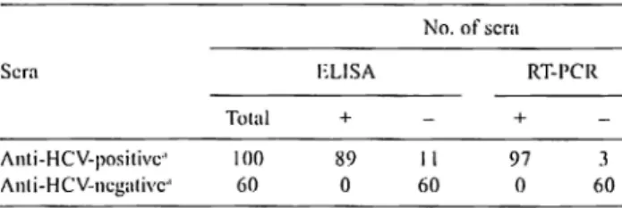
The purified C 178 was used in ELISA. The specificity of the C 178 protein in ELISA was determined by testing 100 HCV EIA-posilive (exclusively la and lb subtypes) and 60 EIA-negativc sera as determined by a 3,t1 generation EIA. For comparison, also RT-PCR was performed to detect HCY RNA in the tested sera. The results are shown in Table I. A negative control serum was previously tested and found HCV RNA-negative by RT-PCR and anti-HCY-negative by Western blot analysis. ELISA readings at least 2.5 times higher than that of the negative control were regarded as positive, while those at most 1.5 times lower than that of the negative control were regarded as negative. No ELISA readings between 1.5-fold and 2.5-fold of that of the negative control were obtained. Of
JOO
E!A-positive sera 89 were positive and 11 were negative in ELISA. All the 60 El A-negative sera were also ELISA-negative. In order to further test lhc 11 El A-positive and ELISA-negative sera we performed Western blot analysis using the C 178 protein as a probe antigen (data not shown). We could not detectTable I. Results of anli-HCV assays b~· EIA and ELISA and IICV RNA assay hy RT-PCR
Sera
Total Anti-HCV-positivc-' 100 /\nl i-HCV-ncgalivc·' 60
"l)ctcrmincd by a 3"' generation El/\. (+), (-) = positive, negative. ELISA + 89 0 No. of scra RT-l'CR + 11 97 3 60 0 60
32
EROGLU, C. et al.: ELISA OF ANTIBODIES TO HCVany reaction for 3 of these sera. These sera were found HCV
RNA-negative in RT-PCR suggesting that they were false
ETA-positive. The remaining 8 sern were found Western
blot-positive and RT-PCR-blot-positive.
In conclusion, 92% of the patients, which were
anti-HCV-positive by ETA, were anti-HCV-anti-HCV-positive also by ELISA
using the C 178 antigen. This antigen showed no false
positive results with the negative control sern suggesting
that it interacted specifically with anti-HCV
Discussion
In this study, the 3 · -truncated con: gene of HCV was
cloned and expressed and its product was purified and used
in testing of human sera in order to assess its ability to
diagnose HCV infection. Since the sequence of this gene
varies among genotypes, we have first aligned the available
GenBank HCY core gene entries and designed the primer
pair to amplify the 178 amino acids long region of the core
gene. This region showed variation. Moreover, the
C-tcrminal end of the core protein (aa 172-191) is highly
hydrophobic and reported to be cleaved which suggests that
its first 171 amino acids contain important domains and
possibly all of the immunodominant epitopes (Hijikata
et
al., 1991 ). Thus were concluded that the 178 amino acids
long core antigen is comparable to the complete one as a
probe in detecting antibodies raised by host against the HCV
core. The amplified core protein (C 178) was cloned into
bacterial expression plasmid pQE30, expressed in£.
coli
as an N-terminal his-tagged fusion protein, and purified by
Ni ion affinity chromatography under denaturing conditions.
The 6-His affinity tag not only enabled to purify the
expressed protein by affinity chromatography but also to
minimize addition of extra amino acids to the C 17 8 protein.
To date, all core region antigens used in detecting HCV
infection have either big polypeptides or small synthetic
oligopeptides fused at their N-terminus. These fused
(attached) polypeptides may exhibit non-specific reactions
and reduce the reaction sensitivity of epitope(s). The core
protein has been previously expressed in different organisms
including insects, yeast,
E.coli and mammalian cells and
found to be useful for early diagnosis of HCV infection
(Chiba
et al., 1991; Harada et al., 1991; Chien et al., 1992;
Yokosuka
et al., 1993). However, in these studies, the
expressed core antigen was not much purified or did not
cover as large as 178 amino acids long portion of the core
protein. The largest core protein that was expressed and
purified to homogeneity (CI 15) contained its first 115 amino
acids (Seki
et al., 1995).
We used the purified His-tagged C 178 in establishing an
ELISA for detection of antibodies against HCV core protein
in human scra. Eighty-nine of the 100 chronic hepatitis
patients, that were previously tested by a 3"
1generation ElA
and found positive, were found positive also in this ELISA,
while 60 healthy (control) subjects were negative both in
EIA and ELISA. The cut off value for a positive resul was
chosen at the 2.5-fold of the average of the negative control
readings. The limit for a negative result was chosen at the
I .5-fold of that average. There were no readings between
these two limits, however, in the future, samples with such
readings should be retested, and if these results would be
confirmed, another method such as Western blot analysis
should be used. In order to decide whether our 11
ELISA-negative sera were due to insensitivity of our ELISA or lack
of antibodies against C 178, we subjected them to Western
blot analysis and RT-PCR. Eight of these sera were positive
in both tests. Three EIA-positive sera were negative in both
ELISA and RT-PCR, which suggested that they were false
EIA-positivc. Therefore it can be concluded that the
sensitivity of our ELISA was 92% (89/97) compared to ETA.
As C 178 was produced in
E. coli, there might he some
impurities in the protein preparation and these could interact
with the sera and give false positive results. In order to
eliminate this possibility, an£.
coli lysate was prepared and
used as an internal control in ELISA. We found that it did
not interact with the sera.
There are I'', 2
1111and 3"
1generation EIAs used worldwide
for the diagnosis ofHCV infection. The sensitivity of these
assays ranges from 80%, to 97%. When compared to them,
the sensitivity of our ELISA is between the 2"'
1and 3"
1generation EIAs. These EIAs use antigens from the core
region as well as NS3, NS4 and NS5 regions. The core
protein is the putative capsid protein of HCV and antibodies
directed to the viral capsicl proteins arc expected to arise
early in infection. Therefore, antibodies against the core
protein most probably arise much earlier than those against
the non-structural proteins. That is why it is important to
include all of the epitopes of the core region into the probe
antigen used in an EIA to increase its efficiency. We believe
that CI 78 protein contains all the epitopes of the core region
oflTCV
Acknowledgement. This work was supported by a grant from ICGEB, Jtaly.
References
Aach R, Stevens C, Hollinger 1'~ Mosley J, Peterson D, 'faylor P, Johnson R ( 199 I): Hepatitis C virus infection in post-transfusion hepatitis. An analysis with first- and second-generation assays. N Engl .. I. Mee/. 325, 1325-1329. Barrera J, Prancis B, Ercilla G, Nelles M,Achord D, Darner J, Lee
S ( 1995): Improved detection of anti-HCV in post-transfusion hepatitis by a third-generation ELISA. 11,x
EROGLU, C. et al.: ELISA OF ANTTBODIES TO HCV
33
Bradford MM ( 1976): A rapid and sensitive method for the quantitation of microgram quantities of protein utilizing the principle of protein-dye binding. Anal. Bioc:l1e111. 7, 248-254.
Bukh J, Purcell RH, Miller R ( 1994): Sequence analysis of the core gene of 14 hepatitis C virus genotypes. Proc. Natl.
A cad. Sci. USA 87, 8898-8902.
Chiba J, Ohba H, Matsuura Y, Watanabe Y, Katayama T, Kikuchi S, Saito I, Miyamura T ( 1991 ): Serodiagnosis of hepatitis C virus (HCV) infection with an I ICV core protein molecularly expressed by a recombinant baculovirus.
Proc. Natl. Acad. Sci. USA 88, 4641-4645.
Chien DY, Choo QL, Tabrizi A, Kuo C, McFarland J, Berger K, Lee C, Shuztcr JR, Nguyen T, Moyer DL, Tong M, Furuta S, Omata M, Tegtmeier G, Alter H, Schiff E, Jeffers L, Houghton M, Kuo G ( 1992): Diagnosis of hepatitis C virus (HCV infection using an immunodominant chimeric polyprotein to capn1re circulating antibodies: Reevaluation of the role of HCV in' liver disease. Proc.
Natl. Acad. Sci. USA 89, 1001110015.
Choo QL, Kuo G, Weiner AJ, Overby LR, Bradley DW, Houghton M ( 1989): Isolation of a cDNA clone derived from a blood-borne non-A, non-B viral hepatitis genome. Science 244, 359-364.
Gretch DR (1997): Diagnostic tests for hepatitis C. Hepa10/ogy
26, 43-47.
Harada S, Watanabe Y, Takeuchi K, Suzuki T. Katayama T, Takabe Y, Saito J, Miyamura T ( 1991 ): Expression of processed core protein ofhcpatitits C virus in mammalian cells . . I.
Virol. 65, 3015-3021.
Heintges T, Wands JR ( 1997) Hepatitis C virus: epidemiology and transmission. Hepatology 26, 521-526.
Hijikata M, Kato N, Ootsuyama Y, Nakagawa M, Shimotohno K (1991): Gene mapping of the putative structural region of the hepatitis C virus genome by in vitro processing analysis. Proc. Natl. Acacl. Sci. USA 88, 5547-555 l. Kao G, Choo QL, Alter HJ, Gitnlck GL, Redeker AG, Purcell RH,
Miyamura T, Dienstag JL, Alter l'vlJ, Stevens CE, Tegtmeier GE, Bonino I-~ Colombo M, Lee WS, Kuo C, Berger K, Shuster JR, Overby LR, Bradley OW, Houghton M ( 1989): An assay for circulating antibodies
to a major etiologic virus of human non-A, 11011-8 hepatitis. Science 244, 362-364.
Kleinman S, Alter H, Busch M, Holland P, Tegtmeier G, Lee S (1992): Increased detection of HCV-infcctcd blood donors by a multiple-antigen HCV enzyme immunoassay. Tra11.\"fi1sion 32, R05-8 I 3.
Mimms L, Vallari D, Ducharme L, Holland P, Kuramato IK, Zcldis J ( 1990): Specificity of anti-HCV ELISA assessed by reactivity to three immunodominant HCV regions. Lancet 336, 925-930.
Murphy EL, Bryzman S, Williams AE, Co-Chien H, Schreiber GB, Ownby HE ( 1996): Demographic determinants of hepatitis C virus scroprevalancc among blood donors.
JAMA 275, 995-1000.
Nagayama R, Tsuda F, Okamoto H, Wang Y, Mitsui T, Tanaka T, Miyakawa Y, Mayumi M ( 1993): Genotype dependence of hepatitits C virus antibodies detectable by the first-generation enzyme-linked immunosorbcnt assay with CI 00-3 protein . . I. Clin. Invest. 92, 1529-1533. Sambrook J, Fritsch EF, Maniatis T ( 1989): Molecular Cloning: A
Lahorato,:v Manual. 2"~--!!d., Cold Spring Harbor
Laboratory Press, Cold Spring Harbor.
Seki M, Honda Y, Kondo J, Fukuda K, Ohta K, Sugimoto J, Yamada E ( 1995): Effective production of the hepatitis C virus core antigen hnving high purity in E. coli . . I. Biotec:/11101. 38, 229-241.
Urden MS, Wucstehube LJ, Laurenson PM, Wilber JC ( 1997): Hepntitis C - dia!,'llosis and monitoring. Cli11. Chem. 43, 1507-1511. Uyttcndaele S, Claeys H, Mertens W, Verhacrt H, Vermylcn C ( 1994):
Evaluation of third-generation screening and confirmatory assays for HCV antibodies. Tvx Sang. 66, 122-129. Wilson JJ, Polyak SJ, Day TD, Gretch DR ( 1995): Characterization
of simple and complex hepatitis C virus quasispecies by heteroduplcx gel shift analysis:correlation with nucleotide sequencing . . I. Gen. Virol. 76, 1763-1771.
Yokosuka 0, Ito Y, Sakuma J, Imazeki F, Ohio M, Omata M ( 1993): Expression of hepatitis C virus core protein as a fusion protein with maltose binding protein. Dig. Dis. Sci. 38,
626-630.
Younossi Z, McHutchison J ( 1996): Serological tests for HCV infection. Viral Hepatitis Rev. 2, 161-173.