Possible Relationship Between Bilateral Elastofibroma Dorsi and
Hypermastia
İlknur Keskin1, Bircan Kolbaşı1, Aslı Akhan2, Hasan Çelik3, Mustafa Hancı3, Mustafa Keskin3
1Department of Histology and Embryology, İstanbul Medipol University School of Medicine, İstanbul, Turkey 2Department of Pathology, İstanbul Medipol University School of Medicine, İstanbul, Turkey
3Department of Plastic Reconstructive and Aesthetic Surgery, İstanbul Medipol University School of Medicine, İstanbul, Turkey
Elastofibroma dorsi (EFD) is a rare, benign, soft tissue tumor with an unclear pathogenesis, typically localized to the subscapular region. It occurs within the periscapular area between the ribs and dorsal chest wall muscles. Repetitive microtrauma by friction be-tween the lower part of the scapula and the thoracic wall may cause reactive hyperproliferation of the fibroblastic tissue. This view has been supported by the higher prevalence of EFD, particularly among individuals who perform hard manual labor. EFD can, however, also be observed in those who have never performed hard manual labor and particularly in women over the age of 50. In this report, we present a case of bilateral EFD in a 52-year old woman with bilateral hypermastia, and examine the possible correlation between hypermastia and EFD.
Keywords: Elastofibroma dorsi, breast hypertrophy, hypermastia
Abstract
DOI: 10.5152/TurkJPlastSurg.2017.2131
INTRODUCTION
Elastofibroma dorsi (EFD) is a rare, benign soft tissue tumor typically localized to the subscapular region. It is mostly seen on the scapular plane, bilaterally or unilaterally, in adults over mid-age. These masses appear poorly circumscribed since they are deeply located in the musculature. They are often asymptomatic and grow slowly. Uncertainties remain about its pathogenesis. Although The World Health Organization has defined elastofibroma as a benign fibroblast tumor, many authors do not accept it as a real tumor, but rather as the hyperplasia of elastic tissues associated with the mechanical irritation caused by the friction between the scapula and the thoracic wall. It is suggested that repetitive trauma caused by friction leads to an overproduction of the collagenous connective tissue, and thereby to the degeneration of the collagen fibers, which are filled with excessive amounts of elastinophilic, polymorphic fibers and hyperplastic adipose tissue.2 This suggestion primarily bases on the early reports of higher prevalence of EFD particularly among individuals who perform hard manual labor. Publications in the following years, however, have reported EFD to be seen in individuals of other types of professions, and even in housewives, although more prevalent in some specific professions.3 Another aspect that has been demonstrated over the years is the female predilection of EFD, with a majority being over 50 years of age.
In our case presentation, we would like to draw attention to the possible correlation between hypermastia and EFD based on a patient who presented to our clinic with large breasts and was diagnosed with bilateral EFD.
CASE PRESENTATION
A 52-year old housewife presented to our clinic with complaints of large breasts, rash and bad smell underneath the breasts between the folds of skin, as well as pain and swelling in her back. The pain increased when she moved her arm. She had received long-term physiotherapy for her back and shoulder pain. Physical examination showed that her breasts were asymmetrically positioned on the
154
www.turkjplastsurg.org
Correspondence Author: Dr. Mustafa Keskin E-mail: [email protected]
Received: 17.08.2016 Accepted: 18.11.2016 Cite this article as: Keskin İ, Kolbaşı B, Akhan A, Çelik H, Hancı M, Keskin M.Possible Relationship Between Bilateral Elastofibroma Dorsi and Hypermastia. Turk J
Plast Surg 2017; 25(3): 154-158.
Content of this journal is licensed under a Creative Commons
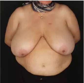
chest wall, and the distance between the nipple-areolar com-plex and the sternal notch measured 37 cm on the right and 36 cm on the left (Figure 1). Examination of her subscapular region revealed soft and mobile bilateral masses in the peri-scapular area of four cm on the left and a more palpable one of 5 cm on the right. Both masses were more palpable with arms abducted. Adenopathy was not observed in the axillary cavity. Neurological examination of the upper extremities was normal. Her laboratory tests and medical history involved no other significant data. Magnetic resonance imaging showed lesions of 38x19x48 mm in the right infrascapular area and of 49x32x45 mm in the left infrascapular area that demonstrat-ed mild contrast uptake following the injection of contrast material. Both had the same intensity as the muscle tissue and were consistent with elastofibroma (Figure 2). Taking into account the patient’s complaints of pain, and to pathological-ly confirm the diagnosis, surgical excision of the subscapular lesions was planned together with a breast reduction surgery.
The patient was taken to operation in prone position under general anesthesia. Surgical exploration was performed over bilateral five cm horizontal skin incisions in the infrascapular area, and 12 cm laterally to the midline. Scapular muscles were cauterized to reach the masses. Mobile, rigidly elastic, ill-defined and non-vascular masses were firmly attached to the costal ribs, advancing toward the subscapula. The masses were dissected and totally excised over the posterior costal plane (Figure 3). Drains were placed and the surgical site was closed. Then the patient was positioned supine, and superior medial pedicle breast reduction was performed to move the areolas from 37 cm and 36 cm to 21 cm, 1.890 g and 1.810 g tissue were resected from the right and left breasts, respec-tively, and the sites were closed. Pressure corset was applied after the operation and her shoulders were immobilized for one week. No events or recurrence was encountered in the one-year follow-up (Figure 4).
Histologic Evaluation
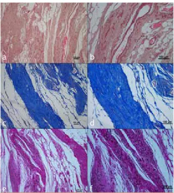
The excised tissue was fixed in 10% formalin, and then em-bedded in paraffin. Microtome sections of 5 µm were cut from the paraffin blocks (Thermo Scientific, HM 340E, Thermo Fish-er Scientific Germany Co. KG, Bonn, Germany). Histopatho-logical evaluation was performed with standard techniques and Hematoxylin-Eosin, Masson Trichrome, and Weigert Van Gieson staining (Bio-Optica, CAT#2311, CAT#5114, CAT#04-010802, CAT#04-053812, Milan, Italy) were used. The material was assessed and visualized with a light microscope. Further, the material was labelled with anti-Heat Shock Protein (HSP) 70 (anti-HSP-70 1:100, sc-24, Sigma-Aldrich, St Louis, USA), to evaluate HSP-70 expression, and visualized with a confocal microscope (Zeiss LSM780, Carl Zeiss, Oberkochen, Germany). Histologic examination revealed macroscopically solid le-sions sporadically covered with fibrous capsules and con-taining fatty tissue, with yellowish-white pigmentation on the surface of the cross-section. In microscopic examination of hematoxylin-eosin sections, increase in collagen fibers of the fibroadipous connective tissues, mature adipocytes, and fibrillous elastic fibers proliferated in a dispersed pattern were observed. Masson Trichrome stain revealed intensive
Figure 2. a, b. Magnetic resonance imaging: T2-weighted (a) and T1-weighted (b) images on axial plane show lesions (arrows) that have the
same intensity as the muscle tissue and are consistent with elastofibroma.
a
b
Figure 1. 52-year old patient presenting with back and neck pain,
and large breasts.
collagen distribution. Elastic fibers stained with elastic van Gieson stain were typically observed to be globule-, disc- and flower-like small degenerate fragments (Figure 5). Positivity of HSP-70 expression in the sections was shown with immuno-fluorescent staining using the standard technique (Figure 6). DISCUSSION
A relatively recently defined tumor, elastofibroma dorsi (EFD) was first described by Jarvi and Saxen in 1961.4 In most of the cases the tumor localizes to the lower end of the scapula.
Sur-Figure 3. Macroscopic view of surgical specimen.
Figure 4. View of patient in first month after the operation.
Figure 6. HSP70 (red) and DAPI (blue) expression in cross-sections (X20, bar = 50 µm)
Figure 5. a-f. (a, b) Hematoxylin and Eosin; (c, d) Masson trichrome;
(e, f) Weigert van Gieson stain (X10, X20, bar = 100µm) Elastic fibers randomly dispersed in collagen matrix. Hypocellular fibrous tissue contains thick, eosinophilic, fragmented elastic fibers. Diffuse blood vessels and adipose tissue. Elastin stain show fibrous tissue to con-tain many thick, fragmented elastic fibers intertwined with adipose tissue. Weigert van Gieson stain show elastic fibers with irregular contours (c).
a
c
e
b
d
f
156
rounded by the subscapular, romboid, latissumus dorsi and serratus anterior muscles, it localizes on the periosteum of the thoracic wall. It does not attach itself to the skin. Very few pa-thologies of this type have been reported in other regions of the body.4,5 Clinically, most of the cases are asymptomatic or patients present with rigid palpable swelling. Especially larger masses can lead to pain in the back and the shoulder region. Most of the reported cases are unilateral and often on the right side. Bilateral cases are mostly asymmetrical, with a more significant mass on one of the sides. In some cases, the patient’s complaints will involve a unilateral mass and the sec-ond one will be revealed radiologically. Other subcutaneous tumors, lipomas, cystic formations and vascular malforma-tions, metastatic or primary sarcomas, desmoid tumors, neu-rofibroma, fibrous histiocytomata, and fibromatosis should be prioritized for differential diagnosis. Needle aspiration or incisional biopsy is not necessary for differential diagnosis; radiological imaging will be sufficient.5 MR imaging is nec-essary for confirming the diagnosis. Well-documented cases through clinical and radiological findings can be surgically excised without biopsy confirmation. Total excision can be easily performed when the rich vascular and neurological anatomy of the region is observed. Postoperative prognosis is good and recurrence rate is very low.6
Since EFD is a rare pathology, there are no clear data on its pathogenesis. Available reports present single or a small number of cases. Genetic predisposition is also being consid-ered since some patients present with positive family histo-ry.1 While recent developments in genetic analysis methods have provided new findings about the genomic changes in the pathogenesis of EFD, the data fall short of demonstrating that EFD is an outcome of chromosomal anomaly.7 The most widely accepted view in explaining the pathogenesis of EFD describes EFD as a reactive formation. According to the classic view proposed by Jarvi et al.4, repetitive microtraumas in the subscapular region caused by the friction between the low-er end of the scapula and the thoracic wall leads to reactive hyperproliferation of the fibroelastic tissue. While the preva-lence of EFD particularly among individuals that perform hard manual labor seems to support this view, the condition is also seen among individuals who have not performed such hard labor in their lifetime or among those who live in different geographies.8 According to Giebel et al., EFD occurs during the physiological aging process which develops secondary to vascular insufficiency.9 In conclusion, pathophysiological determinants for EFD are still unclear and various hypotheses are suggested.
Considering that most of the EFD cases that do not emerge as an occupational disease involve women over 50 years of age, it is beneficial to question the role of large breasts in terms of its pathogenesis in this case group. In an autopsy se-ries, clinically undetected EFD of less than 3 cm in diameter were identified in 11% of the women over 50.10 The back and neck pain often described by patients presenting with large breasts may not only be due to the size of the breasts. Since large breasts often strain the vertebral column, the pain may
also be a symptom of a clinically undiagnosed underlying EFD. Large breasts can place substantial burden on the verte-bral column. The weight on the anterior thoracic wall increas-es the curvature of the vertebral column. Large breasts have been reported to shift the body’s center of gravity forward, away from the spine, which in turn increases cervical lordosis and thoracic kyphosis.11 This forward shift leads to persistent contractions in the posterior spinal muscles, especially the trapezius muscle.12 Increased thoracic kyphosis and flattened lumbar lordosis also affect the relationship between the scapula and the thoracic wall. The close contact between the scapula and the thorax possibly leads to increased irritation. The subacromial gap has been shown to narrow significantly with increased thoracic kyphosis.13 Narrowing of the subacro-mial gap restricts the movement of the scapula and changes its planar position. Thus, large breasts can potentially lead to repetitive microtraumas, hence to the reactive hyperprolifer-ation of fibroelastic tissue of recurrent microtraumas in this region. A breast reduction surgery can significantly reduce the thoracic kyphosis and lumbar lordosis angles, and are also reported to improve poor posture.14-16
It is well-known that large breasts lead to back and neck pain; however, they can also lead to EFD. In such case, a breast re-duction surgery will not relieve the pain. Initially, ultrasound imaging, and in case suspicion persists, magnetic resonance imaging can confirm the diagnosis. To relieve the patient of the pain and to ensure a state of wellness, it may be necessary to identify and resect the EFD localized to the subscapular or infrascapular area.
Informed Consent: : Written informed consent was obtained from
patient who participated in this case.
Peer-review: Externally peer-reviewed.
Author contributions: Concept - M.K., M.H.; Design - M.H.; Supervision -
İ.K.; Resource -H.Çİ.K.; Materials - B.K., İ.K.; Data Collection and/or Proces-sing - İ.K., A.A.; Analysis and/or Interpretation - İ.K., A.A.; Literature Search - A.A., H.Ç.; Writing Manuscript - M.K., İ.K.; Critical Reviews - M.K., M.H.
Conflict of Interest: No conflict of interest was declared by the
aut-hors.
Financial Disclosure: The authors declared that this study has
recei-ved no financial support.
REFERENCES
1. Parratt MT, Donaldson JR, Flanagan AM, Saifuddin A, Pollock RC, Skinner JA, et al. Elastofibroma dorsi: management, outcome and review of the literature. J Bone Joint Surg Br 2010; 92(2): 262-6. [CrossRef]
2. Di Vito A, Scali E, Ferraro G, Mignogna C, Presta I, Camastra C, et al. Elastofibroma dorsi: a histochemical and immunohistochemi-cal study. Eur J Histochem 2015; 59(1): 2459. [CrossRef] 3. Koksel O, Demir Apaydin F, Ayan E, Demir M, Ozdulger A.
Elas-tofibroma dorsi: review of eight cases. Surg Today 2010; 40(5): 423-7. [CrossRef]
4. Jarvi O, Saxen E. Elastofibroma dorse. Acta Pathol Microbiol Scand Suppl 1961; 51(Suppl 144): 83-4.
5. Faccioli N, Foti G, Comai A, Cugini C, Guarise A, Mucelli RP. MR imaging findings of elastofibroma dorsi in correlation with pat-hological features: our experience. Radiol Med 2009; 114(8): 1283-91. [CrossRef]
6. Akçam Tİ, Çağırıcı U, Çakan A, Akın H. Bilateral familial elasto-fibroma dorsi: is genetic abnormality essential? Ann Thorac Surg 2014; 98(2): e31-2. [CrossRef]
7. Karakurt O, Kaplan T, Gunal N, Gulbahar G, Kocer B, Han S, et al. Elas-tofibroma dorsi management and outcomes: review of 16 cases. Interact Cardiovasc Thorac Surg 2014; 18(2): 197-201. [CrossRef] 8. Giebel GD, Bierhoff E, Vogel J. Elastofibroma and
pre-elastofibro-ma: a biopsy and autopsy study. Eur J Surg Oncol 1996; 22(1): 93-6. [CrossRef]
9. Järvi OH, Länsimies PH. Subclinical elastofibromas in the scapu-lar region in an autopsy series. Acta Path Microbiol Scand Sect A 1975; 83(1), 87-108. [CrossRef]
10. Findikcioglu K, Findikcioglu F, Ozmen S, Guclu T. The impact of breast size on the vertebral column: a radiologic study. Aesthetic Plast Surg 2007; 31(1): 23-7. [CrossRef]
11. Letterman G, Schurter M. The effects of mammary hypertrop-hy on the skeletal system. Ann Plast Surg. 1980; 5(6): 425-31. [CrossRef]
12. Gumina S, Di Giorgio G, Postacchini F, Postacchini R. Subacromial space in adult patients with thoracic hyperkyphosis and in healt-hy volunteers. Chir Organi Mov 2008; 91(2): 93-6. [CrossRef] 13. Karabekmez FE, Gokkaya A, Isik C, Saglam I, Efeoglu FB, Gorgu
M. Does reduction mammaplasty revert skeletal disturban-ces in the vertebral column of patients with macromastia? A preliminary study. Aesthetic Plast Surg 2014; 38(1): 104-12. [CrossRef]
14. Berberoğlu Ö, Temel M, Türkmen A. Effects of Reduction Mam-maplasty Operations on the Spinal Column: Clinical and Ra-diological Response. Aesthetic Plast Surg 2015; 39(4): 514-22. [CrossRef]
15. Foreman KB, Dibble LE, Droge J, Carson R, Rockwell WB. The im-pact of breast reduction surgery on low-back compressive for-ces and function in individuals with macromastia. Plast Reconstr Surg 2009; 124(5): 1393-9. [CrossRef]