Original Article
Investigation of dose-dependent neuroprotective effect
of human recombinant erythropoietin in acute spinal
cord injury induced rats
Bülent Özdemir1, Ersegun Batçık1, Ezgi Ayçiçek2, Gökhan Canaz3, Osman Akdemir3, İbrahim Alataş4, Hüseyin Canaz4
1Department of Neurosurgery, Faculty of Medicine, Rize Tayyip Erdogan University, Turkey; 2Department of
Neu-rosurgery, Haydarpasa Numune Reseach and Training Hospital, Turkey; 3Department of Neurosurgery, Haseki
Research and Training Hospital, Tuekey; 4Department of Neurosurgery, Florence Nightingale Hospital, Istanbul
Bilim University, Turkey
Received October 24, 2015; Accepted March 5, 2016; Epub March 15, 2016; Published March 30, 2016
Abstract: The aim of this study was to investigate the dose dependent neuroprotective effect of recombinant human erythropoietin (r-Hu-EPO) in acute spinal cord injury induced rats. The rats were allocated into 4 groups of 8 rats each. Spinal cord injury was produced by Yaşargil aneurysm clip at a pressure of 0.7 N for a duration of 60 seconds. Group I (Controls) received laminectomy only. Group II (The trauma-only group) had no medication. In group III post-operative intraperitoneal (IP) EPO, total 2000 IU/kg, were applied in two doses. Group IV received postpost-operative IP EPO in 3 doses, total 9000 IU/kg. In all groups, neuromotor evaluation using Basso’s locomotor grading test was conducted at the 6th and 24th hours, and every day from the 1st to 15th days following surgery. After the fifteenth
day, all rats were sacrificed and spinal cord samples were obtained for the assessment of caspase-3 activity. The results showed that caspase-3 activity increased to statistically significant higher levels in the spinal cord after in-jury comparing to the control group. Caspase-3 enzyme activity levels were significantly reduced in animals treated either with low dose or high dose of r-Hu-EPO. In addition, we observed significant difference about rate of functional recovery between group 3 and group 4. Our study showed that high dose EPO administration in the early period after spinal cord trauma increases neurological improvement to a greater degree and in a more rapid manner.
Keywords: Erythropoietin, caspase-3, functional recovery, spinal injury, neuroprotective
Introduction
Spinal cord injury causes permanent neurologi-cal deficits and secondary complications in individuals of all ages [1, 2]. The low chance of full neurological recovery after spinal cord inju-ry arouses more interest in posttraumatic bio-chemical and pathological processes and sec-ondary autodestructive mechanisms caused by trauma [3-6]. Although the mechanism is not exactly known, it is thought that pathological processes such as edema, inflammation, decreased blood flow, changes in microvascu-lar permeability contribute to secondary injury development, which arises from various path-ways including lipid peroxidation and free oxy-gen radicals [2-6]. Therefore, there are many studies focused on neuroprotective agents that could be used against secondary injury [3-9].
Apoptosis is just one of the complex processes that cause tissue injury that develops after spi-nal cord damage [10]. Apoptosis is mainly regu-lated by the caspase enzyme family, which is a cysteine protease [11]. The apoptotic role of caspases in injured spinal cord has been dem-onstrated in many experimental studies [12-20]. It is reported that apoptosis also occurs in human spinal cord injuries [21].
Erythropoietin (EPO) is a hematopoietic growth factor that is produced in the renal and fetal liver and stimulates the proliferation and differ-entiation of erythroid precursor cells. Addi- tionally, it is also produced in the central ner-vous system and exhibits non hematopoietic effects [22-24]. In previous studies, it has been reported that it has trophic effects and neuro-protective effects against experimental brain
injury and ischemia in the cholinergic neurons of rats [25-29]. It has neuron protective effects and a vasoconstriction-preventive effect in sub-arachnoid hemorrhage [30-32]. The neuropro-tective effect of EPO is connected to its inhibi-tion of free radical producinhibi-tion and release of excitatory amino acids [28, 33, 34].
The aim of the current study is to investigate dose-dependent neuroprotective effect of EPO in experimental acute spinal cord injury induced rats. In the study, caspase-3 activity was used as a biochemical marker, whereas in the assessment of functional recovery, the locomo-tor grading scale defined by Basso et al. was used.
Material and method Spinal cord injury
Thirty-two male Wistar rats, weighing between 230 and 260 g were used in the study. Anesthesia was performed with xylazine (10 mg/kg) and ketamine hydrochloride (60 mg/ kg) in rats. Following dorsal incision in the prone position, a T6-T9 laminectomy was per-formed. Spinal cord injury was produced by the extradural application of a Yaşargil aneurysm clip at a pressure of 0.7 N for a duration of 60 seconds. Following homeostasis the layers were closed with 3/0 silk in accordance with the anatomy. The studies were conducted with the permission of the Istanbul University Experimental Animals Ethics Committee approval.
Study groups
Group I: (n=8 rats) only laminectomy was performed.
Group II: (n=8 rats) trauma was produced after laminectomy by a Yaşargil aneurysm clip at a pressure of 0.7 N for a duration of 60 seconds.
Group III: (n=8 rats) following trauma produced after laminectomy by a Yaşargil aneurysm clip at a pressure of 0.7 N for a duration of 60 sec-onds; early postoperative IP (intraperitoneal) erythropoietin 1000 IU/kg and postoperative IP erythropoietin 1000 IU/kg (2 doses, total 2000 IU/kg) 24 hours later, were applied.
Group IV: (n=8 rats) following trauma produced after laminectomy by a Yaşargil aneurysm clip
at a pressure of 0.7 N for a duration of 60 sec-onds; early postoperative IP (intraperitoneal) erythropoietin 5000 IU/kg and postoperative IP erythropoietin 3000 IU/kg (3 doses, total 9000 IU/kg) 24 hours later and postoperative IP erythropoietin 1000 IU/kg 48 hours later were applied.
In all rats, neuromotor evaluation using Basso’s locomotor grading test [35] was conducted at the 6th and 24th hours, and the 1st , 2nd, 3rd , 4th, 5th, 6th, 7th, 8th, 9th, 10th, 11th, 12th, 13th, 14th, and 15th days following surgery. After the fif-teenth day, anesthesia was performed with ket-amine hydrochloride (IP) 60 mg/kg and xylazine (IP) 10 mg/kg; spinal cord samples of rats were then obtained and were held at -80°C in a deep freezer for evaluation of caspase-3 activity. Then all rats were sacrificed by perfusion. Evaluation of functional recovery
The neurological motor function of each rat was evaluated by using thelocomotor grading scale defined by Basso et al. [35, 36]. With this grad-ing scale, all joints of the hind leg could be eval-uated in details. The scale is produced from scores ranging between 0 and 21. While there was no observable movement in the 0 score, the 21 score represents a normal, healthy rat. The locomotor grading scale was applied at the 6th and 24th hours, and the 1st, 2nd, 3rd, 4th, 5th, 6th, 7th, 8th, 9th, 10th, 11th, 12th, 13th, 14th, and 15th days to all rats used in the current study. Biochemical assessment
Colorimetric measurement was done with Biovision trademarked caspase-3/CPP 32 ELISA kit. This kit was developed to measure caspase activity recognizing DEVD sequence in an apoptotic pathway. The measurement is based on the detection of light intensity scat-tered by pNA, which is a chromophore and aris-es by the degradation of marked substrate DEVD-pNA (p-nitroanilide). The light intensity scattered by p-nitroanilide was read in microti-ter plaque reader or in the spectrophotomemicroti-ter at 405 nm wave length. The increase in CPP32 activity was determined by comparing the absorbance of pNA in the apoptotic sample with samples in which apoptosis induction did not occur. The tissue samples in which apopto-sis induction has been done were stored at -80°C until the study was done. Homogenization was conducted by studying with kit specific cell
lyses tamponade in a cold environment. The tissue samples were preserved in ice during the study. After homogenization, the tissue samples were centrifuged and a protein analy-sis of supernatant was done by the biuret meth-od in an Abbott architect trademarked autoana-lyzer. Appropriate to the protocol, by adding DEVD-pNA substrate, it was incubated at 37°C for 2 hours. Caspase-3 activity was determined by reading at 405 nm proportional to the amount of protein of the samples.
Statistical analysis
SPSS for Windows 10.0 statistical package pro-gram was used in the assessment of the data.
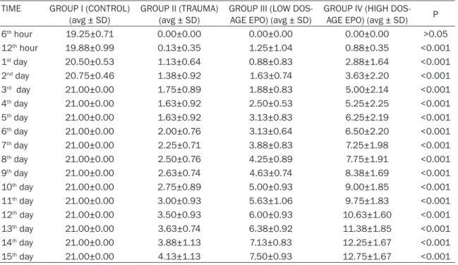
Caspase-3 activities of the groups were deter-mined by obtaining spinal cord samples at the end of the 15th day. Statistically significant dif-ferences were found between all groups in terms of caspase-3 activity at the end of 15th day (Table 2; Figure 2). Caspase-3 activity was significantly higher in the trauma group accord-ing to the other groups (P<0.001). The values of the groups in which low and high dose erythro-poietin was used were also significantly higher than the laminectomy group (P<0.001). It was found that erythropoietin use significantly decreases caspase-3 activity according to the trauma group (P<0.001). It was concluded that low dose EPO use significantly decreases cas-Table 1. Motor function scores according to time
TIME GROUP I (CONTROL)
(avg ± SD) GROUP II (TRAUMA) (avg ± SD) GROUP III (LOW DOS-AGE EPO) (avg ± SD) GROUP IV (HIGH DOS-AGE EPO) (avg ± SD) P 6th hour 19.25±0.71 0.00±0.00 0.00±0.00 0.00±0.00 >0.05 12th hour 19.88±0.99 0.13±0.35 1.25±1.04 0.88±0.35 <0.001 1st day 20.50±0.53 1.13±0.64 0.88±0.83 2.88±1.64 <0.001 2nd day 20.75±0.46 1.38±0.92 1.63±0.74 3.63±2.20 <0.001 3rd day 21.00±0.00 1.75±0.89 1.88±0.83 5.00±2.14 <0.001 4th day 21.00±0.00 1.63±0.92 2.50±0.53 5.25±2.25 <0.001 5th day 21.00±0.00 1.63±0.92 3.13±0.83 6.25±2.19 <0.001 6th day 21.00±0.00 2.00±0.76 3.13±0.64 6.50±2.20 <0.001 7th day 21.00±0.00 2.25±0.71 3.88±0.83 7.25±1.98 <0.001 8th day 21.00±0.00 2.50±0.76 4.25±0.89 7.75±1.91 <0.001 9th day 21.00±0.00 2.63±0.74 4.63±0.74 8.38±1.69 <0.001 10th day 21.00±0.00 2.75±0.89 5.00±0.93 9.00±1.85 <0.001 11th day 21.00±0.00 3.00±0.93 5.63±1.06 9.75±1.83 <0.001 12th day 21.00±0.00 3.50±0.93 6.00±0.93 10.63±1.60 <0.001 13th day 21.00±0.00 3.63±0.74 6.38±0.92 11.38±1.85 <0.001 14th day 21.00±0.00 3.88±1.13 7.13±0.83 12.25±1.67 <0.001 15th day 21.00±0.00 4.13±1.13 7.50±0.93 12.75±1.67 <0.001
Figure 1. Motor function scores of all groups according to time.
Student’s t-test and the Mann-Whitney U-test were used in the comparison of the 1st and 2nd groups; the Mann-Whitney U-test, ANOVA, Turkey HSD, and Kruskal Wallis tests were used in the comparison of the 3rd and 4th groups. P<0.05 was accepted as significant.
Results
Findings of biochemical evaluation
pase-3 activity (P<0.05). It was found that high dose EPO use significantly decreases cas-pase-3 activity at an advanced level (P<0.001). No statistically significant difference was found between low dose EPO use and high dose EPO use in terms of caspase-3 activity (P>0.05). Findings of functional evaluation
There was a significant difference between groups in all postoperative periods. In the post-operative 6th hour comparisons, a statistically significant difference was found between Group I and the others (P<0.001). There was no statis-tically significant difference between Group II, Group III, Group IV at the postoperative 6th hour (P>0.05). Between all traumas groups (Group II, Group III, Group IV) significant differ-ences were initiated beginning from the 3rd day. Upon neurological examination of the groups in which erythropoietin treatment was administered beginning from the 4th post- operative day (Group III, Group IV), the recovery was significantly better than trauma group (Group II) (P<0.001). While a significant recov-ery was found in the low dose recov-erythropoietin group (Group III) beginning from the 5th day when compared to the trauma group (Group II), a significant recovery began at the 12th hour in the high dose erythropoietin group (Group IV) (P<0.05). When both treatment groups (Group III, Group IV) were evaluated, the researchers observed that the significant recovery in the high dose erythropoietin group (Group IV) (P<0.05) developed on the 3rd day.
Discussion
Protection from secondary injury in acute spi-nal cord damage is called neuroprotection. For this purpose, many medical and surgical approaches such as correction of tissue oxy-genation with drug therapies, suppression of spinal cord compression, and stabilization of the vertebral column are experienced. For this purpose, many studies aimed at the pharmaco-logical protection in spinal cord damage have been conducted during the recent 20 years; however, none of these became the standard treatment that would be used in humans. Secondary injury is a process that starts within minutes or hours following the primary injury and continues for weeks. It is not possible to treat the primary injury. The purpose of the
studies aimed at secondary injury is to find and use pharmacological agents and preventions aimed at the protection of the vigor of the neu-rons that are still intact after the primary injury, their connections to distal neurons and the neurons in the area of the lesion, and increas-ing their endurance or terminatincreas-ing the patho-logical processes that could cause harm [37].
In clinical practice, EPO is widely used in treat-ment of anemias caused by renal insufficiency, cancer, and surgery [38]. The presence of the neuroprotective effects of EPO has been dem-onstrated in previous studies [28, 30, 39-42]. Today, the knowledge that is obtained following studies is in support of the finding that endog-enously produced erythropoietin plays an important role in the central nervous system [43].
Although the mechanism of action of erythro-poietin on the central nervous system is not exact, within today’s knowledge, it is thought that it demonstrates the effect on signal sys-tems that are different in hematopoietic and neuroprotective characteristics. The receptor producing neuroprotective effect and the receptor producing hematopoietic effect are different from each other [44].
It is found that the activation of caspase-3 that plays important role in development of apopto-sis is substantially decreased with erythropoie-tin treatment [45].
Furthermore there is a direct anti-inflammatory effect of erythropoietin [29, 46].
Another means of the antiapoptotic effect of erythropoietin is the regulation of gene expres-sion [47]. Wen et al. found that by increasing the Blc/Bax ratio and the Blc2 level of erythro-poietin, it leans toward the antiapoptotic side [48].
Additionally, erythropoietin provides neuropro-tection by triggering the nuclear factor-the KB signal pathway with JAK2 activation [49]. The action mechanisms and clinical results of erythropoietin were studied in different experi-mental models by the investigators. The cur-rent study aimed to functionally and biochemi-cally compare the neuroprotective characte- ristics of erythropoietin, its contribution on
functional improvement, and its antiapoptotic effect using different drug doses. The locomo-tor grading scale defined by Basso et al. for the assessment of functional recovery was used [35, 36]. Upon evaluation, all joints of the hind limb underwent neurological examination in such a way that it obtained 0-21 points. Antiapoptotic activity was evaluated biochemi-cally by measuring caspase-3 activity in spinal cord samples that were obtained at the end of 15 days.
Upon the evaluation that was done using the locomotor grading scale, it was observed that motor score significantly decreased in all experimental groups that were exposed to trau-ma when compared to the control group (P<0.001). In all groups that were exposed to trauma (Group II, Group III, Group IV) flask paralysis was detected at the 6th hour and there was no statistically significant difference in terms of motor function assessments that were done at the 6th hour (P>0.05).
It was found that erythropoietin administration after trauma increased motor function scores. In all treatment groups, erythropoietin adminis-tration significantly improves motor functions (Figure 1; Table 1). This finding is parallel to the previous studies that were conducted using erythropoietin [41, 42, 45, 50, 51].
cord trauma [42]. They have formed trauma, 1×1000 IU/kg EPO, and 3×1000 IU/kg EPO groups in their mounted clip model and formed trauma, 1×5000 IU/kg EPO, 7×5000 IU/kg EPO, and 7×500 IU/kg EPO groups in a contu-sion model. In their observations, Gorio et al. detected significant improvement in motor functions in all treatment groups in which eryth-ropoietin was used [42].
In spinal cord trauma model produced by Okutan et al., in the treatment group in which they administered erythropoietin 1000 IU/kg, they found significant recovery in motor func-tions 24 hours later [45].
In the current study, at the 12th hour a statisti-cally significant recovery was found in motor functions of all trauma groups in which erythro-poietin was administered (P<0.05). This result is parallel to the studies of Vasileios et al. and Gorio et al. The recovery of motor functions in the period between the 4th day and the 15th day was statistically significant at the highest degree (P<0.001). Erythropoietin use acceler-ated motor recovery in the early postoperative period. Gorio et al. demonstrated the recovery of motor functions in the same manner [42]. When compared to the trauma group, a signifi-cant recovery in motor functions was achieved starting from the 5th day in the treatment group Table 2. Caspase-3 activations of all groups
GROUP I
(CON-TROL) (avg ± SD) GROUP II (TRAU-MA) (avg ± SD) GROUP III (LOW DOS-AGE EPO) (avg ± SD) GROUP IV (HIGH DOS-AGE EPO) (avg ± SD) P Caspase 3 activity 0.214±0.042 0.615±0.082 0.471±0.12 0.411±0.087 <0.001
Figure 2. Caspase 3 activity.
In a similar experimental study, Vasileios et al. adminis-tered erythropoietin to two trauma groups with 2×1000 IU/kg and 14×1000 IU/kg (28 days) and they found signifi-cant improvement in motor functions on the postopera-tive 3rd day when compared to the trauma groups [51]. In contusion and clip experi-ment models that were pro-duced by Gorio et al., they investigated the effective-ness of erythropoietin in acute and subacute spinal
in which low dose (2×1000 IU/kg) erythropoie-tin was administered (P<0.05); whereas in the treatment group, in which high dose erythropoi-etin (1×5000 IU/kg, 1×3000 IU/kg, 1×1000 IU/ kg) was used, a significant recovery in motor functions was found after the 12th hour (P<0.05). It was seen that the improvement in motor functions was at a high degree starting from the 4th day (P<0.001).
While in the 1×5000 IU/kg and 7×5000 IU/kg treatment groups, Gorio et al. found motor recovery that is significant to a high degree, in the 7×500 IU/kg treatment group they found motor recovery that is significant at an interme-diate degree at the 4th day of the experimental contusion model [42]. Similarly, in the experi-mental clip model, the difference between erythropoietin 1000 IU/kg and erythropoietin 3×1000 IU/kg gained significance at the 7th day. The treatment group in which erythropoie-tin 3×1000 IU/kg was administered achieved better recovery in motor functions. This differ-ence disappeared on the 2nd week.
Throughout the study until the 19th day, Vasileios et al. found motor function scores higher in the group in which erythropoietin 14×1000 IU/kg was administered and this dif-ference reached the highest level with 2 points on the 13th day. On the 19th day, erythropoietin 1×1000 IU/kg and erythropoietin 14×1000 IU/ kg treatment groups showed equal recovery in motor functions. After this date until the 41st day, higher motor function values could be observed in the erythropoietin 1×1000 IU/kg group. On the 41st day, motor recovery was sig-nificantly higher in the low dose erythropoietin group when the two groups were compared (Figure 1; Table 1).
Upon the evaluation of the treatment groups in which low dose erythropoietin and high dose erythropoietin were administered, the recovery in motor functions gained a significant differ-ence on the 3rd day (P<0.05). Between these two groups, a statistically significant difference at a high degree was found from the 6th until the 15th day (P<0.001). High dose erythropoie-tin treatment provided faster recovery in motor functions in the early periods.
The current study revealed that erythropoietin has significant contributions to the recovery of motor functions. When compared to low dose
erythropoietin use, high dose erythropoietin use improved motor functions more and in a significantly faster manner. The erythropoietin and erythropoietin receptors in the central ner-vous system reach peak level on the 8th day, after which they gradually decrease and con-tinue until the 14th day [52]. The administration of high dose erythropoietin during this time period may provide further contribution to the recovery of motor functions.
The caspase-3 activity obtained in the bio-chemical evaluation at the end of the 15th day according to groups was found to be: 0.21±0.042 in Group I, 0.615±0.082 in Group II, 0.471±0.12 in Group III, and 0.411±0.087 in Group IV (Figure 2; Table 2). There was a signifi-cant difference between experiment groups in terms of caspase-3 activity (P<0.001). Trauma also significantly increased caspase-3 activity. This finding was similar to the findings of Okutan et al. [45]. Similarly, the spinal cord trauma model developed by Yoshiya et al. demonstrat-ed that apoptosis develops in oligodendrocytes and there is caspase-3 activation in these cells [53].
In the study by Okutan et al., caspase-3 activity significantly decreased after the administration of erythropoietin 1000 IU/kg (P<0.05) [45]. Yoshiya et al. administered erythropoietin 5000 IU/kg and found the decrease in cas-pase-3 activity statistically significant at a high degree (P<0.001). In the current study, it was observed that low dose erythropoietin adminis-tration decreased caspase-3 activity (P<0.05). The high dose erythropoietin group also decreased caspase-3 activity at a high degree (P<0.001). Our findings are convenient with Okutan and Yoshiya [45, 53]. When the cas-pase-3 activity of high dose and low dose eryth-ropoietin groups were examined no statistically significant difference was found between groups (P>0.05).
In the current study as already mentioned in the previous literatures we observed that trau-ma increases caspase-3 activity. Erythropoietin use decreases caspase-3 activity after trauma. Decreasing caspase-3 activity is an effective step in apoptosis cell formation and is an important step in the prevention of apoptosis. If the significance of different doses of erythro-poietin in terms of caspase-3 activity is investi-gated in a larger population of groups it would give more accurate results.
Conclusion
High dose erythropoietin administration in the early period after spinal cord trauma increases neurological improvement to a greater degree and in a more rapid manner. With our present knowledge, for erythropoietin to be an agent that could be used in the management of neu-ral trauma, there is a need to study the man-agement of dose and time of treatment in a larger series and for a longer period.
Disclosure of conflict of interest None.
Address correspondence to: Dr. Gökhan Canaz, Department of Neurosurgery, Haseki Research and Training Hospital, Fatih, Istanbul 34087, Turkey. Tel: +90 212 529 44 00; E-mail: gokhancanaz@gmail. com
References
[1] Hall ED and Springer JE. Neuroprotection and acute spinal cord injury: a reappraisal. Neu-roRx 2004; 1: 80-100.
[2] Aslan A, Cemek M, Eser O, Altunbas K, Buyu-kokuroglu ME, Cosar M, Bas O, Ela Y and Fidan H. Does dexmedetomidine reduce secondary damage after spinal cord injury? An experi-mental study. Eur Spine J 2009; 18: 336-344. [3] Kaptanoglu E, Tuncel M, Palaoglu S, Konan A,
Demirpence E and Kilinc K. Comparison of the effects of melatonin and methylprednisolone in experimental spinal cord injury. J Neurosurg 2000; 93 Suppl: 77-84.
[4] Gul S, Celik SE, Kalayci M, Tasyurekli M, Cokar N and Bilge T. Dose-dependent neuroprotec-tive effects of melatonin on experimental spi-nal cord injury in rats. Surg Neurol 2005; 64: 355-361.
[5] Hoffman WE, Kochs E, Werner C, Thomas C and Albrecht RF. Dexmedetomidine improves neurologic outcome from incomplete ischemia in the rat. Reversal by the alpha 2-adrenergic antagonist atipamezole. Anesthesiology 1991; 75: 328-332.
[6] Gaviria M, Privat A, d’Arbigny P, Kamenka J, Ha-ton H and Ohanna F. Neuroprotective effects of a novel NMDA antagonist, Gacyclidine, after experimental contusive spinal cord injury in adult rats. Brain Res 2000; 874: 200-209. [7] Cosar M, Eser O, Fidan H, Sahin O, Buyukbas
S, Ela Y, Yagmurca M and Ozen OA. The neuro-protective effect of dexmedetomidine in the hippocampus of rabbits after subarachnoid hemorrhage. Surg Neurol 2009; 71: 54-59; discussion 59.
[8] Ates O, Cayli S, Altinoz E, Gurses I, Yucel N, Ko-cak A, Yologlu S and Turkoz Y. Effects of resve-ratrol and methylprednisolone on biochemical, neurobehavioral and histopathological recov-ery after experimental spinal cord injury. Acta Pharmacol Sin 2006; 27: 1317-1325.
[9] Kayali H, Ozdag MF, Kahraman S, Aydin A, Gonul E, Sayal A, Odabasi Z and Timurkaynak E. The antioxidant effect of beta-Glucan on oxi-dative stress status in experimental spinal cord injury in rats. Neurosurg Rev 2005; 28: 298-302.
[10] Solaroglu I, Kaptanoglu E, Okutan O, Beskon-akli E, Attar A and Kilinc K. Magnesium sulfate treatment decreases caspase-3 activity after experimental spinal cord injury in rats. Surg Neurol 2005; 64 Suppl 2: S17-21.
[11] Cohen GM. Caspases: the executioners of apoptosis. Biochem J 1997; 326: 1-16. [12] Bao F and Liu D. Peroxynitrite generated in the
rat spinal cord induces apoptotic cell death and activates caspase-3. Neuroscience 2003; 116: 59-70.
[13] Citron BA, Arnold PM, Sebastian C, Qin F, Mal-ladi S, Ameenuddin S, Landis ME and Festoff BW. Rapid upregulation of caspase-3 in rat spi-nal cord after injury: mRNA, protein, and cellu-lar localization correlates with apoptotic cell death. Exp Neurol 2000; 166: 213-226. [14] Lee SM, Yune TY, Kim SJ, Park DW, Lee YK,
Kim YC, Oh YJ, Markelonis GJ and Oh TH. Mino-cycline reduces cell death and improves func-tional recovery after traumatic spinal cord in-jury in the rat. J Neurotrauma 2003; 20: 1017-1027.
[15] Nottingham S, Knapp P and Springer J. FK506 treatment inhibits caspase-3 activation and promotes oligodendroglial survival following traumatic spinal cord injury. Exp Neurol 2002; 177: 242-251.
[16] Nottingham SA and Springer JE. Temporal and spatial distribution of activated caspase-3 af-ter subdural kainic acid infusions in rat spinal cord. J Comp Neurol 2003; 464: 463-471. [17] Springer JE, Azbill RD and Knapp PE. Activation
of the caspase-3 apoptotic cascade in trau-matic spinal cord injury. Nat Med 1999; 5: 943-946.
[18] Springer JE, Azbill RD, Nottingham SA and Ken-nedy SE. Calcineurin-mediated BAD dephos-phorylation activates the caspase-3 apoptotic cascade in traumatic spinal cord injury. J Neu-rosci 2000; 20: 7246-7251.
[19] Stirling DP, Khodarahmi K, Liu J, McPhail LT, McBride CB, Steeves JD, Ramer MS and Tet-zlaff W. Minocycline treatment reduces de-layed oligodendrocyte death, attenuates axo-nal dieback, and improves functioaxo-nal outcome after spinal cord injury. J Neurosci 2004; 24: 2182-2190.
[20] Takagi T, Takayasu M, Mizuno M, Yoshimoto M and Yoshida J. Caspase activation in neuronal and glial apoptosis following spinal cord injury in mice. Neurol Med Chir (Tokyo) 2003; 43: 20-29; discussion 29-30.
[21] Emery E, Aldana P, Bunge MB, Puckett W, Srini-vasan A, Keane RW, Bethea J and Levi AD. Apoptosis after traumatic human spinal cord injury. J Neurosurg 1998; 89: 911-920. [22] Jelkmann W. Erythropoietin: structure, control
of production, and function. Physiol Rev 1992; 72: 449-489.
[23] Koury MJ and Bondurant MC. The molecular mechanism of erythropoietin action. Eur J Bio-chem 1992; 210: 649-663.
[24] Dame C, Juul SE and Christensen RD. The biol-ogy of erythropoietin in the central nervous system and its neurotrophic and neuroprotec-tive potential. Biol Neonate 2001; 79: 228-235.
[25] Konishi Y, Chui DH, Hirose H, Kunishita T and Tabira T. Trophic effect of erythropoietin and other hematopoietic factors on central cholin-ergic neurons in vitro and in vivo. Brain Res 1993; 609: 29-35.
[26] Brines ML, Ghezzi P, Keenan S, Agnello D, de Lanerolle NC, Cerami C, Itri LM and Cerami A. Erythropoietin crosses the blood-brain barrier to protect against experimental brain injury. Proc Natl Acad Sci U S A 2000; 97: 10526-10531.
[27] Bernaudin M, Marti HH, Roussel S, Divoux D, Nouvelot A, MacKenzie ET and Petit E. A poten-tial role for erythropoietin in focal permanent cerebral ischemia in mice. J Cereb Blood Flow Metab 1999; 19: 643-651.
[28] Kawakami M, Sekiguchi M, Sato K, Kozaki S and Takahashi M. Erythropoietin receptor-me-diated inhibition of exocytotic glutamate re-lease confers neuroprotection during chemical ischemia. J Biol Chem 2001; 276: 39469-39475.
[29] Siren AL, Fratelli M, Brines M, Goemans C, Casagrande S, Lewczuk P, Keenan S, Gleiter C, Pasquali C, Capobianco A, Mennini T, Heu-mann R, Cerami A, Ehrenreich H and Ghezzi P. Erythropoietin prevents neuronal apoptosis af-ter cerebral ischemia and metabolic stress. Proc Natl Acad Sci U S A 2001; 98: 4044-4049.
[30] Alafaci C, Salpietro F, Grasso G, Sfacteria A, Passalacqua M, Morabito A, Tripodo E, Calapai G, Buemi M and Tomasello F. Effect of recom-binant human erythropoietin on cerebral isch-emia following experimental subarachnoid hemorrhage. Eur J Pharmacol 2000; 406: 219-225.
[31] Buemi M, Grasso G, Corica F, Calapai G, Salpi-etro FM, Casuscelli T, Sfacteria A, Aloisi C, Ala-faci C, Sturiale A, Frisina N and Tomasello F. In
vivo evidence that erythropoietin has a neuro-protective effect during subarachnoid hemor-rhage. Eur J Pharmacol 2000; 392: 31-34. [32] Grasso G. Neuroprotective effect of
recombi-nant human erythropoietin in experimental subarachnoid hemorrhage. J Neurosurg Sci 2001; 45: 7-14.
[33] Bany-Mohammed FM, Slivka S and Hallman M. Recombinant human erythropoietin: possi-ble role as an antioxidant in premature rabbits. Pediatr Res 1996; 40: 381-387.
[34] Sakanaka M, Wen TC, Matsuda S, Masuda S, Morishita E, Nagao M and Sasaki R. In vivo evidence that erythropoietin protects neurons from ischemic damage. Proc Natl Acad Sci U S A 1998; 95: 4635-4640.
[35] Basso DM, Beattie MS and Bresnahan JC. A sensitive and reliable locomotor rating scale for open field testing in rats. J Neurotrauma 1995; 12: 1-21.
[36] Basso DM, Beattie MS and Bresnahan JC. Graded histological and locomotor outcomes after spinal cord contusion using the NYU weight-drop device versus transection. Exp Neurol 1996; 139: 244-256.
[37] Fernando L Vale, Jennifer Burns, Amie B. Jack-son and Mark N. Hadley. Combined medical and surgical treatment after acute spinal cord injury: results of a prospective pilot study to as-sess the merits of aggressive medical resusci-tation and blood pressure management. J Neurosurg 1997; 87: 239-246.
[38] Kaptanoglu E, Solaroglu I, Okutan O, Surucu HS, Akbiyik F and Beskonakli E. Erythropoietin exerts neuroprotection after acute spinal cord injury in rats: effect on lipid peroxidation and early ultrastructural findings. Neurosurg Rev 2004; 27: 113-120.
[39] Akisu M, Kullahcioglu Girgin F, Baka M, Hussey-inov A and Kultursay N. The role of recombi-nant human erythropoietin in lipid peroxida-tion and platelet-activating factor generaperoxida-tion in a rat model of necrotizing enterocolitis. Eur J Pediatr Surg 2001; 11: 167-172.
[40] Akisu M, Tuzun S, Arslanoglu S, Yalaz M and Kultursay N. Effect of recombinant human erythropoietin administration on lipid peroxida-tion and antioxidant enzyme(s) activities in preterm infants. Acta Med Okayama 2001; 55: 357-362.
[41] Celik M, Gokmen N, Erbayraktar S, Akhisaroglu M, Konakc S, Ulukus C, Genc S, Genc K, Sagi-roglu E, Cerami A and Brines M. Erythropoietin prevents motor neuron apoptosis and neuro-logic disability in experimental spinal cord isch-emic injury. Proc Natl Acad Sci U S A 2002; 99: 2258-2263.
[42] Gorio A, Gokmen N, Erbayraktar S, Yilmaz O, Madaschi L, Cichetti C, Di Giulio AM, Vardar E, Cerami A and Brines M. Recombinant human
erythropoietin counteracts secondary injury and markedly enhances neurological recovery from experimental spinal cord trauma. Proc Natl Acad Sci U S A 2002; 99: 9450-9455. [43] Grasso G, Sfacteria A, Passalacqua M,
Morabi-to A, Buemi M, Macri B, Brines ML and Toma-sello F. Erythropoietin and erythropoietin re-ceptor expression after experimental spinal cord injury encourages therapy by exogenous erythropoietin. Neurosurgery 2005; 56: 821-827; discussion 821-827.
[44] Bernaudin M, Bellail A, Marti HH, Yvon A, Vivi-en D, Duchatelle I, MackVivi-enzie ET and Petit E. Neurons and astrocytes express EPO mRNA: oxygen-sensing mechanisms that involve the redox-state of the brain. Glia 2000; 30: 271-278.
[45] Okutan O, Solaroglu I, Beskonakli E and Taskin Y. Recombinant human erythropoietin de-creases myeloperoxidase and caspase-3 activ-ity and improves early functional results after spinal cord injury in rats. J Clin Neurosci 2007; 14: 364-368.
[46] Agnello D, Bigini P, Villa P, Mennini T, Cerami A, Brines ML and Ghezzi P. Erythropoietin exerts an anti-inflammatory effect on the CNS in a model of experimental autoimmune encepha-lomyelitis. Brain Res 2002; 952: 128-134. [47] Chong ZZ, Kang JQ and Maiese K.
Hematopoi-etic factor erythropoietin fosters neuroprotec-tion through novel signal transducneuroprotec-tion cas-cades. J Cereb Blood Flow Metab 2002; 22: 503-514.
[48] Wen TC, Sadamoto Y, Tanaka J, Zhu PX, Nakata K, Ma YJ, Hata R and Sakanaka M. Erythropoi-etin protects neurons against chemical hypox-ia and cerebral ischemic injury by up-regulat-ing Bcl-xL expression. J Neurosci Res 2002; 67: 795-803.
[49] Digicaylioglu M and Lipton SA. Erythropoietin-mediated neuroprotection involves cross-talk between Jak2 and NF-kappaB signalling cas-cades. Nature 2001; 412: 641-647.
[50] Cetin A, Nas K, Buyukbayram H, Ceviz A and Olmez G. The effects of systemically adminis-tered methylprednisolone and recombinant human erythropoietin after acute spinal cord compressive injury in rats. Eur Spine J 2006; 15: 1539-1544.
[51] Kontogeorgakos VA, Voulgaris S, Korompilias AV, Vekris M, Polyzoidis KS, Bourantas K and Beris AE. The efficacy of erythropoietin on acute spinal cord injury. An experimental study on a rat model. Arch Orthop Trauma Surg 2009; 129: 189-194.
[52] Junk AK, Mammis A, Savitz SI, Singh M, Roth S, Malhotra S, Rosenbaum PS, Cerami A, Brines M and Rosenbaum DM. Erythropoietin admin-istration protects retinal neurons from acute ischemia-reperfusion injury. Proc Natl Acad Sci U S A 2002; 99: 10659-10664.
[53] Arishima Y, Setoguchi T, Yamaura I, Yone K and Komiya S. Preventive effect of erythropoietin on spinal cord cell apoptosis following acute traumatic injury in rats. Spine (Phila Pa 1976) 2006; 31: 2432-2438.