88
Acta Medica Anatolia
Correspondence: Afak Durur Karakaya, Istanbul Medipol University, Department of Radiology, Istanbul, Turkey Conflict of Interest: None
E-mail: [email protected]
Afak Durur Karakaya1, Adem Karaman2, Fatih Alper2
1 Istanbul Medipol University, Faculty of Medicine, Department of Radiology, Istanbul, Turkey 2 Ataturk University, Faculty of Medicine, Department of Radiology, Erzurum, Turkey
Received: 25.11.2015 Accepted: 09.02.2016
Multisystem Imaging Findings of a Case with Relapsing Polichondritis
Dear Editor,
Relapsing polychondritis is a rare immune-mediated systemic inflammatory disease. It is associated with inflammation in cartilaginous tissue and primarily affects cartilaginous structures of the ears, nose and tracheobronchial tree but also the joints, the inner ear, the eyes, and the cardiovascular system (1). With this report we share the multisystem imaging findings of a case with relapsing polychondritis.
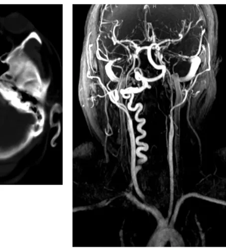
A 26-year-old female had complaints of numbness in right hand referred to our department for computed tomography. During the physical examination stridor and diffuse stiffness at bilateral auricular had been noted. At computed tomography bilateral ossification of both auricula was seen (Figure 1a) and a suspicion of previous infarction of both frontal lobes was aroused. Magnetic resonance (MR) imaging and TOF MR Angiography were performed. MR imaging confirmed previous infarction. Because TOF MR Angiography showed the cerebral circulation fed by only right vertebral artery contrast enhanced MR angiography was performed. At that MR angiography right subclavian artery had irregular walls and advanced narrowing’s. Bilateral internal carotid artery and left vertebral artery were not filled with contrast material. Origin of the right vertebral artery was narrowed apparently. However after that there was a dilated and tortious course of that artery. Cerebral circulation was fed by that artery (Figure 1b). Thorax computed tomography showed diffuse tracheobronchial thickening and left main bronchus narrowing (Figures 2). With these findings relapsing polichondritis associated with ear and tracheobronchial involvement, vasculitis was concluded. At laboratory tests erythrocyte sedimentation rate (57 mm/h) and C-reactive protein (57 mg/L) levels were high. Because she had severe stridor, bronchoscopy was not performed. With this clinical picture, immediate high dose pulse steroid therapy was started.
Volume 4 Issue 2 2016 doi: 10.15824/actamedica.14622
Letter to the Editor
Relapsing polychondritis is an autoimmune connective tissue disease (1). It is generally starts at 5-7. decade of life however it may occur in children also (2). Its incidence is reported to be 3.5 per million population per year (3). Polyarthritis, aortitis or arteritis, uveal inflammation and recurrent cartilage inflammation contribute the clinical picture (1). Looking at the individual systems, variable landscape better understood;
Ear: Auricular involvement is the most common feature. Impaired hearing, tinnitus, and vertigo can be seen (1). During imaging calcification of auricular cartilage, mastoiditis, otitis can be seen.
Nose: Crusting, rhinorrhea and epistaxis can be noted. Cartilage destruction can lead to saddle nose deformity (2). Calcifications of the pinnae, and nasal cartilage collapse may be seen on CT (4).
Airways: Large airway involvement results in hoarseness, aphonia, wheezing, stridor, coughing and dyspnea (5). Laryngeal or tracheal collapse may occur (1). Radiologic manifestations include long segment tracheobronchial strictures, mural thickening, and calcification. Chest radiography can detect tracheal and main bronchial narrowing (6). Subglottic stenosis, tracheobronchial luminal narrowing, densely calcified and thickened tracheal cartilage, peripheral bronchial narrowing and bronchiectasis may be seen on computed tomography (7).
Joints: Typically parasternal joints can be involved (1). Non-erosive and asymmetric peripheral joint disease may be observed (8). Radiographic features are of the non-erosive arthropathy (1).
Heart and vessels: Aortic or mitral valvular disease, pericarditis, heart blocks, valvular disease, aortic dilatation and myocardial infarction may be observed (9). Imaging techniques play a crucial role in securing the diagnosis of a vasculitis, and in demonstrating cerebral involvement. In cerebral vasculitis, both
89
Letter to the Editor
Acta Med Anatol 2016;4(2):88-90
Karakaya et al.
ischemic and hemorrhagic lesions of different ages as well as findings of focal or diffuse inflammation are observed (10).
Figure 2. (a) Coronal reformat computed tomography image demonstrates diffuse tracheobronchial thickening and left main
bronchus narrowing. (b) 3D volume rendering image shows left main bronchus narrowing.
In conclusion relapsing polychondritis is a rare systemic inflammatory disorder affecting cartilaginous tissues. Our case shows the diffuse, multisystem and potentially life-threatening nature of the disease.
Figure 1. (a) Computed tomography image shows bilateral ossification of both auricular cartilage. (b) At contrast enhanced
MR angiography right subclavian artery has irregular walls and advanced narrowing’s. Bilateral internal carotid artery and left vertebral artery are not filled with contrast material. Origin of the right vertebral artery is narrowed apparently. However after that there is a dilated and tortious course of that artery. Cerebral circulation was fed by right vertebral artery.
90
Acta Med Anatol 2016;4(2):88-90
Karakaya et al.
Letter to the Editor
1. Lahmer T, Treiber M, von Werder A, Foerger F, Knopf A, Heemann U, et al. Relapsing polychondritis: an au-toimmune disease with many faces. Autoimmun Rev 2010;9:540–546.
2. Kent PD, Michet CJ, Luthra HS. Relapsing polychondri-tis. Curr Opin Rheumatol 2004;16:56–61.
3. Liu Y, Zhu H, Wang Z, Yu X, Qi Z, Guo S. Airway compro-mise in a special situation--relapsing polychondritis. QJM 2014;107:153-154.
4. Mendelson DS, Som PM, Crane R, Cohen BA, Spiera H. Relapsing polychondritis studied by computed tomog-raphy. Radiology 1985;157:489–490.
5. Tsunezuka Y, Sato H, Shimizu H. Tracheobronchial in-volvement in relapsing polychondritis. Respiration 2000;67:320–322.
6. Crockford MD, Kerr IH. Relapsing polychondritis. Clin Radiol 1988;39:386–390.
7. Davis SD, Berkmen YM, King T. Peripheral bronchial
References
involvement in relapsing polychondritis: demon-stration by thin-section CT. AJR Am J Roentgenol 1989;153:953–954
8. Balsa A, Expinosa A, Cuesta M, MacLeod TI, Gijon-Ba-nos J, Maddison PJ. Joint symptoms in relapsing poly-chondritis. Clin Exp Rheumatol 1995;13:425–430. 9. Lang-Lazdunski L, Hvass U, Paillole C, Pansard Y,
Lan-glois J. Cardiac valve replacement in relapsing poly-chondritis. A review, 4. J Heart Valve Dis 1995. p. 227–235.
10. Pomper MG, Miller TJ, Stone JH, Tidmore WC, Hell-mann DB. CNS vasculitis in autoimmune disease: MR imaging findings and correlation with angiography. Am J Neuroradiol 1999;20: 75-85.