Accepted: 2016.03.18 Published: 2016.04.08
2079
—
5
30
In Vitro Evaluation of Planktonic Growth on
Experimental Cement-Retained Titanium
Surfaces
ABCEF 1
Nur Balci
ABEF 2
Umut Cakan
BEF 3
Burak Aksu
BD 3
Oncu Akgul
ABDE 3
Nurver Ulger
Corresponding Author: Nur Balci, e-mail: [email protected] Source of support: Departmental sources
Background: The purpose of this study was to compare the effects of selected cements, or their combination with titanium, on the growth of two periodontopathic bacteria: Prevotella intermedia (Pi) and Fusobacterium nucleatum (Fn).
Material/Methods: This study was comprised of several experimental groups: 1) Dental luting cements (glass ionomer cement,
methacrylate-based resin cement, zinc-oxide eugenol cement, eugenol-free zinc oxide cement; 2) titanium discs; and 3) titanium combination cement discs. The disks were submerged in bacterial suspensions of either Fn or Pi. Planktonic bacterial growth within the test media was measured by determining the optical density
of the cultures (OD600). Mean and standard deviations were calculated for planktonic growth from three
sepa-rate experiments.
Results: Intergroup comparison of all experimental groups revealed increased growth of Pi associated with cement-ti-tanium specimens in comparison with cement specimens. Regarding the comparison of all groups for Fn, there was an increased amount of bacterial growth in cement-titanium specimens although the increase was not statistically significant.
Conclusions: The combination of cement with titanium may exacerbate the bacterial growth capacity of Pi and Fn in con-trast to their sole effect.
MeSH Keywords: Dental Cements • Fusobacterium nucleatum • Peri-Implantitis • Prevotella intermedia
Full-text PDF: http://www.medscimonit.com/abstract/index/idArt/898274 Authors’ Contribution: Study Design A Data Collection B Statistical Analysis C Data Interpretation D Manuscript Preparation E Literature Search F Funds Collection G
1 Department of Periodontology, Faculty of Dentistry, Istanbul Medipol University, Istanbul, Turkey
2 Department of Prosthodontics, Faculty of Dentistry, Istanbul Medipol University, Istanbul, Turkey
3 Department of Medical Microbiology, Faculty of Medicine, Marmara University, Istanbul, Turkey
Background
Over the last few decades, osseointegrated dental implants have provided a high positive aesthetic impact and improvement in quality of life for completely and partially edentulous patients, substituting one or more missing teeth [1]. Prosthodontic pro-cedures performed in connection with dental implants restore the functional and aesthetic requirements of the oral cavity with good occlusal harmony. Cement retention is mostly pre-ferred in implant-supported fixed restorations versus screw retention due to relatively easier fabrication procedures and for cost containment issues. Dentists are also more familiar with cementation procedures [2].
Even though there is no consensus on a cement selection proto-col for implant restorations [3], the cements designed for tooth-supported restorations are commonly used [4]. Although there are advantages in cement retention, the risk of leaving excess cement in the implant sulcus is a possible cause for peri-implantitis when acting as a mechanical irritant or as a reposi-tory for bacteria or both [5–7]. Peri-implantitis is an inflamma-tory process that leads to the destruction of osseointegrated implant supported tissues such as alveolar bone. Although peri-implantitis is associated with a polymicrobial biofilm that is similar with the microflora in tooth sites, periodontopathogen-ic bacteria including Porphyromonas gingivalis (Pg), Tannerella forsythia (Tf), Prevotella intermedia (Pi), Aggregatibacter acti-nomycetemcomitans (Aa) and Fusobacterium nucleatum (Fn) orchestrate progressive destruction and instigate inflamma-tory disease [8–11]. While some studies reported bacterial ad-herence to implant surfaces [10,12,13] and also documented implant complications as a result of cement extrusion into the subgingival peri-implant tissues [14,15], little is known about the influence of the type of excess cement on peri-implant disease progression.
Methacrylate cements may prepare a ground for the develop-ment of bacterial colonization and promote inflammation in the peri-implant tissue [16,17]. Furthermore, a clinical study on methacrylate cements supported this hypothesis where a methacrylate cement and a zinc oxide-eugenol cement were compared to investigate the clinical effect on the peri-implant tissue [18]. The results of the study showed that methacry-late cement led to more cement excess (62%), while excess cement was not detectable on any of the implant surfaces re-tained with a zinc oxide-eugenol cement. In addition, there were more signs of inflammation such as bleeding on prob-ing and pocket suppuration in the methacrylate cement group. Microbial colonization on the surfaces of dental implants causes infection of implant-supporting tissues. Periodontopathogenic bacteria, including Pg, Fn, Aa, Pi and Tf, have very similar ef-fects on inflammatory peri-implant tissue disease to that
in periodontal disease of residual teeth [19]. Although ex-cess cement around dental implants is reported as a risk fac-tor for peri-implant disease by the American Academy of Periodontology [20], little is known about the influence of ce-ment type on bacterial growth.
This study compared the effects of selected cements or their combination with titanium, on the growth of two periodonto-pathic bacteria: Pi and Fn.
Material and Methods
Specimen fabrication
The study was conducted with three different experimental groups: 1) dental luting cements; 2) titanium (Ti) discs; and 3) titanium combination cement disc group.
1) Dental luting cements
We used two permanent dental luting cements: glass ionomer cement (GI, Ketac Cem, 3M ESPE, MN, USA), and methacrylate-based resin cement (DT, DentoTemp, Itena, Paris France); and two temporary dental luting cements: zinc-oxide eugenol ce-ment (E, Temp-Bond Original, Kerr, CA, USA), and eugenol-free zinc oxide cement (NE, Temp-Bond Non-Eugenol, Kerr, CA, USA). Five stainless steel discs were machined to be 5 mm in diam-eter and 3 mm in thickness. The discs were bonded to a glass slab with wax boxing, and a single-mix impression of these discs was made with a polyether impression material to fab-ricate a mold. The cements were prepared in accordance with the manufacturer’s instructions under aseptic conditions, dis-pensed into the mold and pressed between two glass plates. The glass plate was separated after the cement was set and disc shaped specimens (n=18) were produced for each cement with the same procedure. The cement specimens were steril-ized with ultraviolet light [21].
2) Titanium discs
Titanium Grade 5 (TiAl6V4) rods (Bag-San, Istanbul, Turkey) 5 mm in diameter and 1 mm in length were sectioned into disc spec-imens of 3 mm thickness, using a lathe tool and (n=90) titani-um specimens in total were obtained. The titanititani-um specimens were subjected to long autoclave sterilization for 121°C/20 min.
3) Titanium combination cement discs
Seventy-two of 90 specimens titanium specimens were ran-domly selected and divided into four subgroups. In each sub-group (n=18), one of the diametral surfaces of the specimens
were covered with a thin layer of tested cements using a hand instrument. The cement specimens combined with titanium were also sterilized with U.V. [21]. The remaining 18 specimens out of 90 were used for the titanium group.
Bacterial strains and culture conditions
This study used Pi and Fn (strains isolated from clinical sam-ples and stored at institute’s culture collection). Pi and Fn were grown in Brucella broth containing haemin (10 μg/mL), and menadione (5 μg/mL) in anaerobic conditions (5% H2, 5% CO2, and balance N2) at 37°C for 24–72 h (Figure 1). The concentration of bacteria within the broth was estimated by
measuring optical density (OD) at 600 nm in a spectropho-tometer (Densichek, bioMérieux, France). OD600 measurement directly correlates with the concentration of bacteria in a liq-uid culture.
Analysis of planktonic growth by spectrophotometer
Planktonic bacterial growth was determined by using a micro-plate reader (PR4100, Biorad, USA) as described in [22]. Cement, titanium and cement combined with titanium specimens were placed in individual wells of sterile 24-well plates and sub-merged in 1 mL liquid bacterial culture containing 107 CFU/ml of tested bacteria (Figure 1). All types of materials in a ster-ile liquid broth served as a control to confirm that the cement specimens were not contaminated during experiments. Sterile liquid broth alone served as the negative control. Bacterial planktonic growth was determined by measuring the OD600 of the bacterial culture from each test well after incubation under anaerobic conditions at 37°C for 48 h. All experiments were performed at least twice for verification, each time with a freshly prepared medium and subcultured bacterial strains.
Statistical analysis
Statistical analysis was performed using IBM SPSS Statistics 22 (IBM SPSS, Turkey) program. A Kruskal Wallis test was used for
Figure 1. Tested well plate example after anaerobic incubation for determining planktonic growth of bacteria.
Figure 2. Planktonic growth measurement by OD600 test values for Prevotella intermedia and Fusobacterium
nucleatum in the presence of different cement disks.
No significant difference was observed. GI – Glass Ionomer; DT – Dentotemp; E – Temp-Bond Eugenol; NE – Temp-Bond Non-Eugenol. 0.25 0.20 0.15 0.10 0.05 0.00 Pi Fn Optical density (O D 600) GI DT E NE
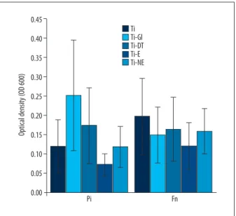
Figure 3. Planktonic growth measurement by OD600 test values for Prevotella intermedia and Fusobacterium nucleatum in the presence of titanium combined different cement disks. Ti-GI displayed the most bacterial growth but the difference was not statistically significant in Pi. Ti – Pure Titanium disc; Ti-GI – Glass Ionomer combined with titanium; DT – Dentotemp combined with titanium; E – Temp-Bond Eugenol combined with titanium; NE – Temp-Bond Non-Eugenol combined with titanium. 0.45 0.40 0.35 0.30 0.25 0.20 0.15 0.10 0.05 0.00 Pi Fn Optical density (O D 600) Ti Ti-Gl Ti-DT Ti-E Ti-NE
the intergroup comparisons of parameters. Significance was evaluated at a level of p<0.05.
Results
Planktonic bacterial growth of Pi and Fn in each cement test group as determined by the OD600 of each sample well are shown in Figure 2. The levels of bacterial growth for both Pi and Fn presented similar levels in all cement groups. GI pre-sented the most prominent bacterial growth in culture me-dia (Figure 2). Although there was no significant difference in bacterial growth among all cement specimens, NE displayed lower Pi and Fn levels in comparison with E for temporary lut-ing cements, whereas DT showed the same effect for perma-nent luting cements (Figure 2).
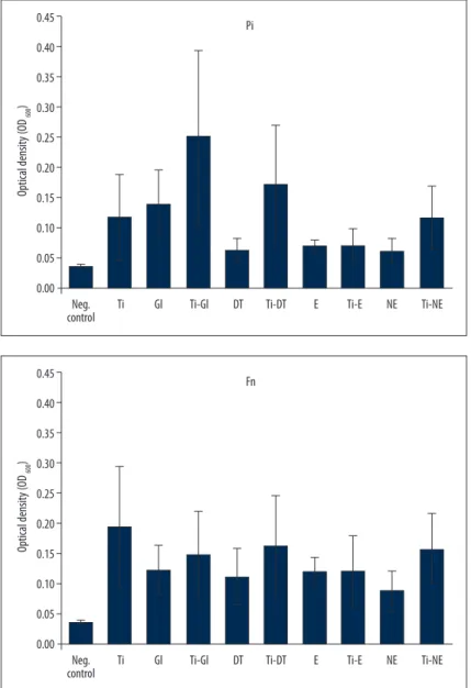
After demonstrating the bacterial growth of cements in different culture media, the effect of cements combined with titanium on planktonic bacterial growth was investigated. According to the spectrophotometer values for Pi, titanium specimens showed less bacterial growth compared to all cement-titanium groups, except for Ti-E. Ti-E displayed the least bacterial growth, where-as Ti-GI displayed the most. The difference, however, wwhere-as not statistically significant (Figure 3). In contrast, for Fn, titanium specimens inhibited the least bacterial growth compared to ce-ment-titanium groups. Similar with Pi groups, Ti-E had the most inhibitory effect on bacterial growth of Fn (Figure 3). The inter-group comparison of all experimental inter-groups revealed increased growth of Pi associated with cement-titanium specimens in parison with cement specimens (Figure 4). Regarding the com-parison of all groups for Fn, there was an increased amount of bacterial growth in cement-titanium specimens although the increase was not statistically significant (Figure 5).
Figure 4. Planktonic growth measurement
by OD600 test values for Prevotella
intermedia in the presence of
cements and different cement discs combined with titanium. There was not significantly different among all groups. Ti – Pure Titanium disc; GI – Glass Ionomer; DT – Dentotemp; E – Bond Eugenol; NE – Temp-Bond Non-Eugenol Ti-GI – Glass Ionomer combined with titanium; DT – Dentotemp combined with titanium; E – Temp-Bond Eugenol combined with titanium; NE – Temp-Bond Non-Eugenol combined with titanium. 0.45 0.40 0.35 0.30 0.25 0.20 0.15 0.10 0.05 0.00 Pi Neg.
control Ti Gl Ti-Gl DT Ti-DT E Ti-E NE Ti-NE
Optical density
(O
D 600
)
Figure 5. Planktonic growth measurement by
OD600 test values for Fusobacterium
nucleatum in the presence of different
cements and cements combined with titanium. There was not significantly different among all groups. Ti – Pure Titanium disc; GI – Glass Ionomer; DT – Dentotemp; E – Temp-Bond Eugenol; NE – Temp-Bond Non-Eugenol; Ti-GI – Glass Ionomer combined with titanium; DT – Dentotemp combined with titanium; E – Temp-Bond Eugenol combined with titanium; NE – Temp-Bond Non-Eugenol combined with titanium. 0.45 0.40 0.35 0.30 0.25 0.20 0.15 0.10 0.05 0.00 Fn Neg.
control Ti Gl Ti-Gl DT Ti-DT E Ti-E NE Ti-NE
Optical density
(O
D 600
Discussion
Gram-negative anaerobic bacteria are frequent pathogen find-ings in peri-implant diseases and other oral infections such as periodontitis. Although oral gram-negative microbiota exhibit a broad heterogeneity, both Pi and Fn can be isolated in the pa-tients with peri-implantitis. Provetella intermedia is indole-posi-tive and moderately saccharolytic [23] and commonly produces immunoglobulin-degrading enzymes [24], whereas Fn is a filamen-tous bacteria that produces serine protease capable of degrad-ing extracellular matrix proteins [25]. As such, they can present different virulence and also growth characteristics as observed in the present study. Fusobacterium nucleatum is the first colo-nized anaerobic species of the mouth, which makes it the most common bacterial species in the mouth [26]. In this study, it was observed that Fn grew easier and also presented more consistent growth than Pi in an in vitro planktonic bacterial growth model. The risk factors for peri-implantitis are classified as poor oral hygiene, prior history of periodontitis, habits like smoking, sys-temic disease that makes host susceptible to inflammation, inaccurate surgical procedures, and improper prosthetic reha-bilitation [1]. Additionally, excess cement is reported as one of the risk factors for inflammatory peri-implant disease [27]. However there is still no consensus about the impact of the ce-ment type on microbial colonization and biofilm formation on peri-implant tissue infection. In a retrospective clinical study, implant restorations cemented with methacrylate-based res-in cement exhibited higher res-inflammatory periodontal res-indices (bleeding on probing, pocket suppuration and alveolar bone loss has been reported as higher than on implants cemented with a zinc-oxide eugenol cement [18].
An in vitro study where five different luting cements of zinc ox-ide-eugenol, eugenol-free zinc oxide, zinc orthophosphate, and two methacrylate-based resin cements were compared in terms of their effect on bacterial growth and biofilm formation associ-ated with peri-implantitis, zinc-oxide cements presented lower growth levels of Aa, Pg, and Fn than methacrylate-based resin cements [22]. In accordance with the aforementioned results, the planktonic growth for both Pi and Fn in the present study, were lower in the presence of zinc-oxide cement although it was not statistically significant. In contrast, both bacteria presented higher growth levels in the presence of a glass-ionomer cement. This comparative study investigated peri-implantitis associated bacteria, Pi and Fn, which are known as late colonizers [10,11]. To the best of the author’s knowledge, this was the first study investigating the planktonic growth of these bacteria not only with cements, but also cements combined with titanium in an
in vitro model. This experimental design was aimed at
consti-tuting the natural environment of a peri-implant region that represented peri-implant sulcus with luting cement residue
on a titanium abutment surface and its microbiological dy-namics. No significant differences in growth of both Pi and Fn were observed among cement-titanium groups, although a decrease was seen in Ti-E cement disks. This may be attrib-uted to the antibacterial activity of zinc-oxide nanoparticles with or without eugenol [28].
Pi growth was enhanced in the presence of Ti-GI. In an in
vitro study, glass ionomer cement inhibited the growth of
Streptococcus mutans and Actinomyces viscosus, whereas it had no antibacterial effect on Enterococcus faecalis [29]. The antibac-terial effect of the cements may be specific to bacteria. For the titanium specimens, Fn demonstrated higher growth levels when compared to cement groups. Both Fn and Pi displayed more in-creased growth in cement groups than compared to cement-tita-nium groups. Sanchez et al. (2014) compared three different sur-face materials of dental implants, hydroxyapatite, titanium and zirconium, in an in-vitro biofilm model [30]. They demonstrated that the number of bacteria on hydroxyapatite was significant-ly higher than titanium and zirconium surfaces, but there was no difference between titanium and zirconium surfaces. They also reported that Fn reached its peak at 24 h whereas V. parvu-la, Aa and Pg reached a peak at 72 h on titanium surfaces, sug-gesting that each bacteria can present diverse growth behavior in different conditions. This in agreement with our data show-ing different growth levels of Pi and Fn with a titanium material. This study was done with four different luting cements and with only two specific bacterial species, and as such, it does not fully represent the complex microbiota of peri-implant dis-eases. In addition, implant restorations in the oral environment are subjected to saliva containing various proteins and en-zymes, food products and beverages, great extremes of tem-perature and functional or parafunctional loading. Therefore, a comprehensive evaluation of these factors may be consid-ered for future research.
Conclusions
Within the limitations of this study, the following conclusions were drawn:
1. Higher Pi and Fn growth levels may be anticipated when us-ing the tested glass ionomer lutus-ing cement.
2. The combination of cement with titanium may exacerbate the bacterial growth capacity of Pi and Fn in contrast to their sole effect.
Acknowledgements
The authors would like to thank Prof. Dr. Guven Kulekci, Istanbul University, School of Dentistry, for her help in reviewing the study design.
References:
1. Belibasakis GN: Microbiological and immuno-pathological aspects of peri-implant diseases. Arch Oral Biol, 2014; 59: 66–72
2. Taylor TD, Agar JR: Twenty years of progress in implant prosthodontics. J Prosthet Dent, 2002; 88: 89–98
3. Tarica DY, Alvarado VM, Truong ST: Survey of United States dental schools on cementation protocols for implant crown restorations. J Prosthet Dent, 2010; 103: 68–79
4. Wadhwani C, Hess T, Piñeyro A et al: Cement application techniques in lut-ing implant-supported crowns: A quantitative and qualitative survey. Int J Oral Maxillofac Implants, 2012; 27: 859–64
5. Callan DP, Cobb CM: Excess cement and peri-implant disease. JIACD, 2009; 1: 61–68
6. Dumbrigue HB, Abanomi AA, Cheng LL: Techniques to minimize excess lut-ing agent in cement-retained implant restorations. J Prosthet Dent, 2002; 87: 112–14
7. Gapski R, Neugeboren N, Pomeranz AZ, Reissner MW: Endosseous implant failure influenced by crown cementation: A clinical case report. Int J Oral Maxillofac Implants, 2008; 23: 943–46
8. Slots J, Bragd L, Wikström M, Dahlen G: The occurrence of Actinobacillus actinomycetemcomitans, Bacteroides gingivalis and Bacteroides intermedi-us in destructive periodontal disease in adults. J Clin Periodontol, 1986; 13: 570–77
9. Teles RP, Haffajee AD, Socransky SS: Microbiological goals of periodontal therapy. Periodontol 2000, 2006; 42: 180–218
10. Egawai M, Miura T, Kato T et al: In vitro adherence of periodontopathic bac-teria to zirconia and titanium surfaces. Dent Mater J, 2013; 32(1): 101–6 11. Rams TE, Degener JE, van Winkelhoff AJ: Antibiotic resistance in human
peri-implantitis microbiota. Clin Oral Implants Res, 2014; 25(1): 82–90 12. Rimondini L, Cerroni L, Carrassi A, Torricelli P: Bacterial colonization of
zir-conia ceramic surfaces: An in vitro and in vivo study. Int J Oral Maxillofac Implants, 2002; 17: 793–98
13. van Brakel R, Cune MS, van Winkelhoff AJ et al: Early bacterial colonization and soft tissue health around zirconia and titanium abutments: An in vivo study in man. Clin Oral Implants Res, 2011; 22: 571–77
14. Pauletto N, Lahiffe BJ, Walton JN: Complications associated with excess ce-ment around crowns on osseointegrated implants: A clinical report. Int J Oral Maxillofac Implants, 1999; 14: 865–68
15. Gapski R, Neueboren N, Pomeranz AZ, Reissner MW: Endosseous implant failure influenced by crown cementation: A clinical case report. Int J Oral Maxillofac Implants, 2008; 23: 943–46
16. Busscher HJ, Rinastiti M, Siswomihardjo W, van derMei HC: Biofilm for-mation on dental restorative and implant materials. J Dent Res, 2010; 89: 657–65
17. Korsch M, Robra BP, Walther W: Predictors of excess cement and tissue re-sponse to fixed implant-supported dentures after cementation. Clin Implant Dent Relat Res, 2013; 17(1): e48–53
18. Korsch M, Walther W: Peri-implantitis associated with type of cement: A retrospective analysis of different types of cement and their clinical corre-lation to the peri-implant tissue. Clin Implant Dent Relat Res, 2015; 17(2): e434–43
19. Sato J, Gomi K, Makino T et al: The evaluation of bacterial flora in progress of peri-implant disease. Aust Dent J, 2011; 56: 201–6
20. Rosen P, Clem D, Cochran D et al: Peri-implant mucositis and peri-implan-titis: A current understanding of their diagnoses and clinical implications. J Periodontol, 2013; 84: 436–43
21. Farrugia C, Cassar G, Valdramidis V, Camilleri J: Effect of sterilization tech-niques prior to antimicrobial testing on physical properties of dental re-storative materials. J Dent, 2015;43: 703–14
22. Raval NC, Wadhwani CP, Jain S, Darveau RP: The interaction of implant luting cements and oral bacteria linked to peri-implant disease: An in vi-tro analysis of planktonic and biofilm growth – a preliminary study. Clin Implant Dent Relat Res, 2015; 17(6): 1029–35
23. Shah HN, Gharbia SE: Biochemical and chemical studies on strains des-ignated Prevotella intermedia and proposal of a new pigmented species, Prevotella nigrescens sp. nov. Int J Syst Bacteriol, 1992; 42: 542–46 24. Jansen HJ, Grenier D, Van der Hoeven JS: Characterization of
immunoglob-ulin G-degrading proteases of Prevotella intermedia and Prevotella nigres-cens. Oral Microbiol Immunol, 1995; 10: 138–45
25. Bachrach G, Rosen G, Bellalou M et al: Identification of a Fusobacterium nu-cleatum 65 kDa serine protease. Oral Microbiol Immunol, 2004; 19: 155–59 26. Könönen E: Oral colonization by anaerobic bacteria during childhood: Role
in health and disease. Oral Dis, 1999; 5: 278–85
27. Wilson TG: The positive relationship between excess cement and peri-im-plant disease: A prospective clinical endoscopic study. J Periodontol, 2009; 80: 1388–92
28. Sirelkhatim A, Mahmud S, Seeni A et al: Review on zinc oxide nanoparti-cles: Antibacterial activity and toxicity mechanism. Nano-Micro Lett, 2015; 7(3): 219–42
29. Davidovich E, Weiss E, Fuks AB, Beyth N: Surface antibacterial properties of glass ionomer cements used in atraumatic restorative treatment. J Am Dent Assoc, 2007; 138(10): 1347–52
30. Sánchez MC, Llama-Palacios A, Fernández E et al: An in vitro biofilm mod-el associated to dental implants: Structural and quantitative analysis of in vitro biofilm formation on different dental implant surfaces. Dent Mater, 2014; 30: 1161–71