R E S E A R C H
Open Access
Rapid maxillary expansion outcomes according
to midpalatal suture maturation levels
Gül
şilay Sayar
1*and Delal Dara K
ılınç
2Abstract
Background: This study aims to compare the relationship between skeletal and dental outcomes of rapid maxillary expansion (RME) treatment on cone beam computed tomography (CBCT) images between pre-pubertal peak (pre-peak) and post-pubertal peak (post-(pre-peak) patients. The null hypothesis was that there was no difference in the outcomes of RME treatment between the groups.
Methods: Thirty-two patients who underwent RME treatment were classified according to midpalatal suture maturation levels and then divided into two groups as pre-peak and post-peak. Skeletal and dental measurements were performed on the CBCT images at T0 (pre-treatment stage) and at T1 (post-treatment stage). Paired samplet test was used to evaluate normally distributed data andP < 0.05 was taken as the significance level.
Results: There were significant differences between T0 and T1 within the groups, but the changes between the pre-peak and post-peak patient groups were not statistically significant.
Conclusion: Non-significant changes were found between the two groups, and the null hypothesis was excepted. Keywords: Rapid maxillary expansion, Cone beam computed tomography, Midpalatal suture, Orthodontics Introduction
Maxillary transversal deficiency can be associated with every types of sagittal malocclusion [1, 2]. Orthodontists prefer to widen the maxillary basal bone by using vari-ous types of maxillary expansion devices such as bonded, banded, or hybrid expanders [3–5].
Skeletal and dental effects were widely investigated in the literature [1, 3–6], but there is still limited informa-tion present about the predicinforma-tion of maxillary expansion results [7]. Clinical outcomes can be different from the pre-treatment prediction. The biological response of midpalatal suture, skeletal tissues, and dental tissues to rapid maxillary expansion was traditionally reported as it was related to patient’s chronological age. As a result, age is usually accepted as the determining factor while planning maxillary expansion.
In cadaveric studies, it was shown that midpalatal su-ture maturation could be observed as non-masu-ture even in adults; therefore, age was not accepted as the determin-ant factor in maturation of the midpalatal suture [8–10].
Besides the treatment method, at the beginning of the treatment, it is very important for an orthodontist to be able to predict whether the enlargement will be more den-tal than skeleden-tal, or contrary. Highly matured midpaladen-tal suture can be seen even though in young patients, and some kind of complications like pain, alveolar bending, and recession in gingiva or failures such as absence of su-tural opening debonding of the expander may be observed during rapid maxillary expansion (RME) treatment [11–13]. Orthopedic correction of maloclussions, as well as RME treatment, is more easily and effectively per-formed in puberty. With an exception of individual differ-ences, it is known that increase of interdigitation in the midpalatal suture causes a decrease in response to skeletal expansion especially after puberty [8,14,15].
Using hand-wrist radiographs [16], cervical vertebral
maturation (CVM) method [17] and statural height
in-crease are reliable methods of evaluating skeletal matur-ation [18]. A new approach to determine skeletal palatal sutural maturation was introduced by Angelieri et al.
[19] which was based on evaluation of the phases of
midpalatal suture maturation on cone beam computed tomography (CBCT) images.
© The Author(s). 2019 Open Access This article is distributed under the terms of the Creative Commons Attribution 4.0 International License (http://creativecommons.org/licenses/by/4.0/), which permits unrestricted use, distribution, and reproduction in any medium, provided you give appropriate credit to the original author(s) and the source, provide a link to the Creative Commons license, and indicate if changes were made.
* Correspondence:[email protected];[email protected]
1Department of Orthodontics, School of Dentistry, Istanbul Medipol
University, Atatürk Bulvarı No: 27, 34083 Unkapanı–Fatih/Istanbul, Turkey Full list of author information is available at the end of the article
In the light of the fact that skeletal maturation level af-fects the biological response to rapid maxillary expan-sion, the aim of this study was to investigate the relationship between skeletal and dental outcomes of RME treatment between pre-peak and post-peak pa-tients who were classified according to midpalatal suture maturation levels.
Materials and methods
Thirty-two patients (11 males, 21 females) between 10 and 18 years of age were included in this retrospective study. Twenty-eight patients (14 patients in each group) were found as sufficient to have the power 80% with a
95% confidence interval (CI) and an α of 0.05 to find a
meaningful difference of 1 mm in interdental width. The Human Ethics Committee of Istanbul Medipol Univer-sity approved this study with the approval number 10840098-604.01.01-E.53516 (19.12.2018-712). Skeletal maturation of each patient was assessed by using the
midpalatal suture maturation (MSM) method [19] which
was performed on CBCT images. Following this, the same subjects were divided into two groups as pre-peak (stage A + B + C) and post-peak (stage D + E) according to the midpalatal maturation stages. The pre-peak group consisted 18 patients (7 males, 11 females), and post-peak group consisted 14 patients (4 males, 10 females).
The CBCT images of the patients were taken at T0 (just before the application of RME device) and T1 (post-treatment stage, at the 6-month retention period, just after the debonding of the device). Inclusion criteria of this study were as follows: patients who have maxil-lary bilateral transverse deficiency, no craniofacial dis-order, and no previous orthodontic treatment were included in the study.
CBCT images were taken by using the i-CAT® dental CBCT device (model 17-19, Imaging Sciences Inter-national, Hatfield, PA, USA). The exposure settings were as follows: 120 kV, 5.0 mA for 26.9 s, and voxel size of 0.2 mm. Field of view of the images was 23 × 17 cm. The dataset was evaluated by using the software of iCAT® device.
Treatment protocol
The routine treatment protocol of RME in this study was as follows: A bonded type of RME device with a hyrax expansion screw (hyrax®, Dentaurum, Ispringen, Germany) was bonded to the teeth with a glass ionomer luting cement (KetacTM Cem radiopaque, 3 M ESPE, Neuss, Germany). The first activation of the bonded RME was performed in the clinic, and then the patients were told to turn the screw 2 turns per day. One week after the first appointment, a control was carried. After 2 weeks of activation (14 days), a 2-mm overcorrection which lasted 5 days was performed, so 3 weeks of
activation in total was obtained. Activation numbers of screw were the same for all of the patients. Retention phase was 6 months, and during this period, the RME appliance was left in the mouth.
CBCT evaluation and method error
Orientation of the software for assessment was pre-sented in Fig.1. The maturation stages of the midpalatal
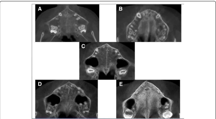
suture were as follows and are shown in Fig. 2. The
evaluation of CBCT images in our study was performed as same as in the study of Angelieri et al. [19].
Stage A: The midpalatal suture is a straight high dens-ity line with no interdigitation. Stage B: The midpalatal suture shows an irregular shape, like as a scalloped high density line. Stage C: The midpalatal suture is like as two parallel, scalloped, high-density lines that are close to each other. Stage D: Fusion of the midpalatal suture has occurred in the palatine bone, and in the maxillary portion of the suture, the fusion has not yet occurred. Stage E: Fusion of the midpalatal suture has also oc-curred in the maxilla. The actual suture is not visible, and the bone density is the same as in whole part of the palate.
Sixteen images were randomly selected and midpalatal suture maturation stage reassessed by the same re-searcher (GS) 2 weeks after the first assessment. Accord-ing to Houston’s formula, the method error was found as 0.91 [20].
Statistical analysis
Statistical analyses were performed using the Statistical Package for Social Science (SPSS for Windows, version 23.0, SPSS Inc., Chicago, IL, USA). The normality of the data was assessed by using one-way ANOVA test.
Normally distributed data were evaluated with paired sample t tests. The significance level was taken as P < 0.05.
Results
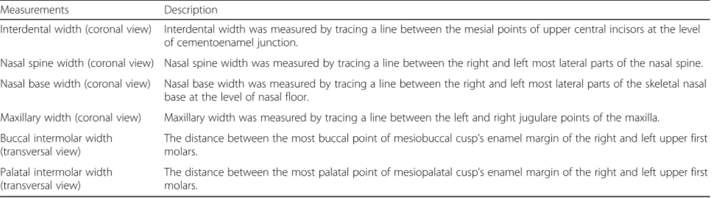
The description of the measurements used in the study
is presented in Table 1. Changes of the measurements
within the groups are shown in Table2. Mean significant increases were found between T0 and T1 in all measure-ments of all groups except interdental width in group E and palatal intermolar width and buccal intermolar width in group B. There were significant differences be-tween all of the measured parameters of pre-peak and post-peak subjects. There were no significant differences
between pre-treatment and post-treatment results
(Table3).
There were no statistically significant differences in mean differences of each parameter according to the pre- and post-peak groups; these results are given in Table4.
Discussion
Orthodontists meet two basic questions while consider-ing rapid maxillary expansion treatment. The first one is the method of expansion (orthopedic or surgical), and the second is the type of expansion device that will be used. The other and perhaps the most important point is skeletal and dental response to the expansion.
Fusion of maxillary sutures is completed at the age
of 14–15 in females and 15–16 in males [12]. It is a
general belief that rapid maxillary expansion is more skeletal in individuals who are younger these ages. However, the expansion is thought to be dental and dentoalveolar rather than skeletal in the patients older
than those ages [20]. Age is usually used as a
param-eter in the evaluation of maturation of patients;
however, skeletal maturation levels are more reliable than chronological age [10, 19].
Pubertal stage of a patient could be determined by using hand-wrist radiography or cervical vertebral maturation method, but when it comes to predict the response to maxillary expansion, the midpalatal suture maturation level was thought to be more related with this treatment. It was previously evaluated in histological and radiological studies, and in histological studies, it was found that vari-ous maturation levels were present in the same age group [21–23]. Also, Angelieri et al. [19] stated that the children in same chronological age could present differences in midpalatal suture maturation levels.
Bacetti et al. [24] used the CVM method and classified the patients by using the said method [23] as pre-peak
Fig. 1 Orientation of the software
Fig. 2 The maturation stages of the midpalatal suture. a The midpalatal suture has no interdigitation. b The midpalatal suture shows an irregular shape. c The midpalatal suture is like as two parallel, scalloped, high-density lines. d Fusion of the midpalatal suture has occurred in the palatine bone. e Fusion of the midpalatal suture has also occurred in the maxilla
and post-peak subjects to compare the outcomes of RME treatment between these two growth phases. Fur-thermore, Angelieri et al. [19] have noticed the need of an assessment method for the individual midpalatal su-ture maturation stage and determined a classification
method. Angelieri et al. [25] noted that pre-pubertal
CVM stages of CS1, CS2, and CS3 are equal to midpala-tal maturational stages of A, B, and C, respectively. This statement was found as reliable for CS5 and stages D and E. Therefore, the aim of this study was to evaluate the dento-skeletal changes after RME in patients who are classified as pre-peak and post-peak based on midpa-latal suture maturation method and to determine if there was a difference between the two groups.
When maxillary expansion is performed, a resistance originates from the neighborhood structures of maxilla such as apertura piriformis, zygomatic buttress, and ptrygoid plate, zygomaticotemporal, zygomaticomaxil-lary, pterygopalatine sutures, and also, the main target is the midpalatal suture itself [26]. The differences in indi-vidual maturation levels of the midpalatal suture pro-duce different treatment results. A few methods were previously used to evaluate the midpalatal suture matur-ation such as midpalatal suture density (MPSD), obliter-ation index (OI), and midpalatal suture morphology (MPSM), and these methods were found more reliable than chronological age [9, 10, 19, 25]. But there were some limitations of these methods. The obliteration
Table 1 Description of the measurements
Measurements Description
Interdental width (coronal view) Interdental width was measured by tracing a line between the mesial points of upper central incisors at the level of cementoenamel junction.
Nasal spine width (coronal view) Nasal spine width was measured by tracing a line between the right and left most lateral parts of the nasal spine. Nasal base width (coronal view) Nasal base width was measured by tracing a line between the right and left most lateral parts of the skeletal nasal
base at the level of nasal floor.
Maxillary width (coronal view) Maxillary width was measured by tracing a line between the left and right jugulare points of the maxilla. Buccal intermolar width
(transversal view)
The distance between the most buccal point of mesiobuccal cusp’s enamel margin of the right and left upper first molars.
Palatal intermolar width (transversal view)
The distance between the most palatal point of mesiopalatal cusp’s enamel margin of the right and left upper first molars.
Table 2 Measurements with changes according to midpalatal suture maturation groups
Measurements Time Stage A (mean ± SD) Stage B (mean ± SD) Stage C (mean ± SD) Stage D (mean ± SD) Stage E (mean ± SD) P Interdental width T0 1.98 ± 0.67 1.55 ± 0.33 2.27 ± 0.56 1.55 ± 0.82 2.11 ± 0.79 0.234
T1 2.79 ± 0.38 2.49 ± 0.29 3.02 ± 0.72 2.43 ± 0.57 2.67 ± 0.92 0.411
P 0.014 0.035 0.001 0.035 0.091
Nasal spine width T0 4.77 ± 1.32 4.13 ± 1.35 4.54 ± 1.34 4.65 ± 1.84 4.25 ± 1.14 0.940 T1 5.57 ± 1.12 5.14 ± 1.14 5.55 ± 1.47 5.67 ± 1.60 5.29 ± 0.98 0.963
P 0.011 0.045 0.002 0.018 0.032
Nasal base width T0 18.91 ± 3.02 17.95 ± 6.1 16.71 ± 1.9 19.12 ± 5 18.59 ± 4.57 0.774 T1 20.33 ± 2.78 19.18 ± 5.84 19.2 ± 2.55 21.52 ± 6.03 19.63 ± 4.46 0.845
P 0.006 0.030 0.012 0.008 0.044
Maxillary width T0 60.19 ± 2.15 55.41 ± 3.40 57.65 ± 4.34 60.33 ± 2.84 58.05 ± 3.906 0.127 T1 63.17 ± 3.05a 57.7 ± 1.83b 60.78 ± 3.11ab 63.68 ± 1.41a 60.35 ± 3.8ab 0.012
P 0.001 0.147 0.001 0.015 0.009
Palatal intermolar width T0 31.16 ± 3.03 29.77 ± 2.20 30.07 ± 2.89 31.24 ± 1.72 30.03 ± 3.88 0.824 T1 34.85 ± 3.84 33.34 ± 3.10 34.57 ± 4.33 35.9 ± 2.51 35.08 ± 4.18 0.862
P 0.005 0.061 0.001 < 0.001 < 0.001
Buccal intermolar width T0 49.75 ± 4.02 48.43 ± 1.97 48.66 ± 2.99 51.27 ± 2.30 48.51 ± 4.73 0.523 T1 55.07 ± 3.67 51.96 ± 2.55 53.75 ± 4.11 55.29 ± 3.34 54.02 ± 4.37 0.631 P < 0.001 0.089 < 0.001 0.002 < 0.001
a, b
There is no difference in the measurements which were marked with the same letter SD standard deviation
index was introduced to make a decision between the treatment alternatives of surgical assisted maxillary expan-sion and orthopedic one, but it was reported that there could be even no obliteration of midpalatal suture in older adult patients [24]. Measurement of the midpalatal suture density was found as a reliable method for estimating the skeletal outcomes of RME, but this method is based on the settings of CBCT machine, and standardization of the different types of the machine is difficult [27].
To overcome standardization errors and determine bone density, Cassetta et al. [28] suggested using a con-version method for transforming the gray density values (voxel values) of CBCT to Hounsfield unit (HU) values of computed tomography.
Angelieri et al. [19] stated that dividing the maturation stages as A–C and D and E could be useful to make a
choice between orthopedic expansion and surgical one. In groups A–C, more skeletal expansion could be man-aged by using RME appliance, but in the more matu-rated groups, D and E, more dental bending might occur and surgical assisted expansion option can be an alterna-tive. To determine the differences in skeletal and dental findings between the pre-peak and post-peak individuals, we also divided the groups as adviced by Angelieri et al [19].
Opening of a median diastema between the upper cen-tral incisors is an expected outcome of RME treatment. It is admitted that a direct relationship is present be-tween the degree of the diastema and the amount of orthopedic expansion, clinically [29].
In this study, significant increase in interdental width
after RME was found in all of the midpalatal
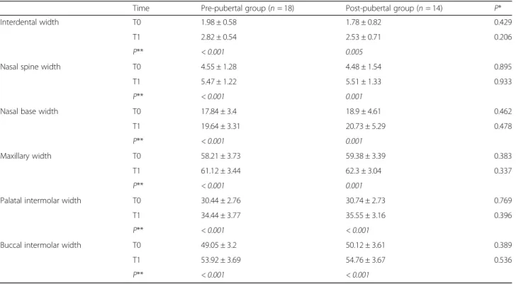
Table 3 Mean differences between the pre-pubertal and post-pubertal groups
Time Pre-pubertal group (n = 18) Post-pubertal group (n = 14) P*
Interdental width T0 1.98 ± 0.58 1.78 ± 0.82 0.429
T1 2.82 ± 0.54 2.53 ± 0.71 0.206
P** < 0.001 0.005
Nasal spine width T0 4.55 ± 1.28 4.48 ± 1.54 0.895
T1 5.47 ± 1.22 5.51 ± 1.33 0.933
P** < 0.001 0.001
Nasal base width T0 17.84 ± 3.4 18.9 ± 4.61 0.462
T1 19.64 ± 3.31 20.73 ± 5.29 0.478
P** < 0.001 0.001
Maxillary width T0 58.21 ± 3.73 59.38 ± 3.39 0.383
T1 61.12 ± 3.44 62.3 ± 3.04 0.337
P** < 0.001 0.001
Palatal intermolar width T0 30.44 ± 2.76 30.74 ± 2.73 0.769
T1 34.44 ± 3.77 35.55 ± 3.16 0.396
P** < 0.001 < 0.001
Buccal intermolar width T0 49.05 ± 3.2 50.12 ± 3.61 0.389
T1 53.92 ± 3.69 54.76 ± 3.67 0.536
P** < 0.001 < 0.001
*
Unpaired sample t test values **
Paired sample t test values
Table 4 Intergroup differences of each parameters
Pre-peak group Post-peak group Test statistics* P
İnterdental width 0.84 ± 0.46 0.75 ± 0.74 0.062 0.675
Nasal spine width 0.93 ± 0.6 1.03 ± 0.76 0.104 0.664
Nasal base width 1.81 ± 1.56 1.83 ± 1.48 0.039 0.960
Maxillary width 2.91 ± 1.68 2.92 ± 2.12 0.001 0.987
Palatal intermolar width 4 ± 2.33 4.82 ± 1.29 0.995 0.211
Buccal intermolar width 4.87 ± 2.22 4.64 ± 1.81 0.143 0.764
maturational stages except the stage E group. In stage E, the maturation of midpalatal suture is completed and this might be the reason of the lower amount of change.
It is presented in some previous studies that size of nasal structures is affected by the expansion of maxilla [30,31]. This was in consistency with our study; further-more, nasal spine width and skeletal nasal base width showed significant increases in all of the sutural matur-ation groups and these increases were present in both of the pre-puberty and post-puberty groups. Similar to our findings, Bacetti et al. [24] noted that the increase in nasal cavity width was significantly higher in the pre-peak group in short time after RME than that of the post-peak patients.
In their study, Pereira et al. [32] found a maxillary width increase with a mean value of 1.76 mm while Capalette et al. [33] presented this value as 3.6 mm. In our study, this value was 2.91 mm for the pre-peak and 2.92 mm for the post-peak group, and the increase in maxillary width was significant in both of the two groups.
In our study, maxillary width, palatal intermolar width, and buccal intermolar width were increased after RME treatment in all of the suture maturation groups except group B. However, this difference was not significant in comparison with the other midpalatal suture maturation groups. Furthermore, the change of these parameters significantly increased in the pre-peak and post peak-groups but the difference was not significant for the two groups. We think that use of the same RME appliance with the same procedure might be resulted similar orthopedic and orthodontic expansion.
The mean increase in skeletal width of the maxilla was found as lower than that of the width of molar teeth in buc-cal and palatinal measurements, but this result might have been related to the inclination of molar and pre-molar teeth. As reported previously, the inclination of posterior teeth is admitted as a normal situation for RME treatment and this was the same with our study [6,32,34–36].
Bacetti et al. [17] concluded that application of RME treatment in the pre-peak phase of puberty resulted more accentuated transversal skeletal changes than that of the post-peak phase. They also stated that application of RME in the post-peak phase resulted in more dentoal-veolar changes; however, we could not find significant differences between the groups in our study. Although RME application resulted as an increase in all of the pa-rameters, there were no differences found between the pre-puberty and post-puberty groups.
Conclusions
There were no differences found between the dental and skeletal changes between the pre-peak and post-peak groups.
Maxillary width was significantly less in group B than that of the other midpalatal suture maturation groups.
It could not be demonstrated that it is possible to pre-dict the amount of maxillary expansion according to the midpalatal suture maturation level.
The results of this study can be assessed as valid or true until 18 years old.
If the distribution of the males and females was equal in each midpalatal suture maturation group, the effect of gender differences on results would be minimized.
Abbreviations
CBCT:Cone beam computed tomography; HU: Hounsfield unit; post-peak: Post-pubertal peak; pre-post-peak: Pre-pubertal peak; RME: Rapid maxillary expansion; T0: Pre-treatment stage; T1: Post-treatment stage
Acknowledgements Not applicable.
Authors’ contributions
GS carried out the data collection of the study, performed the
measurements, and drafted the manuscript. DDK participated in the design of the study and statistical analysis. All authors read and approved the final manuscript.
Funding No funding.
Availability of data and materials
The raw data is present in the CBCT software of our university clinic.
Ethics approval and consent to participate
The Human Research Ethics Committee of Istanbul Medipol University approved the study with the approval number: 10840098-604.01.01-E.53516 (19.12.2018-712).
Consent for publication Not applicable.
Competing interests
The authors declare that they have no competing interests.
Author details
1Department of Orthodontics, School of Dentistry, Istanbul Medipol
University, Atatürk Bulvarı No: 27, 34083 Unkapanı–Fatih/Istanbul, Turkey.
2Department of Orthodontics, School of Dental Medicine, Bahçeşehir
University, Istanbul, Turkey.
Received: 29 March 2019 Accepted: 23 May 2019
References
1. Lione R, Brunelli V, Franchi L, Pavoni C, Quiroga Souki B, Cozza P. Correction to: Mandibular response after rapid maxillary expansion in class II growing patients: a pilot randomized controlled trial. Prog Orthod. 2018;19(1):26.
https://doi.org/10.1186/s40510-018-0231-3.
2. Hwang S, Song J, Lee J, Choi YJ, Chung CJ, Kim KH. Three-dimensional evaluation of dentofacial transverse widths in adults with different sagittal facial patterns. Am J Orthod Dentofac Orthop. 2018;154(3):365–74.https:// doi.org/10.1016/j.ajodo.2017.11.041.
3. Haas AJ. The treatment of maxillary deficiency by opening the mid-palatal suture. Angle Orthod. 1965;35(3):200–17.
4. Canan S,Şenışık NE. Comparison of the treatment effects of different rapid maxillary expansion devices on the maxilla and the mandible. Part 1: evaluation of dentoalveolar changes. Am J Orthod Dentofac Orthop. 2017; 151(6):1125–38.https://doi.org/10.1016/j.ajodo.2016.11.022.
5. Conroy-Piskai C, Galang-Boquiren MT, Obrez A, Viana MG, Oppermann N, Sanchez F, Edgren B, Kusnoto B. Assessment of vertical changes during
maxillary expansion using quad helix or bonded rapid maxillary expander. Angle Orthod. 2016;86(6):925–33.
6. Garrett BJ, Caruso JM, Rungcharassaeng K, Farrage JR, Kim JS, Taylor GD. Skeletal effects to the maxilla after rapid maxillary expansion assessed with cone-beam computed tomography. Am J Orthod Dentofac Orthop. 2008; 134(1):8–9.https://doi.org/10.1016/j.ajodo.2008.06.004.
7. Angelieri F, Franchi L, Cevidanes LH, Bueno-Silva B, McNamara JA Jr. Prediction of rapid maxillary expansion by assessing the maturation of the midpalatal suture on cone beam CT. Dental Press J Orthod. 2016;21(6):115– 25.https://doi.org/10.1590/2177-6709.21.6.115-125.sar.
8. Persson M, Thilander B. Palatal suture closure in man from 15 to 35 years of age. Am J Orthod. 1977;72(1):42–52.
9. Knaup B, Yildizhan F, Wehrbein H. Age-related changes in the midpalatal suture. J Orofac Orthop. 2004;65(6):467–74.
10. Korbmacher H, Schilling A, Puschel K, Amling M, Kahl-Nieke B. Age-dependent three-dimensional micro-computed tomography analysis of the human midpalatal suture. J Orofac Orthop. 2007;68(5):364–76.
11. Angelieri F, Franchi L, LHS C, Gonçalves JR, Nieri M, Wolford LM, McNamara JA Jr. Cone beam computed tomography evaluation of midpalatal suture maturation in adults. Int J Oral Maxillofac Surg. 2017;46(12):1157–561. 12. Haas AJ. Long-term posttreatment evaluation of rapid palatal expansion.
Angle Orthod. 1980;50(3):189–217.
13. Greenbaum KR, Zachrisson BU. The effect of palatal expansion therapy on the periodontal supporting tissues. Am J Orthod. 1982;81(1):12–21. 14. Handelman CS. Nonsurgical rapid maxillary alveolar expansion in adults: a
clinical evaluation. Angle Orthod. 1997;67(4):291–305.
15. Isaacson RJ, Ingram AH. Forces produced by rapid maxillary expansion, II. Forces present during treatment. Angle Orthod. 1964;34(4):261–70. 16. Flores–Mir C, Nebbe B, Major PW. Use of skeletal maturation based on
hand-wrist radiographic analysis as a predictor of facial growth: a systematic review. Angle Orthod. 2004;74(1):118–24.
17. Baccetti T, Franchi L, McNamara JA Jr. The cervical vertebral maturation (CVM) method for the assessment of optimal treatment timing in dentofacial orthopedics. Semin Orthod. 2005;11(3):119–29.
18. Björk A, Helm S. Prediction of the age of maximum pubertal growth in body height. Angle Orthod. 1967;37(2):134–43.
19. Angelieri F, Cevidanes LH, Franchi L, Gonçalves JR, Benavides E, McNamara JA Jr. Midpalatal suture maturation: classification method for individualassessment before rapid maxillary expansion. Am J Orthod Dentofac Orthop. 2013;144(5): 759–69.https://doi.org/10.1016/j.ajodo.2013.04.022.
20. Houston WJB. The analysis of errors in orthodontic measurements. Am J Orthod. 1983;83(5):382–90.
21. Korn EL, Baumrind S. Transverse development of human jaws between the ages of 8.5 and 15.5 years, studied longitudinally with the use of implant. J Dent Res. 1990;69(6):1298–306.
22. Timms DJ, Moss JP. An histological investigation into the effects of rapid maxillary expansion on the teeth and their supporting tissues. Trans Eur Orthod Soc. 1971:263–71.
23. Wertz RA. Skeletal and dental changes accompanying rapid midpalatal suture opening. Am J Orthod. 1970;58(1):41–66.
24. Baccetti T, Franchi L, Cameron CG, McNamara JA Jr. Treatment timing for rapid maxillary expansion. Angle Orthod. 2001;71(5):343–50.
25. Angelieri F, Franchi L, Cevidanes LH, McNamara JA Jr. Diagnostic performance of skeletal maturity for the assessment of midpalatal suture maturation. Am J Orthod Dentofacial Orthop. 2015;148(6):1010–6.https:// doi.org/10.1016/j.ajodo.2015.06.016.
26. Abo Samra D, Hadad R. Midpalatal suture: evaluation of the morphological maturation stages via bone density. Prog Orthod. 2018;19(1):29.https://doi. org/10.1186/s40510-018-0232-2.
27. Grünheid T, Larson CE, Larson BE. Midpalatal suture density ratio: a novel predictor of skeletal response to rapid maxillary expansion. Am J Orthod Dentofac Orthop. 2017;151(2):267–76.
28. Cassetta M, Stefanelli LV, Pacifici A, Pacifici L, Barbato E. How accurate is CBCT in measuring bone density? A comparative CBCT-CT in vitro study. Clin Implant Dent Relat Res. 2014;16(4):471–8.https://doi.org/10.1111/cid. 12027. Epub 2013 Jan 7.
29. Ribeiro GLU, Locks A, Pereira J, Brunetto M. Analysis of rapid maxillary expansion using cone-beam computed tomography. Dental Press J Orthod. 2010;15(6):107–12.
30. Fastuca R, Lorusso P, Lagravère MO, Michelotti A, Portelli M, Zecca PA, Antò VD’, Militi A, Nucera R, Caprioglio A. Digital evaluation of nasal changes
induced by rapid maxillaryexpansion with different anchorage and appliance design. BMC Oral Health. 2017;17(1):113.https://doi.org/10.1186/ s12903-017-0404-3.
31. El H, Palomo JM. Three-dimensional evaluation of upper airway following rapid maxillary expansion: a CBCT study. Angle Orthod. 2014;84(2):265–73. 32. Pereira JDS, Jacob HB, Locks A, Brunetto M, Ribeiro GLU. Evaluation of
the rapid and slow maxillary expansion using cone-beam computed tomography: a randomized clinical trial. Dental Press J Orthod. 2017; 22(2):61–8.
33. Cappellette M Jr, Nagai LHY, Gonçalves RM, Yuki AK, Pignatari SSN, Fujita RR. Skeletal effects of RME in the transverse and vertical dimensionsof the nasal cavity in mouth-breathing growing children. Dental Press J Orthod. 2017; 22(4):61–9.https://doi.org/10.1590/2177-6709.22.4.061-069.oar.
34. Gunyuz Toklu M, Germec-Cakan D, Tozlu M. Periodontal, dentoalveolar, and skeletal effects of tooth-borne and tooth-bone-borne expansion appliances. Am J Orthod Dentofac Orthop. 2015;148(1):97–109.https://doi.org/10.1016/j. ajodo.2015.02.022.
35. Garib DG, Henriques JF, Janson G, Freitas MR, Coelho RA. Rapid maxillary expansion—tooth-tissue-borne vs. tooth-borne expanders: a CT evaluation of dentoskeletal effects. Angle Orthod. 2005;75(4):548–57.
36. Kartalian A, Gohl E, Adamian M, Enciso R. Cone-beam computerized tomography evaluation of the maxillary dentoskeletal complex after rapid palatal expansion. Am J Orthod Dentofac Orthop. 2010;138(4):486–92.
Publisher’s Note
Springer Nature remains neutral with regard to jurisdictional claims in published maps and institutional affiliations.