See discussions, stats, and author profiles for this publication at: https://www.researchgate.net/publication/10692233
The involvement of apoptotic regulators during
in vitro decidualization
Article in European Journal of Endocrinology · August 2003 DOI: 10.1530/eje.0.1490069 · Source: PubMed CITATIONS14
READS21
3 authors: Kamil Can Akcali Ankara University 62 PUBLICATIONS 829 CITATIONS SEE PROFILE Geula Gibori University of Illinois at Chicago 217 PUBLICATIONS 5,297 CITATIONS SEE PROFILE Sohaib Khan University of Cincinnati 60 PUBLICATIONS 1,920 CITATIONS SEE PROFILEAll content following this page was uploaded by Kamil Can Akcali on 05 June 2014.
EXPERIMENTAL STUDY
The involvement of apoptotic regulators during in vitro
decidualization
Kamil C Akcali, Geula Gibori1and Sohaib A Khan2
Department of Molecular Biology and Genetics, Bilkent University, Bilkent, Ankara 06800, Turkey,1Department of Physiology and Biophysics, University of Illinois at Chicago, Chicago, Illinois 60612, USA and2Department of Cell Biology, Neurobiology and Anatomy, College of Medicine,
University of Cincinnati, Cincinnati, Ohio 45267, USA
(Correspondence should be addressed to K C Akcali; Email: [email protected])
Abstract
Objectives: The uterus responds to an implanting blastocyst by undergoing extensive tissue modification leading to decidualization. This modification includes differentiation and apoptosis of epithelial as well as stromal cell compartments. It is generally accepted that the decidual cell regression pattern is similar to the pattern of initial differentiation, suggesting that decidual cell death is the end point of timed differentiation. However, the molecular mechanisms controlling these events are not understood clearly. Therefore, we aimed to investigate the involvement of apop-totic factors using an in vitro cell culture system.
Design: In order to assess the role of apoptotic factors during decidualization, we used a decidual cell line (GG-AD) that had been transformed with a temperature-sensitive SV-40 mutant. At the non-per-missive temperature (39 8C), these cells showed the characteristics of differentiated decidual cells. They dedifferentiated into stromal cells when the temperature was shifted back to 33 8C.
Methods: We performed Northern blot analysis for bax, bcl-xLand bcl-2 at both temperatures. The
onset of apoptosis was examined by Annexin V staining. The expression of p53 protein was also determined by Western blot.
Results: We found an increase in the expression of bax when GG-AD cells were grown at 39 8C. We also showed apoptosis with Annexin V staining at 39 8C. The p53 protein expression was also similar to that of the animal models, suggesting that the programmed cell death of the decidual cells occurred in a p53-independent manner.
Conclusions: These data indicate that a parallelism exists between the increased expression of pro-apoptotic genes and decidual cell death, similar to animal models. Therefore, an in vitro model of GG-AD cells can be used to assess directly the relationship between apoptotic regulators and decidualization and could be used to study the mechanism of decidual cell regression.
European Journal of Endocrinology 149 69–75
Introduction
During placentation, a well-orchestrated plan that includes cell differentiation, proliferation and apoptosis enables remodeling of the uterus (1). Adhesion of the blastocyst results in the differentiation of the endo-metrial stroma into decidual cells, which is an import-ant component of the implimport-antation process. The morphological changes that occur during decidualiza-tion in vivo have been described extensively (2). The majority of these decidual cells regress in a time-depen-dent manner, and their removal is important because they provide space for the growth of the embryo (3).
After inducing decidualization either by blastocyst implantation or by intracervical mineral oil injection in the ovariectomized rat model, it has been shown
that this regression occurs via apoptosis (3, 4). However, molecular mechanisms controlling decidual cell death are poorly understood. Using ovariectomized rat, we previously demonstrated that during deciduali-zation, the expression pattern within the bcl-2 gene family is altered favoring decidual cell apoptosis (5). Bcl-2 family proteins are composed of both pro-apopto-tic and anti-apoptopro-apopto-tic members and can homo- or het-erodimerize and function in a distal apoptotic pathway common to all multicellular organisms. The ratio of death antagonists (Bcl-2, Bcl-xL) to agonists (Bax) has
been shown to have a critical role in determining the fate of the cells. (6, 7). Recently it has been shown that prolactin (PRL) plays an important anti-apoptotic role in the decidua of the pseudopregnant rats and the disappearance of PRL-receptor (PRL-R) allows cell
death and reorganization of the decidua (8). Thus, precise knowledge of Bcl-2 family gene expression is necessary to understand the mechanism of decidual cell death.
Although animal models have provided useful information regarding the remodeling of the uterus during blastocyst implantation, the difficulty of carry-ing out perturbations makes it virtually impossible to define molecular mechanisms. Srivastava et al. have established a temperature-sensitive cell line (GG-AD) from endocrine cells of rat decidua (9). These cells were isolated from the antimesometrial region of rat uterus and were stably transfected with simian virus temperature-sensitive (SV-40 tsA209) mutant, which harbors a temperature-sensitive mutation required for the maintenance of cell transformation. They show temperature-dependent changes in growth and mor-phology. At the permissive temperature of 33 8C, they grow unrestricted and have the characteristics of stro-mal cells. On the other hand, at the non-permissive temperature (39 8C), they differentiate and resemble antimesometrial decidual cells as evidenced by the expression of marker genes such as progesterone recep-tor, PRL-like protein B, activin-bA, and desmin (9).
Furthermore, it has been shown that when GG-AD cells are maintained at 39 8C the expression of estrogen receptor b (ERb) (10), interleukin-6 (IL-6), IL-6 recep-tor and 130 kDa glycoprotein (gp130) (11), basic fibro-blast growth factor (bFGF) and vascular endothelial growth factor (VEGF) (12) are increased. In the present study, we utilized the GG-AD cell line to assess directly the relationship between apoptotic regulators and decidualization. We hypothesized that the balance within the bcl-2 family of genes is altered, and increased expression of pro-apoptotic genes in GG-AD cells at the non-permissive temperature of 39 8C would parallel the onset of decidual cell death. We observed that a paral-lelism exists between the increased expression of pro-apoptotic genes and decidual cell death.
Materials and methods
Cell culture
GG-AD cells were cultured in RPMI-1640 medium (Gibco BRL, Grand Island, NY, USA) supplemented with 1X glutamine (Gibco BRL), 2X antibiotic – antimy-cotic solution (Gibco BRL), 1X non-essential amino acids (Gibco BRL), 1X sodium pyruvate (Gibco BRL), 0.5% D-glucose (Gibco BRL) and 10% fetal bovine
serum (Gibco BRL). Cells were cultured either at 33 8C or 39 8C with 5% CO2. The medium was changed
every third day. For the cell number, 105 cells were plated in a 100 mm tissue culture dish and cultured at 33 8C and 39 8C. Cells were trypsinized and counted at days 4, 7, 12, 17 and 21 (n ¼ 3) using a hemocytometer.
Total RNA isolation and Northern blot
analysis
Total RNA from GG-AD cells was isolated 5 days after starting the culture using RNeasy Mini kit (Qiagen, Santa Clarita, CA, USA) after homogenization with a QIAshredder (Qiagen, Valencia, CA, USA). The concen-tration of total RNA was determined by measuring the optical density at 260 nm. The concentrations and the quality of the RNA preparations were confirmed by comparing 28S and 18S rRNA bands stained with ethi-dium bromide on a 1% agarose – formaldehyde gel. The Northern blot procedure utilized in these experiments has been described previously by Nephew et al. (13). Each Northern blot analysis was repeated three times. Briefly, 30 mg samples of total RNA were electrophor-esed on agarose – formaldehyde gels and then trans-ferred to Duralon (Stratagene, La Jolla, CA, USA) by capillary transfer. Molecular weight marker RNA was run on each gel. Membranes were pre-hybridized for 15 min at 65 8C with Rapid Hybridization Buffer sol-ution (Amersham Life Science, Arlington Heights, IL, USA) and hybridized for 2 h at 65 8C in the same sol-ution with 1.5 £ 106c.p.m./ml 32P-labeled bax, bcl-2, bcl-xLand desmin cDNA probes. After appropriate
wash-ings, the membranes were exposed to Kodak XAR X-ray film at 2 70 8C for 5 days. Densitometric analysis was performed using the Multi-Analyst software. rRNA values were used to correct intensities of mRNA expression.
Detection of apoptosis with Annexin V
GG-AD cells were grown on coverslips at 33 8C and 39 8C for 5 days. The Apo-Alert Annexin Apoptosis Kit (Clontech, Palo Alto, CA, USA) was used to stain cells with FITC-conjugated Annexin V. Cells treated with 5% ethanol were used as a positive control. The staining of GG-AD cells with FITC-conjugated Annexin was examined by fluorescence microscopy (Nikon).
Detection of p53 levels
For detection of p53 expression, GG-AD cells were grown at 33 8C and 39 8C for 5 days, and cell lysates were prepared by adding 0.6 ml of RIPA buffer (1X PBS, 1% Nonidet P-40, 0.5% sodium deoxycholate, 0.1% SDS with 1 mmol/l phenylmethylsulfonyl fluoride (PMSF); 1 mg/ml aprotinin, leupeptin, pepstatin; 1 mmol/l Na3VO4; 1 mmol/l NaF) and 15 mg of protein
were loaded per lane on a minigel electrophoresis apparatus. A 7.5% gel was used for p53 detection. After electrophoresis and transblotting of the proteins onto nitrocellulose membranes, the membranes were washed, blocked with 10% non-fat dry milk in 0.2% Tween 20 in PBS, and then incubated with p53 mono-clonal antibody DO1 (Santa Cruz Biotechnology, Santa Cruz, CA, USA) at a dilution of 1:150 overnight at 4 8C,
70 K C Akcali and others EUROPEAN JOURNAL OF ENDOCRINOLOGY (2003) 149
followed by incubation with anti-mouse horseradish peroxidase-conjugated IgG at a dilution of 1:15 000 for 2 h at room temperature. Immunoreactive bands were visualized by enhanced chemiluminescence. As a positive control, we used u.v.-irradiated normal human melanocytes, which are positive for p53 expression (14).
Results
The expression of apoptotic regulators during
the temperature-induced decidualization of
GG-AD cells
At the permissive temperature (33 8C), GG-AD cells show the characteristics of stromal cells and at the non-permissive temperature (39 8C) they differentiate into decidual cells. Since we have shown that expression of bcl-2 family genes is altered during early pregnancy in our animal model (5), we decided to examine whether these genes were involved during in vitro decidualization. RNA was isolated from cells grown at the permissive temperature (33 8C) or at the non-permissive temperature (39 8C) and bax, bcl-xL
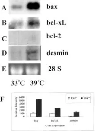
and bcl-2 expression were analyzed using Northern blot analysis. As shown in Fig. 1A, bax mRNA showed a remarkable induction at 39 8C. The expression of bcl-xL(Fig. 1B) also seems to be induced
but the level of increase was not as dramatic as that of bax. The expression of bcl-2 was neither detectable at 33 8C nor at 39 8C (Fig. 1C). Since desmin is a widely accepted decidual cell marker (15), we used desmin expression to monitor the temperature-induced decidualization of GG-AD cells. The expression of desmin shown in Fig. 1D confirmed that GG-AD cells decidualized at 39 8C. Relative expression of these genes was quantitated and normalized using levels of rRNA. The changes in the expression of bax, bcl-xL
and desmin at 33 8C and 39 8C are shown in Fig. 1F. Thus, this cell line seems to mimic what we have observed during the decidualization of stromal cells in vivo.
Monitoring of apoptosis in GG-AD cells
Gu and colleagues have shown that apoptosis plays an important role during decidualization in animal models (3). In order to use this cell line as a model to investi-gate the molecular events during decidualization, we wanted to determine whether these cells undergo apop-tosis at 39 8C. To monitor the apopapop-tosis, we employed Annexin V staining as an indicator of apoptosis. This procedure is based on the observation that soon after the initiation of apoptosis, most cells translocate their phosphotidylserine from the plasma membrane to the cell surface (16). On the cell surface, phosphotidylser-ine can easily be detected by staining with FITC-conju-gated Annexin (17, 18). GG-AD cells were grown at33 8C (Fig. 2A, B) and at 39 8C (Fig. 2C, D). The cells show phosphotidylserine-positive apoptotic staining around their membranes at 39 8C (Fig. 2C) after cultur-ing for 5 days. No staincultur-ing was observed when cells were grown at 33 8C (Fig. 2A). For positive controls, we used 5% ethanol treatment for 90 min to induce early apoptotic signs at 33 8C and 39 8C (Fig. 2B and 2D respectively).
Growth characteristics of GG-AD cells
We reasoned that since GG-AD cells show early signs of apoptosis, the number of viable cells at 39 8C should decrease in due course. In order to test this premise, we plated 105cells into 100 mm tissue culture dishes and maintained them at 33 8C and 39 8C. At different time intervals, cells were trypsinized and counted (n ¼ 3) in a hematocytometer. As depicted in Fig. 3, cells at 33 8C showed an increased growth rate, whereas at 39 8C there was no increase in the
Figure 1 The expression of bax (A), bcl-xL(B), bcl-2 (C) and
desmin (D) during temperature-induced decidualization of GG-AD cells. GG-AD cells show characteristics of stromal cells at 33 8C, whereas at 39 8C they differentiate into decidual cells. At 39 8C, the expression of bax was remarkably induced, whereas the increase in bcl-xLexpression was not as dramatic. No expression
of bcl-2 was observed. Also, the expression of desmin was increased at 39 8C, confirming that the cells decidualized at this temperature. (E) Ethidium bromide-stained 28S band. (F) The results were quantitated by densitometric analysis using rRNA to correct the intensities of mRNA expression.
number of cells. These data confirmed that a substantial number of GG-AD cells died at 39 8C. These results provide further support that the GG-AD cell line could be useful in assessing the relationship between apoptotic regulators and decidualization.
The expression of p53
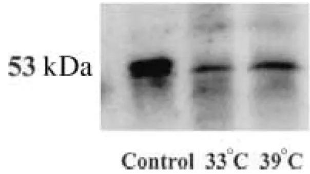
During in vivo decidualization, when decidual cells undergo apoptosis, their p53 levels remain the same (19), suggesting that p53-independent pathways are responsible for the apoptosis. Therefore, we monitored the expression of p53 during in vitro decidualization. Protein extracts obtained from GG-AD cells at 33 8C and 39 8C and were subjected to Western blot analy-sis using p53 monoclonal antibody DO1. As shown in Fig. 4, our results indicated that p53 protein levels did not change, suggesting that apoptosis in GG-AD cells occurs by a p53-independent pathway similar to the in vivo decidualization, observed by Gibori et al. (19).
Discussion
Although there is diversity among species, the purpose of implantation is the same in all species: to attach the embryo to the uterus and to establish a close relation-ship between maternal and fetal tissues. Implantation failure is responsible for 22% of the spontaneous abor-tions that occur early in human pregnancy (20). In farm animals, 80% of embryonic loss occurs during the peri-implantation period (21). Thus, understanding the various signals and molecular pathways known to induce or regulate implantation is of great clinical as well as economic concern.
Use of an animal model for blastocyst implantation has allowed greater insight into the mechanisms
Figure 2 Detection of apoptosis with FITC-conjugated Annexin V in GG-AD cells at 33 8C (A) and 39 8C (C). GG-AD cells were also treated with 5% ethanol at 33 8C (B) and at 39 8C (D) to induce apoptosis and were used as positive controls. Arrows in (B), (C) and (D) show the Annexin V staining at the cell membranes. Observing the staining in (C) (39 8C) but not (A) (33 8C) suggested that cells at 39 8C showed the early signs of apoptosis
(magnification: 40 £ ).
Figure 3 Growth curve of GG-AD cells that were grown at 33 8C (W) and 39 8C (X) (n ¼ 3). Cells showed an increased growth rate at 33 8C but not at 39 8C.
72 K C Akcali and others EUROPEAN JOURNAL OF ENDOCRINOLOGY (2003) 149
of implantation. However, due to the unavailability of a suitable decidual cell line, appropriate experiments could not be designed to probe the molecular mechan-isms of blastocyst implantation. Cell lines were devel-oped from endometrial uterine cells (22), but did not prove useful because these cells differed profoundly from decidual cells and failed to decidualize or express genes that encode decidual hormones (22). Attempts to use primary antimesometrial decidual cells also failed because of their inability to proliferate in primary cell culture. However a temperature-sensitive cell line (GG-AD) from the endocrine cells of rat decidua has been successfully established, circumventing many of the obstacles (9). These cells can be induced to undergo decidualization at the non-permissive temperature of 39 8C. Interestingly, when the temperature is shifted back to 33 8C, GG-AD cells dedifferentiate and regain features of the endometrial stromal cells. Several studies have already used this cell line to study the pro-cess of decidualization and observed similarities between this model and in vivo models of decidualiza-tion (10 – 12). Encouraged by these observadecidualiza-tions, we set out to examine the mechanism of apoptotic regression of decidual cells using the temperature-sen-sitive GG-AD cells as a model.
First we sought to explore whether this in vitro system is really a reflection of the in vivo animal model. Since we have shown that the expression of bcl-2 family genes was altered during early pregnancy in our animal model (5), we decided to examine whether these genes were involved during in vitro decidualization. It has been proposed that the ratio of bcl-2 to bax expression determines the fate of a cell, i.e. whether it should live or undergo programmed cell death (6 – 7). Also these three genes have been shown to form homo- or heterodimers in the mamma-lian system (23). Therefore, we chose to detect the expression level of bax, bcl-2 and bcl-xL amongst the
bcl-2 family of genes. Based on the Northern data for bax, bcl-2 and bcl-xLexpression, this cell line seems to
mimic what we observed previously during the decid-ualization of stromal cells in vivo (5). However, unlike the in vivo system, only the 1.0 kb transcript of bax
was identified in this cell line. We did not detect the other alternatively spliced transcript, 1.5 kb baxb, in GG-AD cells. The functional difference between these two transcripts is not known but Bax protein is trans-lated from the 1.0 kb transcript (baxa), whereas a protein corresponding to baxb has not been identified (S. Korsmeyer, personal communication). Since we used the whole uteri in our animal study, it is likely that baxb is contributed by the uterine cells, which are different from the parental GG-AD cells.
If the in vitro system of GG-AD cells is a true model for in vivo decidualization, then plausibly these cells must undergo apoptosis at the non-permissive temperature of 39 8C. Therefore, we wished to determine whether GG-AD cells show signs of apoptosis when they are grown at 39 8C. We explored this line of reasoning by measuring apoptosis in the GG-AD cells at 39 8C using the Annexin V staining method that has been optimized to detect the initial stages of apoptosis. It is believed that during apoptosis, phosphotidylserine is externalized prior to the known nuclear changes associated with apoptosis (24). Thus an assessment of the externalized phosphotidylserine by Annexin V pro-vides a sensitive tool to measure early events in apopto-sis (16 – 18). Our results with Annexin V indicated that, indeed, the GG-AD cells at 33 8C did not show any cell membrane staining, however at 39 8C, they showed the characteristic staining at their membrane after cultur-ing 5 days, indicatcultur-ing that they underwent apoptosis at 39 8C. It is worth noting that the source of GG-AD cell line (antimesometrial decidual cells from day 10 of pseudopregnant rat) also showed signs of apoptosis as measured by DNA fragmentation. However, the extent of DNA fragmentation showed a marked increase at day 13/14 during in vivo decidualization (3). It is therefore likely that the antimesometrial decid-ual cells used by Srivastava et al. (9) in establishing the GG-AD cell line represent the decidual cells undergoing early stages of apoptosis. Our results using FITC-conju-gated Annexin V staining support this contention.
Since the GG-AD cells show signs of early apoptosis at 39 8C, we reasoned that the number of viable cells at 39 8C should decrease during culture. In contrast, the GG-AD cells maintained at 33 8C should continue to divide and increase in number. Indeed, the GG-AD cells at 33 8C showed an increased growth rate, whereas at 39 8C there was no increase and the cells died 3 weeks after they were plated. Our observation that when maintained at 39 8C the number of GG-AD cells remained unchanged for the first 10 days would suggest that during this phase cell proliferation and cell death are occurring in parallel, resulting in a steady state. However, after 10 days the proportion of cells undergoing apoptosis takes over and results in the gradual loss of all cells at 39 8C by 3 weeks. Our data provide support for using the GG-AD cell line as a good model to explore the relationship between apoptotic regulators and decidual cell death.
Figure 4 p53 expression during temperature-induced
decidualization. The expression of p53 protein did not show any change at 33 8C and 39 8C. Protein extracted from u.v.-irradiated human melanocytes was used for the control group.
Another similarity between in vitro and in vivo decidualization models is the status of p53 expression levels. p53 protein functions at least in part as a transcriptional regulator and can transactivate several cellular genes (25, 26). Two important events are regulated by p53 in connection with its function as a tumor suppressor. p53 has been shown to induce cell cycle arrest at the G1/S border, and to induce apoptosis (27, 28). Members of the bcl-2 family genes, bax and bcl-2 are known to be regulated by p53 (29). p53 activates bax (30), but inhibits the expression of bcl-2 in certain cell lines (31). During in vivo and in vitro decidualization, it was expected that the expression of p53 would increase since in both model systems apoptotic cell death is evident. However, both during in vivo decidual cell regression (19) and during in vitro decidualization of GG-AD cells at 39 8C (this study), the p53 levels did not change. We collected these cells after 5 days of culture because this was the first day of detecting the early changes accompanied with apoptosis with our Annexin V staining. Based on this, we did not expect to see any changes in p53 expression at earlier days of GG-AD cell culture at 39 8C. These observations support a p53-independent pathway for the apoptosis of the decidual cells that is seen during early pregnancy. Though the bax gene promoter clearly has the potential to respond to p53 (30), it is also possible that other cis-acting elements within the bax promoter can mediate p53-independent transactivation of this gene or can modulate the influence of p53 on it. The promoter region of bax has been shown to have potential binding sites for different transcription factors, including Myc (32). Since c-Myc has been reported functionally to induce apoptosis (33), it may act as another transactivator of bax.
Other than transcriptional regulation, the Bax protein is also post-translationally modified (34 – 36). Recently, 16 – 18 kDa proteolytic fragments of the Bax protein were shown to promote apoptosis independent of p53 expression (34 – 36). It is probable that a 16 – 18 kDa proteolytic fragment of Bax promotes apoptosis in the decidual cells. Results presented in this study as well as those described previously by Gibori et al. (19) indicate that apoptotic events during placentation are independent of p53.
In conclusion, we have characterized the expression pattern of the bcl-2 gene family in a T-antigen-trans-formed rat decidual cell line, GG-AD, at the permissive (33 8C) and non-permissive temperatures (39 8C). Our results established a relationship between the expression pattern of the bcl-2 family of genes, and the onset of apoptosis with the decidualization and pro-grammed cell death of GG-AD cells. Furthermore, our findings indicated that the programmed cell death of the decidual cells occurred in a p53-independent manner. Since these in vitro observations correlated well with the reported observations in animal models,
the in vitro model of GG-AD cells could be used to study the mechanism of decidual cell regression.
Acknowledgements
The authors wish to acknowledge the support from NIH HD 29773, U54 HD 40093 and HD 12356.
References
1 Welsh AO. Uterine cell death during implantation and early pla-centation. Microscopy Research and Technique 1993 25 223 – 245. 2 De Feo VJ. Decidualization. In Cellular Biology of Uterus, pp 191 – 290. Ed. RM Wynn. New York: Appleton Century Crofts, 1967.
3 Gu Y, Jow GM, Moulton BC, Lee C, Sensibar JA, Park-Sarge OK et al. Apoptosis in decidual tissue regression and reorganization. Endocrinology 1994 135 1272 – 1279.
4 Moulton BC. Transforming growth factor-b stimulates endo-metrial stromal apoptosis in vitro. Endocrinology 1994 134 1055 – 1060.
5 Akcali KC, Khan SA & Moulton BC. Effect of decidualization on the expression of bax and bcl-2 in the rat uterine endometrium. Endocrinology 1996 137 3123 – 3130.
6 Farrow SN & Brown R. New members of the Bcl-2 family and their protein partners. Current Opinions in Genetics and Develop-ment 1996 6 45 – 49.
7 Sedlak TW, Oltvai ZN, Yang Em, Wang K, Boise LW, Thompson CB & Korsmeyer SJ. Multiple Bcl-2 family members demonstrate selective dimerizations with Bax. PNAS 1995 92 7834 – 7838. 8 Tessier C, Prigent-Tessier A, Ferguson-Gottschall S, Gu Y &
Gibori G. PRL antiapoptotic effect in the rat decidua involves the PI3K/protein kinase B-mediated inhibition of caspase-3 activity. Endocrinology 2001 142 4086 – 4094.
9 Srivastava RK, Gu Y, Zilberstein M, Ou JS, Mayo KE, Chou JY et al. Development and characterization of a simian virus 40trans-formed, temperature sensitive rat antimesometrial decidual cell line. Endocrinology 1995 136 1913 – 1919.
10 Tessier C, Deb S, Prigent-Tessier A, Ferguson-Gottschall S, Gibori GB, Shiu RPC et al. Estrogen receptors a and b in rat decidua cells: cell-specific expression and differential regulation by steroid hormones and prolactin. Endocrinology 2000 141 3842 – 3851.
11 Deb S, Tessier C, Prigent-Tessier A, Barkai U, Ferguson-Gottschall S, Srivastava SK et al. The expression of interleukin-6 (IL-6), IL-6 receptor, and gp130-kilodalton glycoprotein in the rat decidua and a decidual cell line: regulation by 17b-estradiol and prolac-tin. Endocrinology 1999 140 4442 – 4450.
12 Srivastava RK, Gu Y, Ayloo S, Zilberstein M & Gibori G. Develop-mental expression and regulation of basic fibroblast growth factor and vascular endothelial growth factor in rat deciduas and in a decidual cell line. Journal of Molecular Endocrinology 1998 21 355 – 362.
13 Nephew KP, Webb DK, Akcali KC, Moulton BC & Khan SA. Hormonal regulation and expression of the jun-D protooncogene in specific cell types of the rat uterus. Journal of Steroid Biochemis-try and Molecular Biology 1993 46 281 – 287.
14 Im S, Moro O, Peng F, Medrano EE, Cornelius J, Babcock G et al. Activation of the cyclic AMP pathway by a-melanotropin mediates the response of human melanocytes to ultraviolet B radiation. Cancer Research 1998 58 47 – 54.
15 Glasser SR, Lampelo S, Munir MI & Julian J. Expression of desmin, laminin and fibronectin during in situ differentiation (decidualiza-tion) of rat uterine stromal cells. Differentiation 1987 35 132 – 142.
16 Fadok VA, Savill JS, Haslett C, Bratton DL, Doherty DE, Campbell PA et al. Different populations of macrophages use
74 K C Akcali and others EUROPEAN JOURNAL OF ENDOCRINOLOGY (2003) 149
either the vitronectin receptor or the phosphotidylserine receptor to recognize and remove apoptotic cells. Journal of Immunology 1992 149 4029 – 4035.
17 Dachary-Prigent J, Freyssinet JM, Pasquet JM, Carron JC & Nurden AT. Annexin V as a probe of aminophospholipid exposure and platelet membrane vesiculation: a flow cytometry study showing a role for free sulfhydryl groups. Blood 1993 81 2554 – 2565.
18 Raynal P & Pollard HB. Annexins: the problem of assessing the biological role for a gene family of multifunctional calcium- and phospholipid-binding proteins. Biochimica et Biophysica Acta 1994 1197 63 – 93.
19 Gibori G, Gu Y & Srivastava RK. Differential gene expressions and the programmed cell death in the two cell populations forming the rat decidua. In Molecular and Cellular Aspects of Periimplanta-tion Processes, pp 67 – 83. Ed. SK Dey. New York: Springer, 1995. 20 Wilcox AJ, Weinberg CR, O’Connor JF, Baird DD, Schlatterer JP, Armstrong EG et al. Incidence of early loss of pregnancy. New England Journal of Medicine 1988 319 189 – 194.
21 Cross JC, Werb Z & Fisher SJ. Implantation and the placenta: key pieces of the development puzzle. Science 1994 266 1508 – 1518. 22 Helftenbein G, Wiehle RD & Beato M. Establishment of a tempera-ture-dependent cell line from rat endometrium by retroviral infection. European Journal of Cell Biology 1991 56 49 – 57. 23 Sato T, Hanada M, Bodrug S, Irie S, Iwama N, Boise LH et al.
Interactions among members of the Bcl-2 protein family analyzed with a yeast two-hybrid system. PNAS 1994 91 9238 – 9242. 24 Martin SJ, Reutelingsperger CP, McGahon AJ, Rader JA,
van Schie RC, LaFace DM et al. Early redistribution of plasma membrane phosphotidylserine is a general feature of apoptosis regardless of the initiating stimulus: inhibition by overexpression of Bcl-2 and Abl. Journal of Experimental Medicine 1995 182 1545 – 1556.
25 Bouvard V, Zaitchouk T, Vacher M, Duthu A, Canivet M, Choisy-Rossi C et al. Tissue and cell-specific expression of the p53-target genes: bax, fas, mdm2 and waf1/p21, before and following ionising irradiation in mice. Oncogene 2000 19 649 – 660.
26 Kern SE, Pietenpol JA, Thiagalingam S, Seymour A, Kinzler KW & Vogelstein B. Oncogenic forms of p53 inhibit p53-regulated gene expression. Nature 1992 256 827 – 830.
27 Choisy-Rossi C & Yonish-Rouach E. Apoptosis and the cell cycle: the p53 connection. Cell Death and Differentiation 1998 5 129 – 131.
28 Zornig M, Hueber AO, Baum W & Evan G. Apoptosis regulators and their role in tumorigenesis. Biochimica et Biophysica Acta 2001 1551 F1 – F37.
29 Miyashita T, Krajewski S, Krajewska M, Wang HG, Lin HK, Hoff-man B et al. Tumor suppressor p53 is a regulator of bcl-2 and bax in gene expression in vitro and in vivo. Oncogene 1994 9 1799 – 1805.
30 Miyashita T & Reed JC. Tumor suppressor p53 is a direct tran-scriptional activator of the human bax gene. Cell 1995 80 293 – 299.
31 Miyashita T, Harigai M, Hanad M & Reed JC. Identification of a p53-dependent negative response element in bcl-2 gene. Cancer Research 1994 54 3131 – 3135.
32 Mitchell KO, Ricci MS, Miyashita T, Dicker DT, Jin Z, Reed JC et al. Bax is a transcriptional target and mediator of c-myc-induced apoptosis. Cancer Research 2000 60 6318 – 6325.
33 Juin P, Hunt A, Littlewood T, Griffiths B, Swigart LB, Korsmeyer S et al. cMyc functionally cooperates with Bax to induce apoptosis. Molecular and Cellular Biology 2002 22 6158 – 6169.
34 Goldberg Y, Qiao L, Krajewski S, Reed JC, Rigen B & Schiff SJ. Post-translational modification of Bax is associated with the induction of apoptosis. 1998 Proceedings of AACR Special Conference on Molecular Mechanisms of Apoptosis Regulation. Philadelphia: American Association for Cancer Research, A49. 35 Wood DE & Newcomb EW. Cleavage of Bax enhances its cell death
function. Experimental Cell Research 2000 256 375 – 382. 36 Wood DE, Thomas A, Levi LA, Berman Y, Beavis RC, Reed JC et al.
Bax cleavage is mediated by calpain during drug-induced apoptosis. Oncogene 1998 17 1069 – 1078.
Received 11 December 2002 Accepted 17 March 2003