Anti-neuronal and stress-induced-phosphoprotein 1 antibodies in
neuro-Behçet's disease
Burçak Vural
a, Elif U
ğurel
a, Erdem Tüzün
b,⁎
, Murat Kürtüncü
c, Luigi Zuliani
d, Filiz Çavu
ş
a,
Sema
İçöz
b, Ece Erda
ğ
a, Ahmet Gül
e, Ali O. Güre
f, Angela Vincent
d, U
ğur Özbek
a,
Mefkure Eraksoy
b, Gül
şen Akman-Demir
baDepartment of Genetics, Institute for Experimental Medicine, Istanbul University, Istanbul, Turkey bDepartment of Neurology, Istanbul Faculty of Medicine, Istanbul University, Istanbul, Turkey c
Department of Neurology, Acibadem University School of Medicine, Istanbul, Turkey
dNuffield Department of Clinical Neurosciences, Neuroimmunology Group, University of Oxford, United Kingdom e
Division of Rheumatology, Department of Internal Medicine, Istanbul Faculty of Medicine, Istanbul University, Istanbul, Turkey
f
Department of Molecular Biology and Genetics, Bilkent University, Ankara, Turkey
a b s t r a c t
a r t i c l e i n f o
Article history: Received 8 May 2011
Received in revised form 12 July 2011 Accepted 10 August 2011 Keywords: Behçet's disease Neuro-Behçet's disease Anti-neuronal antibody Stress-induced-phosphoprotein 1 Antibody Autoimmunity
No disease-specific neuronal antibodies have so far been defined in neuro-Behçet's disease (NBD). Immuno-histochemistry and immunocytochemistry studies showed antibodies to hippocampal and cerebellar molec-ular layers and the surface antigens of cultured hippocampal neurons in sera and/or cerebrospinalfluids (CSF) of 13 of 20 NBD and 6 of 20 BD patients but not in multiple sclerosis or headache controls. Screening with a protein macroarray led to identification of stress-induced-phosphoprotein-1 (STIP-1) as an antigenic target. High-titer STIP-1-antibodies were detected in 6 NBD patients' sera but not in controls. These results suggest that neuronal antibodies could be useful as diagnostic biomarkers in NBD.
© 2011 Elsevier B.V. All rights reserved.
1. Introduction
Behçet's disease (BD) is a chronic, recurrent and inflammatory dis-order characterized with oral and genital aphthous ulcerations, uve-itis, skin lesions and skin pathergy reaction. Several tissues such as blood vessels, eyes, skin, joints, lungs and brain may be affected during the course of this disease. The involved tissues generally show a non-specific mononuclear and neutrophilic inflammatory reaction (Gül, 2005; Yurdakul and Yazici, 2008). Although the etiology of BD remains unknown, the presence of inflammatory lesions and identification of antibodies directed against antigens shared between microorganisms and the involved tissues of the patients [e.g. heat shock proteins (HSPs)] have suggested an autoimmune nature (Lehner et al., 1991) but efforts to demonstrate autoantibodies that are highly sensitive and specific to BD have so far failed.
The central nervous system (CNS) involvement, named as
neuro-Behçet's disease (NBD), develops in 5–10% of BD patients and
generally afflicts the brain parenchyma and less frequently the brain vessels and meninges (Akman-Demir et al., 1999). In an attempt to identify disease and tissue specific (i.e. anti-neuronal) antibodies, serum and cerebrospinalfluid (CSF) samples of NBD patients and controls were screened using immunohistochemistry, immunocyto-chemistry and protein macroarray and the results were compared with demographic and clinical features of the patients.
2. Materials and methods 2.1. Patients and samples
Twenty consecutive NBD patients (9 women, 11 men; mean age ± standard deviation, 43.6 ± 12.1), who were followed in our outpatient clinic and whose serum and CSF samples were available were included. The average NBD duration of these patients was 9.1 ± 5.1 years. Con-trols included twenty age- and gender-matched BD patients with pri-mary headache disorders without neurological involvement (BD), (10 women, 10 men; mean age ± standard deviation, 43.1 ± 10.4), 20 with multiple sclerosis (MS) (11 women, 9 men; mean age ± standard deviation, 41.9 ± 11.7) and 20 with primary headache disorders only (10 women, 10 men; mean age ± standard deviation, 42.5 ± 9.2).
⁎ Corresponding author at: Department of Neurology, Istanbul University, Istanbul Faculty of Medicine, 34390, Çapa, Istanbul, Turkey. Tel.: + 90 212 4142000x32580; fax: + 90 212 5334393.
E-mail address:[email protected](E. Tüzün).
0165-5728/$– see front matter © 2011 Elsevier B.V. All rights reserved. doi:10.1016/j.jneuroim.2011.08.008
Contents lists available atSciVerse ScienceDirect
Journal of Neuroimmunology
None of the participants had a history of an autoimmune disease other than BD or MS.
All NBD and BD patients fulfilled the diagnostic criteria for BD (International Study Group for Behçet's Disease, 1990) and all MS pa-tients fulfilled the McDonald's criteria for definite MS (Polman et al., 2005). Thirteen of the NBD patients had parenchymal lesions in the brain (parenchymal NBD) and 7 NBD patients had dural sinus thrombo-sis (vascular NBD). All NBD and BD patients underwent a pathergy test and a positive result was obtained in 14 and 16 of NBD and BD patients, respectively. The Expanded Disability Status Scale (EDSS) scores of NBD patients were calculated during serum and CSF sampling.
An informed consent was obtained from all participants before the blood and CSF samples were obtained. Sera were kept frozen at −80 °C until assayed. All CSF samples were tested for protein, glucose and cell content and for the presence of oligoclonal bands. Serum and CSF samples of NBD and MS patients were obtained as a part of routine diagnostic work-up, whereas those of BD and headache patients were obtained during the diagnostic work-up of severe headache. BD pa-tients and headache controls had no other neurological complaints, nor-mal neurological examination, nornor-mal CSF protein, glucose and cell content and no abnormalfindings or lesions in cranial magnetic reso-nance imaging (MRI). CSF sampling had been done during a neurologi-cal attack in 15 NBD patients. The study was approved by the Ethics Committee of Istanbul Faculty of Medicine of Istanbul University. 2.2. Immunohistochemistry on rat brain sections
Whole rat brain was treatedfirst with 4% paraformaldehyde over-night at 4 °C, immersed in 40% sucrose overover-night at 4 °C and
subsequently snap frozen in liquid nitrogen. Seven micrometer-thick frozen sections were serially incubated with 0.3% H2O2for 20 min, 10% goat serum for 1 h and serum/CSF samples (1:200 and 1:2, respec-tively) overnight at 4 °C. They were then incubated in biotinylated goat anti-human IgG (1:2000, Vector Laboratories, Burlingame, CA), and the immunoreactivity developed by serial incubation with avi-din–biotin peroxidase (Vector Laboratories) for 1 h and diaminoben-zidine (Irani et al., 2010a). The immunohistochemistry results were assessed by two independent observers (E.T. and S.İ.), who were blind to patients' identities. Moderate to strong diaminobenzidine-induced brown color that could be localized to a discrete anatomical and/or subcellular location (e.g. cytoplasm, nucleus, cerebellar molec-ular layer etc.) was considered as anti-neuronal antibody positivity (Fig. 1A,B). The absence (Fig. 1C) or very weak presence of brown staining that cannot be robustly distinguished from non-specific back-ground staining was accepted as negative. The IgG binding patterns were classified as neuronal nuclear, cytoplasmic and neuropil (immu-nolabeling of the neuronal axons and dendrites located in cerebellar and/or hippocampal molecular layers) staining.
2.3. Immunofluorescence on live neurons
Antibodies to neuronal surface antigens were detected as described previously (Irani et al., 2010a). Primary cultures of hippocampal neu-rons were prepared from P1 rat pups that were killed by decapitation. Hippocampi were isolated from the brain and collected in chilled HBSS (Hanks' Balanced Salt Solution), with antibiotic–antimytotic, and incubated in 1% trypsin-EDTA solution at 37 °C for 30 min. The solution was aspirated and the hippocampi triturated using a 1 ml and 200μl
Fig. 1. Immunolabeling of frozen rat brain sections and cultured live hippocampal cells with the cerebrospinalfluid (CSF) of neuro-Behçet's disease (NBD) patients. CSF IgG of a NBD patient shows intense reactivity with the cytoplasms of Purkinje cells and cerebellar molecular layer (A). In an adjacent brain section, cerebellar molecular layer corresponding to the area of the cerebellum included in the box (B) shows no immunoreactivity with the CSF sample of a multiple sclerosis patient (C). Double immunolabeling of cultured rat hip-pocampal neurons (D–F) using a NBD patient's serum sample (D, green) and an antibody to the neuronal marker microtubule-associated protein 2 (F, red); note the co-localization of reactivities (E) (original magnification for panels A–C is ×100 and for Panel D–F is ×400). Staining for panels A–C was performed with the avidin–biotin–peroxidase technique with hematoxylin counterstaining.
pipette in 2–3 ml of complete MEM (Minimal Essential Medium) with 10% fetal calf serum and penicillin/streptomycin. After low speed centri-fugation (1000 rpm, 4 min) the supernatant was discarded and the cells were resuspended in complete MEM and plated onto 13 mm di-ameter glass coverslips coated with poly-L-lysine in 6-well plates. Cul-tures were grown at 37 °C in a humidified 95% O25% CO2atmosphere. Twenty four hours after plating, and then twice weekly, half of the me-dium was replaced with neurobasal culture meme-dium with added glutamine, antibiotic–antimytotic and B27 (Invitrogen, UK). For immu-nofluorescence experiments, after at least 7 days in vitro, the coverslips were transferred into 24-well plates and incubated with patients' sera diluted 1:250 in 1% BSA-Hepes-Neurobasal for 1 h at room temperature, followed byfixation (3% formaldehyde for 15 min) and by incubation with Alexa Fluor 488 conjugated anti-human IgG (Invitrogen, UK) for 45 min. Subsequently the cells were permeabilized with 0.3% PBST (0.3% Triton X-100 in PBS) for 15 min at room temperature and incubat-ed with mouse monoclonal microtubule-associatincubat-ed protein 2 (MAP2) antibody (Sigma-Aldrich, UK) (a marker of axonal and dendritic pro-cesses) diluted 1:1000 in 1% BSA for 1 h at room temperature, followed by incubation with Alexa Fluor 568 conjugated anti-mouse IgG (Invitro-gen, UK) at 1:1000 dilution for 45 min. Cells were mounted and
images were photographed under a Zeiss fluorescence microscope
with a digital camera using the Zeiss Axiovision software. The immuno-fluorescence results were assessed by two independent observers (E.T. and E.E.), who were blind to patients' identities. Moderate to strong Alexa Fluor 488-conjugated anti-human IgG-induced green color that colocalized with Alexa Fluor 568-conjugated anti-mouse IgG-induced red color (Fig. 1D–F) was considered as positive.
2.4. Autoantibodies to ion channels
Serum antibodies to voltage-gated potassium channel (VGKC)-complex autoantigens were measured by radioimmunoassay using whole rat brain homogenate and125I-dendrotoxin, as described pre-viously (Vincent et al., 2004; Majoie et al., 2006; Irani et al., 2010b). For N-methyl-D-aspartate receptor (NMDAR) antibody detection, human embryonic kidney (HEK293) cells were grown on glass coverslips in Dulbecco's modified Eagle's medium with 10% fetal calf serum and penicillin, streptomycin and amphotericin. After 24 h, cells were transfected, using polyethylenimine and glucose, with untagged-NR1 and NR2B cDNA at a ratio of 3:1. An EGFP ex-pression vector was co-transfected to visualize cells taking-up cDNAs. To prevent cytotoxicity as a result of glutamate in the
medi-um activating the NMDARs, cells were supplemented with 500μM
ketamine 16 h post-transfection. Transfected cells were then bated with patients' serum (1:400) for 1 h followed by 30 min incu-bation with Alexa Fluor 568 anti-human IgG. Cells were subsequently washed three times in phosphate buffered saline and
mounted on slides influorescent mounting medium
(DakoCytoma-tion, Cambridge, UK) containing DAPI (4 ′,6′-diamidino-2-phenylin-doledichloride, 1:1000). They were visualized with a Zeiss fluorescence microscope with a digital camera using the Zeiss Axio-vision software (Irani et al., 2010a).
2.5. Protein macroarray, sequencing of cDNA inserts and protein expression Sera of NBD patients were screened by using a high-density protein macroarray derived from human fetal brain cDNA expression library (hEX1), which contains approximately 24,000 clones (ImaGenes, Berlin, Germany). To identify the target antigens of the neuronal anti-bodies, hEX1 arrays were prepared and incubated with pooled serum samples of 4 NBD patients with the strongest anti-neuronal antibody staining obtained by immunohistochemistry and immunofluorescence studies, as described previously (Büssow et al., 1998; Preuss et al., 2009). Images were captured and analyzed for signal intensity (Visual-Grid, GPC Biotech, Martinsried, Germany). The arrays were scored
between 0 (absent), 1 (weak) and 3 (strong) confirmed by matched du-plicates. Selected expression clones were obtained (ImaGenes). Plasmid DNA from clones was isolated for DNA sequencing (Qiagen, Hilden, Ger-many) according to the manufacturer's instructions. Cloned cDNAs in the purified plasmid DNA were sequenced by Iontek Laboratory (Istan-bul, Turkey). Nucleotide and translated amino acid sequences were compared with known sequences using BLAST algorithms (National Center for Biotechnology Information, Bethesda, MD). Following the confirmation of the selected clones, His-tagged proteins were recombi-nantly expressed in E. coli, purified by affinity chromatography and the purity of the proteins was documented by SDS-PAGE analysis (Fig. 2), as reported previously (Preuss et al., 2009).
2.6. Enzyme-linked immunosorbent assay
Detection of antibodies to the purified recombinant human proteins in the sera and CSF of study subjects was performed with an enzyme-linked immunosorbent assay (ELISA). The purified proteins (50 μl at 10μg/ml) were added to the wells of a 96-well high-binding-capacity plate and incubated overnight at 4 °C. Wells coated with the E. coli ly-sate or only with bovine serum albumin were used as controls. The plates were washed with Tris-buffered saline (TBS) containing 0.1% Tween 20 (TBS-T) and blocked for 2 h with 5% skim milk in TBS. A 60μl aliquot of each serum (diluted 1:100) and CSF (undiluted) sample in TBS-T was added to protein coated wells and incubated for 2 h at room temperature. The plates were washed six times with TBS-T fol-lowed by the addition of 60μl of alkaline phosphatase (AP)-conjugated goat anti-human IgG (Southern Biotech, Birmingham, AL, USA) diluted 1:2000 in TBS-T and then incubated at room temperature for 1 h. After washing, 60μl of 2-(2-benzothiazoyl)-6-hydroxybenzothiazole phosphate (BBTP) was added for 45 min at room temperature followed by addition of the stopping solution (3 N NaOH). Fluorescent signals were measured at 450/50 excitation and 580/50 emission with a micro-plate reader. For each sample, the value obtained from the protein-coated well was subtracted from the non-protein-coated well. The obtained re-sults were expressed as signal ratios (sample signal/mean signal of the headache controls). Positivity was defined as 2 standard deviations above the mean of headache controls.
Fig. 2. Silver-stained 10% SDS-PAGE analysis of purification of stress-induced-phosphoprotein 1 (STIP-1), heat-shock protein 70 (Hsp-70), stathmin-like 4 (STMN4) and inhibitor of growth family, member 4 (ING4). Molecular masses of puri-fied proteins STIP-1, Hsp-70, STMN4 and ING4 were found to be about 63, 70, 22 and 29 kDa respectively, consistent with the predicted values.
2.7. Immunoblotting analyses
The purified STIP-1 protein was denatured (100 °C, 5 min), 1 μg purified protein was loaded in each lane, electrophoresed (10% acryl-amide gel) and transferred to 0.45-μm polyvinylidene fluoride mem-branes (100 V, 80 min). Memmem-branes were blocked (5% milk in TBS-T; 90 min) and incubated with individual human sera (diluted 1:2000) or rabbit anti-human stress-induced-phosphoprotein 1 (STIP-1, 1:200 dilution) (Santa Cruz Biotechnology, Santa Cruz, CA) followed by HRP-conjugated goat anti-human IgG or goat anti-rabbit IgG (Jack-son ImmunoResearch Laboratory) at 1:1000 dilutions. Immunoreac-tivity was visualized on chemiluminescentfilm using ECL Western blotting substrate (Pierce, Thermo Scientific, USA) according to the manufacturer's instructions.
2.8. Statistics
The demographic and clinical features of NBD patients were com-pared using chi-square, Student's t or Mann–Whitney U tests, as appro-priate. Signal ratios obtained in ELISA experiments were compared among groups by ANOVA. A p value smaller than 0.05 was considered to be statistically significant.
3. Results
3.1. Anti-neuronal antibodies in NBD and BD patients
Immunohistochemistry studies revealed serum and/or CSF IgGs immunoreacting with the neuronal nucleus, cytoplasm or neuropil antigens in 14 NBD (70%) and 7 (35%) BD patients (p = 0.01 by
chi-square test). The IgGs primarily reacted with the neuropil antigens in 13 (65%) NBD and 6 (30%) BD patients (p = 0.01 by chi-square test), whereas one NBD patient and one BD patient had serum and CSF antibodies against neuronal nuclear or cytoplasmic antigens, re-spectively. Anti-neuropil antibodies were detected only in the sera of 6 NBD and 5 BD patients and in both serum and CSF samples of 7 NBD patients and 1 BD patient. None of the NBD or BD patients had neuropil antibodies in the CSF only. While no neuropil antibodies were detected in the sera or CSF of MS and headache patients, anti-neuronal nuclear antibodies were identified in the sera of 1 headache and 2 MS patients (Fig. 1A–C,Table 1). The diffuse molecular layer staining obtained with NBD patients' serum and CSF IgGs was remi-niscent of those previously obtained with ion-channel antibodies (Ances et al., 2005; Lancaster et al., 2010).
Antibodies to neuronal surface antigens of the cultured hippocam-pal neurons were identified in the sera of 10 (50%) NBD but only 5 (25%) BD patients (p = 0.05 by chi square test) and not in those of MS and headache patients (Fig. 1D, Table 1). All positive samples had also shown neuropil staining by immunohistochemistry. More-over, double immunolabeling of hippocampal neuronal cultures with NBD and BD patients' serum antibodies and MAP-2 antibody (a marker of neuronal axonal and dendritic processes) showed co-localization (Fig. 1D–F), indicating that the staining identified was re-lated to neuronal processes rather than glial cells and consistent with the presence of neuropil antibodies.
Since neuropil antibodies have generally signified the presence of antibodies directed against cell surface antigens, including various ion-channels (Vincent et al., 2004; Irani et al., 2010a), we examined serum antibodies against well-characterized neuronal surface auto-antigens, the VGKC-complex autoantigens and NR1/NR2 subunits of
Table 1
Distribution of immunohistochemistry (IHC) staining patterns and anti-neuronal antibodies among patient groups.
IHC staining patterns with serum samples IHC staining patterns with CSF samples Serum antibodies against
Neuropil Nucleus Cytoplasm Neuropil Nucleus Cytoplasm Neurons VGKC NMDAR
NBD (n = 20) 13 1 0 7 1 0 10 1 0
BD (n = 20) 6 0 1 1 0 1 5 0 0
MS (n = 20) 0 2 0 0 0 0 0 0 0
HC (n = 20) 0 1 0 0 0 0 0 0 0
CSF, cerebrospinalfluid; neuropil, staining of axon terminals and dendritic projections located in cerebellar and/or hippocampal molecular layers; neurons, antibodies reacting with axonal and dendritic projections of cultured rat hippocampal neurons detected by immunocytochemistry; VGKC, voltage-gated potassium channel; NMDAR, N-methyl-D-aspartate receptor; NBD, neuro-Behçet's disease; BD, Behçet's disease patients with no neurological involvement; MS, multiple sclerosis; HC, headache controls.
Table 2
Comparison of clinical and demographic features of neuro-Behçet's disease (NBD) patients with and without neuronal or stress-induced-phosphoprotein 1 (STIP-1) antibodies. Neuronal-Ab negative
(n = 6)
Neuronal-Ab positive (n = 14)
p value STIP-1 Ab negative (n = 14) STIP-1 Ab positive (n = 6) p value Gender (women/men) 3/3 6/8 0.38a 5/9 4/2 0.11a Age (mean ± SD) 45 ± 12.7 43 ± 12.2 0.42b 43.3 ± 12.9 44.3 ± 8.5 0.42b
Patients with a positive pathergy test 4 10 0.41a 9 5 0.19a
Patients with samples obtained during an attack 3 12 0.04a
10 5 0.29a
EDSS during sampling (mean ± SD) 5.8 ± 1.5 5.2 ± 1.6 0.39c
4.1 ± 0.9 4.4 ± 2.1 0.35c
Patients with relapsing remitting/progressive clinical course 4/2 12/2 0.16a
11/3 5/1 0.41a
Patients with parenchymal/vascular NBD 5/1 8/6 0.14a
8/6 5/1 0.14a
Duration of BD during sampling (years; mean ± SD) 18 ± 6.9 14 ± 9.8 0.18b
16.1 ± 8.9 12.8 ± 8.8 0.22b
Duration of NBD during sampling (years; mean ± SD) 10 ± 7.6 8 ± 3.6 0.25b 9.4 ± 6.1 7.8 ± 5.1 0.17b
Patients with high (N5/mm3
) CSF cell count during sampling 3 9 0.27a
9 3 0.27a
Patients with high CSF protein (N45 mg/dl) during sampling 5 10 0.29a
11 4 0.29a
Patients with CSF oligoclonal bands during sampling 1 3 0.41a
2 2 0.16a
Patients with serum/CSF neuropil antibodies NA NA NA 8 5 0.14a
Ab, antibody; SD, standard deviation; EDSS, The Expanded Disability Status Scale; CSF, cerebrospinalfluid; NA, not applicable.
a
Chi-square test.
b
Student's t-test.
the NMDAR. Only one NBD patient exhibited moderately raised anti-bodies to VGKC-complex autoantigens (396 pM) (Table 1).
NBD patients with and without neuronal antibodies did not signi fi-cantly differ in terms of gender, age, neurological disability, clinical course, disease duration and laboratoryfindings. However, a signifi-cantly higher antibody positivity rate was observed in samples obtained during a neurological attack (p= 0.04, chi-square test) (Table 2). 3.2. Differential autoantibody expression between NBD and control patients
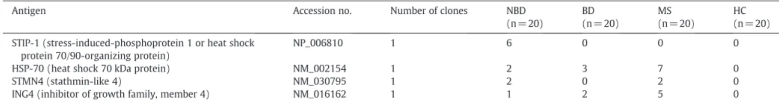
To identify the target antigens of NBD-associated neuronal auto-antibodies, we screened the sera of NBD patients by a protein macro-array derived from a human fetal brain cDNA expression library. This analysis identified four clones that had the highest signal inten-sity (3) and number of duplicates (more than 2): STIP-1 (or HSP 70/90-organizing protein), heat shock 70 kDa protein (HSP-70), stathmin-like 4 (STMN4) and inhibitor of growth family, member 4 (ING4). ELISA studies performed with the corresponding recombi-nant proteins revealed high-titer autoantibodies in varying numbers of NBD, BD and MS patients. MS patients displayed an increased fre-quency of high-titer autoantibodies to HSP-70 (7 patients, 35%) and ING4 (5 patients, 25%) than NBD and BD patients (0–3 patients, 0– 15%). Alternatively, high-titer STIP-1 autoantibodies were only detected in the sera of NBD patients (6 patients, 30%) (Table 3,
Fig. 3). None of the patients' sera gave high-titer antibody values with the lysate of the E. coli strain used to express the proteins, sup-porting the specificity of the autoantibody measurements and sug-gesting that the positivities were not due to crossreaction with
E. coli proteins (Fig. 3E). None of the CSF samples of NBD, BD, MS and headache patients had detectable antibodies directed against the tested recombinant proteins (data not shown). The binding to STIP-1 was confirmed by immunoblotting comparing with an anti-body to STIP-1 (Santa Cruz Biotechnology). Both the commercial and the positive NBD sera bound a band at 63 kDa (Fig. 4).
There were no significant differences between the demographic and clinical parameters of STIP-1 antibody positive and negative NBD patients. Also, there was no evident association between the presence of STIP-1 and neuropil antibodies (p = 0.14 by chi-square test) (Table 2).
4. Discussion
Although autoimmunity has often been considered in BD and NBD, previous studies have not succeeded in identifying useful biomarkers or potentially pathogenic autoantibodies. Here, we have shown that there are autoantibodies to neuronal cell surface antigens in a propor-tion of NBD patients, greater than in BD controls, and that there are also antibodies to STIP-1.
Previously identified neuropil antibodies have been shown to mostly react with the conformational epitopes of neuronal ion-channels. The best examples are NMDAR and components of the VGKC-complex (Vincent et al., 2004; Majoie et al., 2006; Irani et al., 2010a; Irani et al., 2010b). Antibodies to these cell-surface proteins are now being identified routinely in patients who have immunother-apy responsive forms of limbic encephalitis and encephalopathy. Unfortunately, screening of NBD patients' sera with a protein macro-array failed to reveal the antigenic targets of these neuropil
Table 3
Frequencies of serum antibodies to antigens isolated by protein macroarray in patient and control groups.
Antigen Accession no. Number of clones NBD
(n = 20) BD (n = 20) MS (n = 20) HC (n = 20) STIP-1 (stress-induced-phosphoprotein 1 or heat shock
protein 70/90-organizing protein)
NP_006810 1 6 0 0 0
HSP-70 (heat shock 70 kDa protein) NM_002154 1 2 3 7 0
STMN4 (stathmin-like 4) NM_030795 1 2 0 2 0
ING4 (inhibitor of growth family, member 4) NM_016162 1 1 2 5 0
NBD, neuro-Behçet's disease; BD, Behçet's disease patients with no neurological involvement; MS, multiple sclerosis; HC, headache controls; NA, not available.
Fig. 3. ELISA detection of IgG antibodies directed against stress-induced-phosphoprotein 1 (STIP-1) (A), heat-shock protein 70 (Hsp70) (B), inhibitor of growth family, member 4 (ING4) (C), stathmin-like 4 (STMN4) (D), and the E. coli lysate (E) in sera of neuro-Behçet's disease (NBD) patients, Behçet's disease patients with no neurological involvement (BD), multiple sclerosis patients (MS) and headache controls (HC). The dashed lines represent 2 standard deviations above the mean of the HC samples (cut-off values for positiv-ity). Horizontal lines indicate the mean value of each group. ***, pb0.001 by ANOVA.
antibodies since, although 5 out of 6 of the STIP-1 antibody positive sera were also positive for neuropil antibodies, STIP-1 antibody
could not be identified in 8 neuropil antibody positive sera
(Table 2). Nevertheless, considering that STIP-1 is not only confined to the cytoplasm but is also expressed at the cell surface (Zanata et al., 2002), at least one of the antigenic targets of the neuropil anti-bodies might be STIP-1. Given that STIP-1 has primarily an intracellu-lar location, it is intriguing that only one NBD patient's serum sample yielded an intracellular staining pattern with immunohistochemistry (Table 1). Also, the commercial STIP-1 antibody used in immunoblot-ting experiments did not give an appreciable staining with the frozen rat brain sections despite the fact that human and rat STIP-1 are 100% identical (data not shown). These results might be explained with the fact that rat brain STIP-1 expression levels are very low during phys-iological conditions and below the detection threshold of our immu-nohistochemistry method and perhaps attain detectable levels only under stress conditions. Moreover, our immunohistochemistry pro-cessing techniques might have destroyed the STIP-1 proteins.
Demonstration of antibodies to the neuronal axons and dendritic pro-cesses in the majority of NBD patients and emergence of these antibodies during neurological exacerbations suggest that antibody-mediated path-ogenic mechanisms might contribute to the development of neurological symptoms in BD. Whether these antibodies passively develop following blood–brain barrier breach during the attacks or occur prior to attacks and are genuinely involved in disease mechanisms remain to be elucidat-ed. Presence of neuropil antibodies in BD patients with no neurological signs and normal MRIfindings supports the latter assumption.
Protein macroarray enables simultaneous screening of thousands of potential autoantigens regardless of the expression levels of these antigens in their native host tissues. However, it might fail to deter-mine the antigenic targets of the antibodies recognizing conforma-tional epitopes, since the utilized proteins are expressed by bacteria rather than mammalian cells. Therefore, further investigation of the targets of the neuropil antibodies by immunoassays that preserve the conformation of the native proteins is warranted.
BD-associated autoantibody responses directed against diverse autoantigens have been reported by several investigators (Dinc et al., 2003; Fresko et al., 2005; Koca et al., 2007; Lee et al., 2009; Vural et al., 2009), studied by a variety of methods in different BD cohorts (e.g. BD with vascular, rheumatological or neurological symptoms). Previous attempts to look for antibodies in BD have had variable success and the antibodies identified were found in only a small fraction of the patients and were not BD specific. We also identified novel autoantibodies that were only detectable in 5–35% of NBD or BD patients. Since these were intracellular proteins the antibodies are unlikely to be pathogenic
but their presence supports the notion that BD might be an autoin flam-matory disorder and specific antigen-induced immune responses de-velop against random epitopes expressed by the involved tissues merely as an epiphenomenon of enhanced inflammation (Gül, 2005).
The protein macroarray screening did however identify four novel antigenic targets. Among these, STIP-1 is a recently discovered HSP (Zanata et al., 2002; Ji et al., 2007) and thus the identification of STIP-1 antibodies adds a new member to the list of BD-associated anti-bodies to stress-induced proteins, such as HSP-60, HSP-65, HSP-70 and αB-crystallin (Taşçi et al., 1998; Tanaka et al., 1999; Celet et al., 2000; Birtas-Atesoglu et al., 2008). Although HSP-65 andαB-crystallin anti-bodies are more prevalent in NBD patients, they might also be detected in BD patients with no neurological involvement or in MS patients (Gao et al., 1994; Prabhakar et al., 1994; Vojdani et al., 2003; Yokota et al., 2010). By contrast, our results suggest that high-titer STIP-1 antibodies seem to indicate NBD and are more frequently detected in its parenchy-mal subtype. However, STIP-1 antibodies have also been determined in ovarian cancer and rheumatoid arthritis patients (Goëb et al., 2009; Kim et al., 2010), reducing the specificity value of this protein as a er. Nevertheless, STIP-1 antibody might still be used as a novel biomark-er candidate in the diffbiomark-erential diagnosis of inflammatory CNS disorders, which closely mimic the symptoms and signs of NBD. Other autoanti-gens identified by protein macroarray are not specific to NBD and HSP-70 and ING4 antibodies appear to be more prevalent in MS pa-tients. While the association of HSP-70 antibodies with BD and MS has long been demonstrated (Birtas-Atesoglu et al., 2008; Yokota et al., 2010), STMN4 antibodies have only been reported in patients with pseudoexfoliation glaucoma (Dervan et al., 2010) and, to our knowl-edge, ING4 antibodies have not been previously described.
Further studies are needed to evaluate whether the autoanti-bodies or corresponding antigens identified in our study take part in the pathogenesis of NBD. In any case, these antibodies could serve as useful biomarkers to aid in the diagnosis and relapse prediction of NBD. They might also provide novel therapeutic targets for the spe-cific prevention of inflammatory CNS disorders.
Acknowledgment
This work was supported by TUBITAK (Project No: 108S053).
References
Akman-Demir, G., Serdaroglu, P., Tasçi, B., 1999. Clinical patterns of neurological in-volvement in Behçet's disease: evaluation of 200 patients. The Neuro-Behçet Study Group. Brain 122, 2171–2182.
Ances, B.M., Vitaliani, R., Taylor, R.A., Liebeskind, D.S., Voloschin, A., Houghton, D.J., Galetta, S.L., Dichter, M., Alavi, A., Rosenfeld, M.R., Dalmau, J., 2005. Treatment-responsive limbic encephalitis identified by neuropil antibodies: MRI and PET correlates. Brain 128, 1764–1777.
Birtas-Atesoglu, E., Inanc, N., Yavuz, S., Ergun, T., Direskeneli, H., 2008. Serum levels of free heat shock protein 70 and anti-HSP70 are elevated in Behçet's disease. Clin. Exp. Rheumatol. 26, S96–S98.
Büssow, K., Cahill, D., Nietfeld, W., Bancroft, D., Scherzinger, E., Lehrach, H., Walter, G., 1998. A method for global protein expression and antibody screening on high-densityfilters of an arrayed cDNA library. Nucleic Acids Res. 26, 5007–5008. Celet, B., Akman-Demir, G., Serdaroğlu, P., Yentür, S.P., Taşci, B., van Noort, J.M., Eraksoy,
M., Saruhan-Direskeneli, G., 2000. Anti-alpha B-crystallin immunoreactivity in in-flammatory nervous system diseases. J. Neurol. 247, 935–939.
Dervan, E.W., Chen, H., Ho, S.L., Brummel, N., Schmid, J., Toomey, D., Haralambova, M., Gould, E., Wallace, D.M., Prehn, J.H., O'Brien, C.J., Murphy, D., 2010. Protein macro-array profiling of serum autoantibodies in pseudoexfoliation glaucoma. Invest. Ophthalmol. Vis. Sci. 51, 2968–2975.
Dinc, A., Takafuta, T., Jiang, D., Melikoglu, M., Saruhan-Direskeneli, G., Shapiro, S.S., 2003. Anti-endothelial cell antibodies in Behçet's disease. Clin. Exp. Rheumatol. 21, S27–S30.
Fresko, I., Ugurlu, S., Ozbakir, F., Celik, A., Yurdakul, S., Hamuryudan, V., Yazici, H., 2005. Anti-Saccharomyces cerevisiae antibodies (ASCA) in Behçet's syndrome. Clin. Exp. Rheumatol. 23, S67–S70.
Gao, Y.L., Raine, C.S., Brosnan, C.F., 1994. Humoral response to hsp 65 in multiple scle-rosis and other neurologic conditions. Neurology 44, 941–946.
Goëb, V., Thomas-L'Otellier, M., Daveau, R., Charlionet, R., Fardellone, P., Le Loët, X., Tron, F., Gilbert, D., Vittecoq, O., 2009. Candidate autoantigens identified by mass Fig. 4. Representative immunoblots for Western blot analysis of recombinant
stress-induced-phosphoprotein 1 (STIP-1) protein. While both rabbit anti-human STIP-1 an-tibody (Ab) (1) and neuro-Behçet's disease (NBD) patients' sera that were found to be positive for STIP-1 Ab by ELISA (2) yielded ~ 63 kDa bands at the STIP-1 protein loaded gels, NBD sera that were seronegative for STIP-1 Ab by ELISA (3) did not.
spectrometry in early rheumatoid arthritis are chaperones and citrullinated glyco-lytic enzymes. Arthritis. Res. Ther. 11, R38.
Gül, A., 2005. Behçet's disease as an autoinflammatory disorder. Curr. Drug Targets Inflamm. Allergy 4, 81–83.
International Study Group for Behçet's Disease, 1990. Criteria for diagnosis of Behçet's disease. Lancet 335, 1078–1080.
Irani, S.R., Bera, K., Waters, P., Zuliani, L., Maxwell, S., Zandi, M.S., Friese, M.A., Galea, I., Kullmann, D.M., Beeson, D., Lang, B., Bien, C.G., Vincent, A., 2010a. N-methyl-D-as-partate antibody encephalitis: temporal progression of clinical and paraclinical ob-servations in a predominantly non-paraneoplastic disorder of both sexes. Brain 133, 1655–1667.
Irani, S.R., Alexander, S., Waters, P., Kleopa, K.A., Pettingill, P., Zuliani, L., Peles, E., Buckley, C., Lang, B., Vincent, A., 2010b. Antibodies to Kv1 potassium channel-complex proteins leucine-rich, glioma inactivated 1 protein and contactin-associated protein-2 in lim-bic encephalitis, Morvan's syndrome and acquired neuromyotonia. Brain 133, 2734–2748.
Ji, Q., Huang, C.H., Peng, J., Hashmi, S., Ye, T., Chen, Y., 2007. Characterization of STIP, a multi-domain nuclear protein, highly conserved in metazoans, and essential for embryogenesis in Caenorhabditis elegans. Exp. Cell Res. 313, 1460–1472. Kim, S., Cho, H., Nam, E.J., Kim, S.W., Kim, Y.T., Park, Y.W., Kim, B.W., Kim, J.H., 2010.
Au-toantibodies against stress-induced phosphoprotein-1 as a novel biomarker candi-date for ovarian cancer. Genes Chromosomes Cancer 49, 585–595.
Koca, S.S., Akbulut, H., Dag, S., Artas, H., Isik, A., 2007. Anti-cyclic citrullinated peptide anti-bodies in rheumatoid arthritis and Behçet's disease. Tohoku J. Exp. Med. 213, 297–304. Lancaster, E., Lai, M., Peng, X., Hughes, E., Constantinescu, R., Raizer, J., Friedman, D., Skeen, M.B., Grisold, W., Kimura, A., Ohta, K., Iizuka, T., Guzman, M., Graus, F., Moss, S.J., Balice-Gordon, R., Dalmau, J., 2010. Antibodies to the GABA(B) receptor in limbic encephalitis with seizures: case series and characterisation of the antigen. Lancet Neurol. 9, 67–76.
Lee, J.H., Cho, S.B., Bang, D., Oh, S.H., Ahn, K.J., Kim, J., Park, Y.B., Lee, S.K., Lee, K.H., 2009. Human anti-alpha-enolase antibody in sera from patients with Behçet's disease and rheumatologic disorders. Clin. Exp. Rheumatol. 27, S63–S66.
Lehner, T., Lavery, E., Smith, R., van der Zee, R., Mizushima, Y., Shinnick, T., 1991. Asso-ciation between the 65-kilodalton heat shock protein, Streptococcus sanguis, and the corresponding antibodies in Behçet's syndrome. Infect. Immun. 59, 1434–1441. Majoie, H.J., de Baets, M., Renier, W., Lang, B., Vincent, A., 2006. Antibodies to
voltage-gated potassium and calcium channels in epilepsy. Epilepsy. Res. 71, 135–141. Polman, C.H., Reingold, S.C., Edan, G., Filippi, M., Hartung, H.P., Kappos, L., Lublin, F.D.,
Metz, L.M., McFarland, H.F., O'Connor, P.W., Sandberg-Wollheim, M., Thompson,
A.J., Weinshenker, B.G., Wolinsky, J.S., 2005. Diagnostic criteria for multiple sclero-sis: 2005 revisions to the“McDonald Criteria”. Ann. Neurol. 58, 840–846. Prabhakar, S., Kurien, E., Gupta, R.S., Zielinski, S., Freedman, M.S., 1994. Heat shock
pro-tein immunoreactivity in CSF: correlation with oligoclonal banding and demyelin-ating disease. Neurology 44, 1644–1648.
Preuss, K.D., Pfreundschuh, M., Ahlgrimm, M., Fadle, N., Regitz, E., Murawski, N., Grass, S., 2009. A frequent target of paraproteins in the sera of patients with multiple my-eloma and MGUS. Int. J. Cancer 125, 656–661.
Tanaka, T., Yamakawa, N., Koike, N., Suzuki, J., Mizuno, F., Usui, M., 1999. Behçet's dis-ease and antibody titers to various heat-shock protein 60s. Ocul. Immunol. Inflamm. 7, 69–74.
Taşçi, B., Direskeneli, H., Serdaroglu, P., Akman-Demir, G., Eraksoy, M., Saruhan-Direskeneli, G., 1998. Humoral immune response to mycobacterial heat shock protein (hsp)65 in the cerebrospinal fluid of neuro-Behçet patients. Clin. Exp. Immunol. 113, 100–104.
Vincent, A., Buckley, C., Schott, J.M., Baker, I., Dewar, B.K., Detert, N., Clover, L., Parkinson, A., Bien, C.G., Omer, S., Lang, B., Rossor, M.N., Palace, J., 2004. Potassium channel antibody-associated encephalopathy: a potentially immunotherapy-responsive form of limbic encephalitis. Brain 127, 701–712.
Vojdani, A., Vojdani, E., Cooper, E., 2003. Antibodies to myelin basic protein, myelin ol-igodendrocytes peptides, alpha-beta-crystallin, lymphocyte activation and cyto-kine production in patients with multiple sclerosis. J. Intern. Med. 254, 363–374. Vural, B., Demirkan, A., Ugurel, E., Kalaylioglu-Wheeler, Z., Esen, B.A., Gure, A.O., Gül, A.,
Ozbek, U., 2009. Seroreactivity against PTEN-induced putative kinase 1 (PINK1) in Turkish patients with Behçet's disease. Clin. Exp. Rheumatol. 27, S67–S72. Yokota, S., Chiba, S., Furuyama, H., Fujii, N., 2010. Cerebrospinalfluids containing
anti-HSP70 autoantibodies from multiple sclerosis patients augment anti-HSP70-induced proin-flammatory cytokine production in monocytic cells. J. Neuroimmunol. 218, 129–133. Yurdakul, S., Yazici, H., 2008. Behçet's syndrome. Best Pract. Res. Clin. Rheumatol. 22,
793–809.
Zanata, S.M., Lopes, M.H., Mercadante, A.F., Hajj, G.N., Chiarini, L.B., Nomizo, R., Freitas, A.R., Cabral, A.L., Lee, K.S., Juliano, M.A., de Oliveira, E., Jachieri, S.G., Burlingame, A., Huang, L., Linden, R., Brentani, R.R., Martins, V.R., 2002. Stress-inducible protein 1 is a cell surface ligand for cellular prion that triggers neuroprotection. EMBO J. 21, 3307–3316.