The in
fluence of blood and/or hemostatic agent contamination on
Micro-TBS to dentin
E. Karamana*, D. Tuncerb, T. Tozc, M. Kusdemircand G. Gencc
aDepartment of Restorative Dentistry, School of Dentistry, Ondokuz Mayıs University, Samsun, Turkey;bDepartment of Restorative Dentistry, School of Dentistry, Baskent University, Istanbul, Turkey;cDepartment of Restorative Dentistry, School of Dentistry, Istanbul Medipol University,
Istanbul, Turkey
(Received 9 January 2015;final version received 25 February 2015; accepted 28 February 2015) Aim: The purpose of this study was to evaluate the effects of blood contamination and hemostatic agent Ankaferd Blood Stopper (ABS: Ankaferd Drug Inc, Istanbul, Turkey) on the microtensile bond strength of a self-etching adhesive. Material and methods: Flat dentin surfaces were created from 40-M teeth and randomly divided to four groups according to contamination and adhesive procedure. The specimens of Group 1 are contaminated with blood; ABS was applied to the specimens of Group 2 after blood contamination and applied to the specimens of Group 3 without blood contamination. Group 4 is control group and self-etching adhesive was applied to all specimens. Teeth were restored with a nanohybrid composite. The specimens were sectioned to the beams and microtensile testing was carried out and the data were statistically analyzed with analysis of variance test. Tukey’s honestly significant difference post hoc test was also performed for multiple comparisons to compare subgroups. Results: Group 4 had the highest strength value, followed by Group 3, while Group 1, which contacts only with blood, had the lowest strength value. Conclusion: ABS has a negative effect on the bond strength of one-step self-etching adhesive system.
Keywords: blood contamination; hemostatic agent contamination; Micro-TBS
Introduction
Gingival bleeding caused by cavity preparation is a common problem in dentistry. While using resin-based materials, any contamination of prepared tooth surface by saliva, blood, or gingival fluids should be avoided in order to achieve a successful bonding. Especially, when the caries extends closely to the gingival margin, contamination control is difficult and isolation with rubber dam is not feasible.[1,2]
Bond strength of adhesive systems to dentin has been reported to be negatively influenced by blood contamination.[3,4] Using topical hemostatic agents, such as hydrogen peroxide (H2O2), aluminum chloride, ferric sulfate, trichloroacetic acid, and Ankaferd Blood Stopper (ABS) (Ankaferd Drug Inc, Istanbul, Turkey) is the most common procedure used to control bleeding.[4–6]
ABS is a standardized mixture of medicinal plant extract, each of which has different effects on blood cell tissues, endothelium, angiogenesis, cellular proliferation,
*Corresponding author. Email:[email protected]
© 2015 Taylor & Francis
http://dx.doi.org/10.1080/01694243.2015.1025465
vascular dynamics, or cell mediators. It contains Thymus vulgaris, Glycyrrhiza glabra, Vitis vinifera, Alpinia officinarum, and Urtica dioica plants, that have been approved in Turkey for the management of post-surgery dental bleedings and external hemor-rhage.[4,7,8] Several studies have investigated the effects of hemostatic agents after blood contamination, on bond strength to dentin. However, there is limited data about the effects of ABS on bond strength to dentin.
Nowadays one-step self-etching (all-in-one) adhesive systems have attracted the attention of most dentists as they simplify the bonding process and decrease the chair-side time for restorative procedures. As a result of no requirement to separate etching, rinsing, and drying steps, technique sensitivity and dentin desiccation risk are elimi-nated with these adhesive systems.[9,10] These advantages of one-step self-etching adhesive systems make them one of most popular adhesives used in case of bleeding in operationfield.
In clinical practice, when gingival bleeding occurred, irrespective of contamination field, hemostatic agent is applied to all cavity walls. But what about the cavity sides which were not contaminated with blood? Does not hemostatic agent affect the bond strength to these non-contaminated tooth tissues? To the knowledge of authors, the literature is limited with regard to evaluations of the effects of only hemostatic agent contamination on dentin. So this study aimed to investigate the effects of blood and/or ABS contamination on microtensile bond strength to dentin and compare with only ABS-contaminated dentin. The hypothesis of study was that contamination with blood and/or ABS will reduce bond strength to dentin.
Materials and methods Tooth preparation
Forty freshly extracted, caries-free human molar teeth were disinfected in 0.5% chlora-mine, stored in distilled water, and used within six months after extraction. Flat dentin surfaces were created on mid-coronal dentin using a slow-speed diamond saw (Isomet, Buehler Ltd, IL, USA) under water cooling. The exposed dentin was inspected with a stereomicroscope (Olympus SZ 61, Olympus Corporation, Japan) to ensure that all of the occlusal enamel had been removed and the pulp horns had not been perforated, then abraded with wet 600 grid silicon carbide papers to provide a uniform dentin surface for bonding. The teeth were then rinsed with distilled water to remove any debris.
Experimental design
The teeth were randomly assigned into four groups (n = 10) and adhesive system was used under four protocols, these protocols were: Group 1 ‘control,’ Group 2 ‘Blood contamination,’ Group 3 ‘Blood + ABS contamination,’ and Group 4 ‘ABS contamination.’
Restorative procedures and specimen preparation
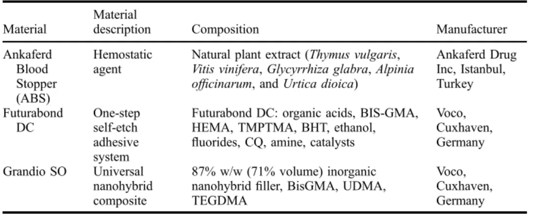
The materials used in the current study are listed in Table1. All of the materials were applied strictly in accordance with manufacturer’s instructions.
Group 1 (Blood contamination)
The blood collected from a single individual (needle-prick of an alcohol-wiped forefinger) at the time of experiment was applied to the prepared dentin surfaces. It has been mentioned in the literature that freshly drawn capillary blood is more suitable in laboratory investigations involving blood contamination than heparinized blood.[11] The surfaces were washed with distilled water following the contamination (20 s) and air-dried. Then, self-etching adhesive Futurabond DC (Voco, Cuxhaven, Germany) was applied to the dentin surface and rubbed for 20 s, dried for 5 s with an air syringe, and light-cured (Guilin Woodpecker Medical Instrument Co., Ltd, China) for 10 s.
Group 2 (Blood + ABS contamination)
One drop of hemostatic agent ABS (Ankaferd Drug Inc, Istanbul, Turkey) was applied to the blood-contaminated surfaces for 20 s, rinsed for 10 s with water spray, and blot-ted with absorbent paper. Adhesive procedures were then applied as mentioned in Group 1 according to the manufacturer’s instructions.
Group 3 (ABS contamination)
Self-etching adhesive procedures (Futurabond DC) were applied according to the manu-facturer’s acknowledgements after the hemostatic agent contamination and rinsing of the surfaces as explained above.
Group 4 (Control)
Futurabond DC was applied to the dentin and light-cured (10 s).
After the bonding procedure, a composite crown was built up by layering of Grandio SO (Voco, Cuxhaven, Germany) to a height of 4–5 mm. Each increment was not exceeding 2 mm in height and cured for 20 s. The restored teeth were stored in distilled water at 37 °C for 24 h, the specimens were sectioned using a slow-speed diamond saw (Isomet, Buehler Ltd, Lake Bluff, IL, USA) to obtain resin-dentin sticks from a tooth with a cross-sectional area of approximately 1 mm2 measured with a digital caliper (Sylvac, Fred V. Fowler Co., Swiss). Only beams from the central region of each tooth were used.
Table 1. Materials used in the study.
Material
Material
description Composition Manufacturer
Ankaferd Blood Stopper (ABS) Hemostatic agent
Natural plant extract (Thymus vulgaris, Vitis vinifera, Glycyrrhiza glabra, Alpinia officinarum, and Urtica dioica)
Ankaferd Drug Inc, Istanbul, Turkey Futurabond DC One-step self-etch adhesive system
Futurabond DC: organic acids, BIS-GMA, HEMA, TMPTMA, BHT, ethanol, fluorides, CQ, amine, catalysts
Voco, Cuxhaven, Germany Grandio SO Universal nanohybrid composite 87% w/w (71% volume) inorganic nanohybridfiller, BisGMA, UDMA, TEGDMA
Voco, Cuxhaven, Germany
Microtensile bond strength testing
The resin-bonded specimens were stored in water at 37 °C for a further 24 h. The sticks were then attached with cyanoacrylate adhesive (Zapit, Dental Ventures of America, Corona, CA, USA) to a testing apparatus (Bencor-Multi-T-testing: Danville Engineering, Danville, CA, USA), and a tensile load was applied using a universal test-ing machine (Instron 5566 series 5000: Instron Corporation, London, UK) at a cross-head speed of 1.0 mm/min. The μTBS was derived by dividing the force imposed at the time of fracture by the bond area (mm2). When specimens failed before actual test-ing, a bond strength of 0 MPa was included in the calculation of the mean μTBS. The mean microtensile bond strength for each material was calculated from the values obtained with 14 slices and expressed in MPa.
Statistical analyses
Statistical analyses were performed using SPSS for Windows Release 16.0 (SPSS Corporation, Chicago, IL). Descriptive statistics, including the mean and Standard deviation (SD) were calculated for each of the four groups. The parametric one-way analysis of variance (ANOVA) test was used to determine when significant differences were present in Micro-TBS between the four groups. The Tukey’s honestly significant difference post hoc test was also performed for multiple comparisons to compare subgroups. The results were evaluated with a 95% confidence interval. Significance for all statistical tests was predetermined at p < 0.05.
Results
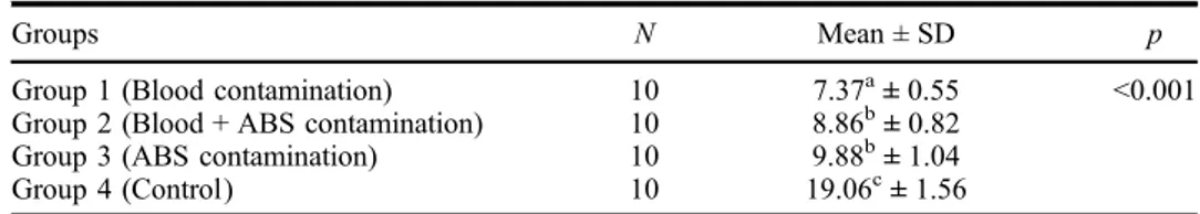
The results of the ANOVA indicated significant differences in the Micro-TBS between the four groups (p < 0.01). The Tukey’s honestly significant difference post hoc test indicated that the mean Micro-TBS values of Groups 2 and 3 were similar, while Groups 1 and 4 were significantly different from each other and Groups 2 and 3 (p < 0.01). Group 4 had the highest strength value, followed by Group 3, while Group 1 had the lowest strength value (Table2).
Discussion
In this study, the effect of blood and/or ABS contamination on Micro-TBS of a one-step self-etching adhesive systems to dentin was evaluated. The results obtained in this study supported the hypothesis that blood and/or ABS contamination reduced the bond strength value of self-etching adhesive, so the test hypothesis was accepted.
Freshly drawn blood was used to contaminate prepared dentin surfaces for this study and blood was drawn immediately prior to adhesive application to simulate
Table 2. Mean Micro-TBS values (MPa) ± SDs for each group examined in the current study.
Groups N Mean ± SD p
Group 1 (Blood contamination) 10 7.37a± 0.55 <0.001
Group 2 (Blood + ABS contamination) 10 8.86b± 0.82
Group 3 (ABS contamination) 10 9.88b± 1.04
Group 4 (Control) 10 19.06c± 1.56
Note: Mean values exhibiting different letters were significantly different.
gingival bleeding in clinical conditions. Dietrich et al. [11] reported that blood coagula-tion might be accepted as an important factor in the relacoagula-tion of blood and dentin on bonding prognosis. The authors investigated the effect of heparinized or native blood on marginal adaptation of adhesive restorations, and observed high percentages of mar-ginal gaps after contamination with fresh capillary blood than anti-coagulated blood.
Appropriate cleansing of blood-contaminated dental tissue is an important step in obtaining better adhesion. In this study, the blood-contaminated dentin surfaces were washed with distilled water following the contamination (20s) and air-dried. Tachibana et al. [12] evaluated the efficacy of cleansing agents on contaminated enamel and
den-tin, and they reported that the use of a water stream for 10 s was enough to remove blood contamination on enamel and dentin before the application of a self-etch adhesive.
In the current study, Micro-TBS of the blood-contaminated group was lower than that for control group, which is similar to the previous studies.[13,14] It has been reported that high protein content of blood with macromolecules like fibrinogen can form a film on dentin surfaces [15] and damage the penetration of the adhesive through dentin tubules.[2] This could explain the lowest Micro-TBS values obtained in Group 1.
In Group 2, ABS application after blood contamination resulted in higher Micro-TBS values compared with only blood contamination group, while these values were statistically lower than control group values. In this study, ABS appeared to be a physical barrier that breaks down the mechanical retention of adhesive resins. Similarly, Ulusoy et al. [16] reported lower Micro-TBS values of ABS groups than non-contami-nated groups but higher than that of blood-contaminon-contami-nated groups’.
In clinical practice, bleeding mostly occurs around the gingival margin but the clini-cian applies the hemostatic agent to all cavity walls. Therefore, a large part of cavity surface is contaminated just by the hemostatic agent without blood contamination. In this study, ABS was applied without blood contamination in Group 3 in order to simu-late this situation and investigate the effects of hemostatic agent alone on bond strength to dentin. The results showed that ABS contamination decreased the bond strength to dentin. In contrast to ourfindings, Harnirattisai et al. [17] reported no significant
differ-ences between the dentin bond strengths to normal dentin and astringent containing 25% aluminum chloride-contaminated dentin surfaces, although authors conclude that these results were limited to the materials used in their study and other materials might exhibit differently.
This result was consistent with previous studies of Ulusoy et al. and Arslan et al. that used ABS as hemostatic agent.[16,18] Finally, our study was effective in proving the negative interference of blood and/or ABS contamination on adhesion to dentin using a one-step adhesive system. However, it is important not to forget that this is an in vitro study, and in vivo studies are necessary to provide relevant knowledge to dental professionals.
Conclusion
Although our study findings have shown that ABS has a negative effect on the bond strength of one-step self-etching adhesive system to the non-contaminated dentin sur-face, results also have shown that in the presence of blood, washing with water is not sufficient and use of a hemostatic agent still is necessary. In vivo studies are necessary to prove the safety and longevity of this adhesiveness.
ORCID
G. Genc http://orcid.org/0000-0002-8310-2410
References
[1] Yazici AR, Tuncer D, Dayangaç B, Ozgünaltay G, Onen A. The effect of saliva contamina-tion on microleakage of an etch-and-rinse and a self-etching adhesive. J. Adhes. Dent. 2007;9:305–309.
[2] de Carvalho Mendonça EC, Vieira SN, Kawaguchi FA, Powers J, Matos AB. Influence of blood contamination on bond strength of a self-etching system. Eur. J. Dent. 2010;4: 280–286.
[3] Chang SW, Cho BH, Lim RY, Kyung SH, Park DS, Oh TS, Yoo HM. Effects of blood contamination on microtensile bond strength to dentin of three self-etch adhesives. Oper. Dent. 2010;35:330–336.
[4] Trakyali G, Oztoprak MO. Plant extract ankaferd blood stopper effect on bond strength. Angle Orthod. 2010;80:570–574.
[5] Kuphasuk W, Harnirattisai C, Senawongse P, Tagami J. Bond strengths of two adhesive systems to dentin contaminated with a hemostatic agent. Oper. Dent. 2007;32:399–405. [6] Ercan E, Erdemir A, Zorba YO, Eldeniz AU, Dalli M, Ince B, Kalaycioglu B. Effect of
different cavity disinfectants on shear bond strength of composite resin to dentin. J. Adhes. Dent. 2009;11:343–346.
[7] Barka EA, Belarbi A, Hachet C, Nowak J, Audran JC. Enhancement of in vitro growth and resistance to gray mould of Vitis vinifera co-cultured with plant growth-promoting rhizobac-teria. FEMS Microbiol. Lett. 2000;186:91–95.
[8] Goker H, Haznedaroglu IC, Ercetin S, Kirazli S, Akman U, Ozturk Y, Firat HC. Haemostatic actions of the folkloric medicinal plant extract ankaferd blood Stopper®. J. Int. Med. Res. 2008;36:163–170.
[9] Perdigao J, Gomes G, Lopes MM. Influence of conditioning time on enamel adhesion. Quintessence Int. 2006;37:35–41.
[10] Aguilera FS, Osorio E, Toledano M, Osorio R. Ultra-structure characterization of self-etching treated cementum surfaces. Med. Oral Patol. Oral Cir. Bucal. 2009;16:e265–e270. [11] Dietrich T, Kraemer ML, Roulet JF. Blood contamination and dentin bonding effect of
anticoagulant in laboratory studies. Dent. Mater. 2002;18:159–162.
[12] Tachibana A, Castanho GM, Vieira SN, Matos AB. Influence of blood contamination on bond strength of a self-etching adhesive to dental tissues. J. Adhes. Dent. 2011;13:349–358. [13] Kaneshima T, Yatani H, Kasai T, Watanabe EK, Yamashita A. The influence of blood
con-tamination on bond strengths between dentin and an adhesive resin cement. Oper. Dent. 2000;25:195–201.
[14] Kilic K, Arslan S, Demetoglu GA, Zararsiz G, Kesim B. Do blood contamination and haemostatic agents affect microtensile bond strength of dual cured resin cement to dentin? J. Appl. Oral Sci. 2013;21:85–91.
[15] Botta SB, Vieira SN, Cordon R, Marques MM, Matos AB. Can the method of primer application influence adhesion to Er:YAG-laser irradiated dentin? J. Contemp. Dent. Pract. 2009;10:49–57.
[16] Ulusoy AT, Bayrak S, Tunc ES, Tuzuner T. Effect of new haemostatic agent on microtensile bond strength of two adhesive systems to dentin. Mater. Res. Innovations. 2011;15:330–334. [17] Harnirattisai C, Kuphasuk W, Senawongse P, Tagami J. Bond strengths of resin cements to
astringent-contaminated dentin. Oper. Dent. 2009;34:415–422.
[18] Arslan S, Ertas H, Zorba YO. Influence of Ankaferd Blood Stopper on shear bond strength of bonding systems. Dent. Mater. J. 2012;31:226–231.
Downloaded by [Baskent Universitesi] at 00:15 26 March 2015
View publication stats View publication stats