Full Terms & Conditions of access and use can be found at
https://www.tandfonline.com/action/journalInformation?journalCode=yscm20
The Journal of Spinal Cord Medicine
ISSN: 1079-0268 (Print) 2045-7723 (Online) Journal homepage: https://www.tandfonline.com/loi/yscm20
A new lumbar fixation device alternative to
pedicle-based stabilization for lumbar spine: In
vitro
cadaver investigation
Cengiz Gomleksiz, Deniz Ufuk Erbulut, Halil Can, Manoj Kumar Kodigudla,
Amey V. Kelkar, Eser Kasapoglu, Ali Fahir Ozer & Vijay K. Goel
To cite this article: Cengiz Gomleksiz, Deniz Ufuk Erbulut, Halil Can, Manoj Kumar Kodigudla, Amey V. Kelkar, Eser Kasapoglu, Ali Fahir Ozer & Vijay K. Goel (2020) A new lumbar fixation device alternative to pedicle-based stabilization for lumbar spine: In�vitro cadaver investigation, The Journal of Spinal Cord Medicine, 43:1, 98-105, DOI: 10.1080/10790268.2018.1495932
To link to this article: https://doi.org/10.1080/10790268.2018.1495932
Published online: 16 Jul 2018.
Submit your article to this journal
Article views: 216
View related articles
A new lumbar fixation device alternative to
pedicle-based stabilization for lumbar spine:
In vitro cadaver investigation
Cengiz Gomleksiz
1, Deniz Ufuk Erbulut
2, Halil Can
1, Manoj Kumar Kodigudla
2,
Amey V. Kelkar
2, Eser Kasapoglu
3, Ali Fahir Ozer
4, Vijay K. Goel
21
Department of Neurosurgery, Biruni University, Istanbul, Turkey,2Engineering Center for Orthopaedic Research Excellence (ECORE), University of Toledo, Toledo, Ohio, USA,3Department of Computer Assisted Design and Animation Program, Istanbul Medipol University, Istanbul, Turkey,4Department of Neurosurgery, Koc University, Istanbul, Turkey
Context:To evaluate the stability provided by a new bilateral fixation technique using anin vitro investigation for posterior lumbar segmental instrumentation.
Design:Experimental cadaver study. In this study, we propose an alternative technique for a posterior lumbar fixation technique called“inferior-oblique transdiscal fixation” (IOTF).
Setting: Study performed at Engineering Center for Orthopedic Research Exellence (ECORE) in Toledo University-Ohio.
Participants:Six human lumbar cadaveric specimen used in this study.
Interventions:In this study, we propose an alternative technique for a posterior lumbar fixation technique called “inferior-oblique transdiscal fixation” (IOTF). As a novel contribution to the classical technique, the entry point of the screw is the supero-lateral point of the intersecting line drawn between the corpus and the pedicle of the upper vertebra. This approach enables the fixation of two adjacent vertebrae using a single screw on each side without utilizing connecting rods.
Outcome Measures:Flexion (Flex), extension (Ext), right and left lateral bending (LB & RB), and right and left axial rotation (LR & RR), and the position data were captured at each load step using the Optotrak motion measurement system and compared for IOTF and posterior transpedicular stabilization.
Results: The Posterior stabilization system (PSS) and IOTF significantly reduced the ROM of L4-L5 segment compared to intact segment’s ROM. During axial rotation (AR) IOTF fused index segment more than PSS. Besides this, addition of transforaminal lumbar interbody fusion (TLIF) cage improved the stabilization of IOTF system during flexion, extension and lateral bending. Whereas, PSS yielded better fusion results during extension compared to IOTF with and without interbody fusion cages.
Conclusions: We hypothesized that the new posterior bilateral system would significantly decrease motion compared to the intact spine. This cadaver study showed that the proposed new posterior fusion technique IOTF fused the index segment in a similar fashion to the classical pedicle screw fusion technique.
Keywords: Lumbar fusion, Posterior fixation, Pedicle screw fixation, Bilateral system,In vitro lumbar, Lumbar spine
Introduction
Posterior lumbar segmental instrumentation using pedicle screws has long been used by spine surgeons for fixation in degenerative spine diseases, trauma and congenital/acquired spinal deformities. Posterior
stabilization was first described by Albee for the treat-ment of Pott disease in 1911, and Hibbs described the same technique for degenerative instability in the same year.1,2 This technique could be described as interspi-nous posterior fixation since autografts were placed between the decorticated spinous processes. In 1934, Mixter and Barr3 hypothesized that lumbo-sciatalgia was caused by disc herniation and that the most durable and effective treatment would be “a crude
Correspondence to: Cengiz Gomleksiz, Department of Neurosurgery, Medical Faculty, University of Biruni, Istanbul/TURKEY. Email: [email protected]; Gsm:+905321634938
Color versions of one or more of the figures in the article can be found online atwww.tandfonline.com/yscm.
pure fusion technique”. In 1962, Harrington4proposed a novel method for treating scoliosis. Harrington’s method involved the use of metal rods and hooks for providing stability to the spine along with correcting the curvature of the spine. Later, these rods became known as “Harrington rods”. These rods were com-monly used during the subsequent 30 years for scoliosis, degenerative spine and instability surgeries. A technical paradigm shift in spinal instrumentation was achieved by the introduction of transpedicular screws. Today’s commonly used contemporary transpedicular screwing technique was first used in 1970 by Roy-Camille et al.5 and in 1984 by Cotrel and Dubousset.6 Anatomical landmarks are well defined for pedicle screw insertion, and real-time image guidance by intrao-perative computed tomography has improved the accu-racy of screw trajectory. However, significant malposition or inadvertent course of screws remains a major concern among spine surgeons.7,8
Lumbar pedicle screw-based fixation is usually accomplished by open surgery; however, percutaneous lumbar surgery is also performed, as reported in the lit-erature.9–12The pedicle screw fixation technique, despite being the“gold standard”, carries the risk of violation of the facet joint and excessive degeneration of the adjacent vertebrae.13–16In addition, pedicle screws also carry the risk of screw loosening/failure, as reported in the litera-ture.17–20Over time, pedicle screw designs have varied. Currently, there are rigid screws and dynamic screws that can be used in conjunction with dynamic and/or rigid rods. However, posterior stabilization with dynamic screws in conjunction with rigid rods led to insufficient flexibility, and dynamic rods in conjunction with rigid screws led to screw loosening.21
The aim of this study was to compare a new bilateral fusion technique with the traditional pedicle screw fix-ation technique. Hence, we developed a new screw inser-tion technique as an alternative to the classical pedicle screw technique. We named this new technique “inferior-oblique transdiscal fixation” because this approach enabled fixation of two adjacent vertebrae
using a single screw on each side without utilizing con-necting rods. In the current study, the biomechanics and limitations of this new screwing technique were tested and compared to those of the standard posterior transpedicular screw insertion technique with and without transforaminal interbody fusion cages using an in vitro cadaver testing method.
Materials and methods
Description of the“inferior-oblique transdiscal fixation” technique (IOTF)
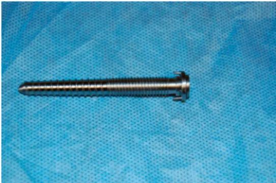
The entry point of the lag screw in this new technique is the supero-lateral point of the intersection line between the corpus and the pedicle of the upper vertebra (Figure 1). From this point on, the lag screw (Figure 2) is directed toward approximately 30 degrees in the sagittal plane (antero-inferior) and 45 degrees in the coronal plane (medio-inferior) bilaterally; thus, the overall trajectory is oblique and antero-infero-medial (AIM) (Figure 3). This oblique inclination enables the screw to pass through the entire corpus of the upper vertebra, the inter-vertebral disc and the upper half of the lower vertebra.
As described earlier by Wiltse et al.22a lumbar bilat-eral para-median skin incision five centimeters in length and bilateral paravertebral muscular blunt dissection between the plane of the multifidus and longissimus muscles are performed. The facet joints and transverse processes are exposed on both sides. As a novel contri-bution to the classical technique, the entry point of the screw is the supero-lateral point of the intersection line between the corpus and the pedicle of the upper verte-bra. To expose this entry point, the lateral border of the pars interarticularis and the transverse process of the upper vertebra should be well visualized. Subsequently, the intertransverse ligament is incised over the attachment of the transverse process medially up to the lateral border of the pars interarticularis and then superiorly up to the inferior border of the upper transverse ligament to create an L-shaped flap reflected laterally. From this point on, the pedicle-corpus junction
Figure 1 Picture of entry point demonstration. Figure 2 Picture of prototype of screw used in study.
Gomleksiz et al. A new lumbar fixation device alternative to pedicle-based stabilization for lumbar spine
is identified by following the pedicle starting from the lateral aspect of the pars interarticularis. Then, the exiting nerve root is gently retracted laterally and the screw (Figure 1) is directed under the guidance of the O-arm or C-arm. When a transforaminal lumbar inter-body fusion (TLIF) procedure is attempted in addition to these screws, the transverse process of the two lower vertebrae should also be exposed. For example, if we plan to stabilize L4-L5 segment using this new technique, L3 and L4 transverse processes should be exposed bilat-erally; if we plan to add TLIF, we should additionally expose the L5 transverse processes bilaterally. When the plane passing through the vertebral end plates is assumed to be a reference horizontal plane (axis), the screw inclination is approximately 30 degrees in sagittal plane (antero-inferior) and 45 degrees in coronal plane (medio-inferior); thus, the overall trajectory is oblique and antero-infero-medial (AIM) (Figure 2). This oblique inclination enables the screw to pass through the entire corpus of the upper vertebra, the intervertebral disc and the upper half of the lower vertebra
The new proposed surgical technique offers superior-ity in some aspects over the classical pedicle screw tech-nique in terms of the following: easy revision surgery, absence of need for domino placement, tri-cortical
fixation and less risk of loosening of screws, safer trajec-tory with regard to the risks of dural sac and root inju-ries, shorter incision, less paravertebral muscle and soft tissue dissection; possibility of interbody fusion and shorter duration of hospitalization. The other advantage of this new technique is the fact that it does not require a longer learning curve.
A disadvantage of this new technique is the potential need of exiting nerve root retraction during the screw placement. On the other hand, this technique is not useful for deformity surgery because of the absence of correction maneuvers.
Biomechanical testing
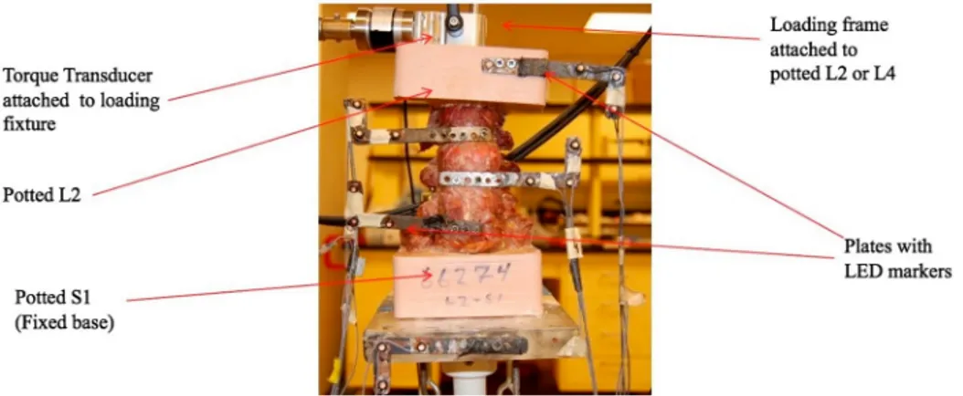
We tested the efficacy of new technique (IOTF) for L4-5 fusion and compared the results with those of the classi-cal pedicle screw technique biomechaniclassi-cally at E-CORE (Engineering Center for Research Excellence, The University of Toledo). Six L2-S1 cadaveric specimens were obtained and tested in this study. Detailed infor-mation regarding the specimens is listed in Table 1. The DEXA T scores for these specimens ranged from -2.8 to -5.1. The spines were thawed, and the tissue sur-rounding the specimen, except for ligaments and discs, were carefully removed. The cranial (L2) and caudal (sacrum) ends of each specimen were potted using bondo (a 2-part epoxy resin). Potted sacra of each speci-men were fixed on the testing apparatus located in the field of view of the Optotrak (NDI, Waterloo, Canada) motion capture system. LED (Light emitting diode) marker sets were attached to L2, L3, L4, L5 and the potted sacrum for recording the spatial locations in response to the applied moment using the Optotrak motion capture system. A loading fixture was attached to the potted cranial (L2) end and a torque transducer was attached to that fixture (Figure 4).
Each intact specimen was subjected to pure moments applied by the torque transducer in steps of 0, 2.5, 5, 7.5,
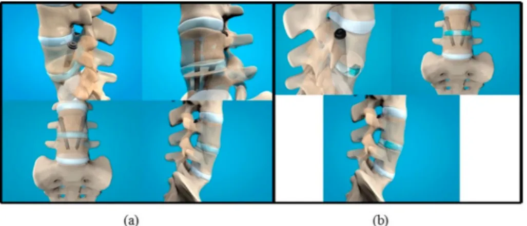
Figure 3 3-D illustration of application of new system screws (a) without TLIF and (b) with TLIF.
Table 1 Detailed specimen information. Specimen ID Age Sex T Score Condition Cause of death (COD) 66568 72 Female -2.8 Osteoporosis Lung cancer,
COPD, Hypertension, Hyperlipidemia 66814 84 Male -2.8 Osteoporosis -66310 77 Female -5.1 Osteoporosis COPD, Resp.
failure 68153 87 Male 2.7 Normal -66267 57 Male -3 Osteoporosis Gunshot wound 65177 62 Female -3.8 Osteoporosis Cardiac arrest
10 and 0 Nm in flexion (Flex), extension (Ext), right and left lateral bending (LB & RB), and right and left axial rotation (LR & RR), and the position data were cap-tured at each load step using the Optotrak motion measurement system (Figure 3). This system uses infra-red cameras to track the spatial locations of three light-emitting diodes affixed to each vertebral body in response to the applied loads.
Following the testing of intact specimens, each L4-L5 was stabilized using a pedicle screw system (PSS) and was tested for range of motion (ROM). In the third step, the pedicle screws were removed and the IOTF screws were inserted bilaterally using the new technique and were tested for ROM. In the fourth step, both the IOTF screws were partially removed and TLIF cages were inserted bilaterally. The lag screws were reinserted (IOTF+ TLIF cages) and tested for ROM. Finally, the IOTF screws were removed and pedicle screws of larger size were inserted and fixed, keeping the cages (PSS+ TLIF cages), and were tested for ROM.Table 2shows the implantation information for each specimen tested. Figure 5shows the X-ray images of implantation in each case. Range of motion for each segment (L2-3, L3-4, L4-5, and L5-S1) was measured independently for flexion, exten-sion, right and left lateral bending, and for right and left rotation under 10 Nm pure moment. The mean and standard deviation of ROM for various cases tested were computed (n= 6). Statistical analysis using the paired T-test (Minitab) was performed on the kinematic data to determine the significance of multiple pair-wise comparisons. Testing hypotheses were considered statistically significant at P values below 0.05. Using statistical data, the following two hypotheses were investigated:
Hypothesis 1: The implanted cases have significantly decreased motion compared to intact cases.
Hypothesis 2: The bilateral approach using lag screws with or without cages provided less stability than did the traditional pedicle screw approach.
Results
Results of the biomechanical tests are summarized in
Figures 6–8, and statistical analysis data are shown in
Table 3. Figure 6 shows the range of motion (ROM) data in response to the applied 10 Nm moments. They represent the mean data of six specimens. We showed that the instrumented cases reduced the motion across the segment more than did the intact cases. The percen-tage of reduced motion was more for flex, ext and lateral bending cases but was less for left and right rotations.
Compared to the intact, the PSS at L45 decreased the motion of the segments in flex (68%), ext (58%), lb (53%), rb (62%), lr (22%) and rr (15%). Compared to the intact, the IOTF at L45 decreased the motion of the segments in flex (47%), ext (34%), lb (33%), rb (42%), lr (29%) and rr (25%). Compared to the intact, the IOTF with TLIF cages at L45 decreased the motion of the segments in flex (56%), ext (46%), lb (50%), rb (52%), lr (26%) and rr (25%).
Compared to the intact, the PSS with TLIF cages at L45 decreased the motion of the segments in flex (73%), ext (58%), lb (56%), rb (61%), lr (38%) and rr (36%).
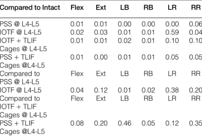
The motion at L4-L5 was significantly reduced in all loading modes except right rotation (Table 3), when the segment was instrumented with the standard pedicle screw system (PSS). The motion at L4-L5 was signifi-cantly reduced in all loading modes except in left rotation, when the segment was instrumented with IOTF (Table 3). The motion at L4-L5 was significantly reduced in all loading modes except in rotations, when the segment was instrumented with IOTF+ TLIF cages (Table 3). The motion at L4-L5 was significantly reduced in all loading modes when the segment was instrumented with PSS+ TLIF cages (Table 3). There
Figure 4 L2-Sacrum lumbar cadaveric specimen test setup.
Gomleksiz et al. A new lumbar fixation device alternative to pedicle-based stabilization for lumbar spine
was no significant increase/decrease in ROM at the other levels due to implantation at L4-L5 (Figures 5and7).
On overall comparison, both systems yielded similar biomechanical results. However, when the outcomes
were analyzed separately, the new system (IOTF) without interbody fusion offered more rigidity in rotation than did the classical system. When both PSS and IOTF cases without cages were compared, the
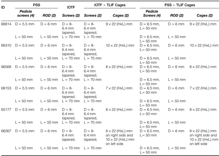
Table 2 Implants information showing the sizes of different implants used for each specimen (H: height, L: length, D: diameter).
ID PSS IOTF IOTF+ TLIF Cages PSS+ TLIF Cages
Pedicle
screws (4) ROD (2) Screws (2) Screws (2) Cages (2)
Pedicle
Screws (4) ROD (2) Cages (2) 66814 D= 5.5 mm D= 6 mm D= 8-6.4 mm tapered, D= 8-6.4 mm tapered, 9× 22 (HxL) mm D= 6.5 mm, L= 50 mm D= 6 mm 9× 22 (HxL) mm L= 50 mm L= 50 mm L = 70 mm L = 70 mm D= 6.5 mm, L= 50 mm L= 50 mm 66310 D= 5.5 mm D= 6 mm D= 8-6.4 mm tapered, D= 8-6.4 mm tapered, 10× 22 (HxL) mm D= 6.5 mm, L= 50 mm D= 6 mm 10× 22 (HxL) mm L= 50 mm L= 50 mm L = 70 mm L = 70 mm D= 6.5 mm, L= 50 mm L= 50 mm 66568 D= 5.5 mm D= 6 mm D= 8-6.4 mm tapered, D= 8-6.4 mm tapered, 8× 22 (HxL) mm D= 6.5 mm, L= 50 mm D= 6 mm 8× 22 (HxL) mm L= 50 mm L= 50 mm L = 70 mm L = 70 mm D= 6.5 mm, L= 50 mm L= 50 mm 68153 D= 5.5 mm D= 6 mm D= 8-6.4 mm tapered, D= 8-6.4 mm tapered, 7× 22 (HxL) mm D= 6.5 mm, L= 50 mm D= 6 mm 7× 22 (HxL) mm L= 50 mm L= 50 mm L = 70 mm L = 70 mm D= 6.5 mm, L= 50 mm L= 50 mm 65177 D= 5.5 mm D= 6 mm D= 8-6.4 mm tapered, D= 8-6.4 mm tapered, 8× 22 (HxL) mm D= 6.5 mm, L= 50 mm D= 6 mm 8× 22 (HxL) mm L= 50 mm L= 50 mm L = 70 mm L = 70 mm D= 6.5 mm, L= 50 mm L= 50 mm 66267 D= 5.5 mm D= 6 mm D= 8-6.4 mm tapered, D= 8-6.4 mm tapered, 8× 22 (HxL) mm on right side and 10× 22 (HxL) mm on left side
D= 6.5 mm, L= 50 mm
D= 6 mm 8× 22 (HxL) mm on right side and 10× 22 (HxL) mm on left side L= 50 mm L= 50 mm L = 70 mm L = 70 mm D= 6.5 mm,
L= 50 mm
L= 50 mm
PSS performed better than did the IOTF in flexion and lateral bending (Table 3). When PSS and IOTF cases with cages were compared, there was no significant difference between the techniques (Table 3).
The results offered by our new system seem encoura-ging, and the biomechanical efficacy would be even higher in live human subjects since most of the cadaveric specimens were osteoporotic and pedicle-corpus junc-tions would have been weakened severely due to mul-tiple trials in the course of the experiment.
Discussion
Spine surgeons have performed posterior lumbar segmen-tal instrumentation using pedicle screwing techniques for posterior vertebral column fixation and fusion in degen-erative spine diseases, traumatic and congenital/acquired spinal deformities. Adjacent segment disease is one of the most challenging conditions after fusion surgery. If revi-sion surgery is needed; there are two surgical options: first, total exploration of the existing system, adding two screws with replacement of rods; and second, adding two screws and rods with a domino. However, there are some restrictions of these options, including extensive incision, more muscle dissection and longer operation time for the former option, and displacement of rods from a domino for the latter option.
Currently, spinal instrumentation is most commonly performed using the transpedicular screw and rod
system that was popularized by Cotrel and Dubousse6. This same technique with some minor modifications has also widely been used for the correction of spinal deformities. Nevertheless, despite advanced surgical technique and neuromonitoring capabilities, malposi-tion of screws and the risk of a resultant neurological deficit remains a major concern. In short, the classical transpedicular system is associated with the following drawbacks: screw loosening, particularly at the L5-S1 level; extensive paravertebral muscle dissection; malpo-sition of screws and resulting dural sac and nerve root injuries and CSF fistulae; difficulty in revision surgeries; and potential need for domino placement in case of adjacent segment involvement.7,8 In this context, a safer technique with a reasonably long trajectory from dural sac and nerve roots is needed. Thus, we aimed to develop such a technique that may fulfill this crucial requirement. We named this new technique “inferior oblique transdiscal fixation”, as this approach enabled fixation of two adjacent vertebrae using a single screw on each side.
In this study, we compared the biomechanical efficacy of the classical posterior transpedicular technique with a
Figure 6 Mean range of motion of L3-L4 segment.
Figure 7 Mean range of motion of L4-L5 segment.
Figure 8 Mean range of motion of L5-S1 segment.
Table 3 P-values showing statistical significance (P< 0.05). Compared to Intact Flex Ext LB RB LR RR PSS @ L4-L5 0.01 0.01 0.00 0.00 0.00 0.06 IOTF @ L4-L5 0.02 0.03 0.01 0.01 0.59 0.04 IOTF+ TLIF Cages @ L4-L5 0.01 0.01 0.02 0.01 0.10 0.10 PSS+ TLIF Cages @L4-L5 0.01 0.00 0.01 0.01 0.05 0.05 Compared to PSS @ L4-L5 Flex Ext LB RB LR RR IOTF @ L4-L5 0.04 0.12 0.01 0.02 0.38 0.20 Compared to IOTF+ TLIF Cages @ L4-L5 Flex Ext LB RB LR RR PSS+ TLIF Cages @L4-L5 0.08 0.20 0.46 0.05 0.12 0.35
Gomleksiz et al. A new lumbar fixation device alternative to pedicle-based stabilization for lumbar spine
novel surgical technique (IOTF) with and without inter-body fusion on fresh human lumbar cadaveric speci-mens. IOTF was equally effective to classical transpedicular fixation in terms of stabilization of instable spinal segment.
The new proposed surgical technique offered super-iority in some aspects to the classical pedicle screw tech-nique, including easy revision surgery, no need for domino placement, tri-cortical fixation and less risk of loosening of screws, safer trajectory with regard to the risks of dural sac and root injuries, shorter incisions, less paravertebral muscle and soft tissue dissection; possibility of interbody fusion and shorter duration of hospitalization. The other advantage of this new tech-nique was the fact that it did not require a longer learn-ing curve.
A disadvantage of this new technique would be the potential need for exiting nerve root retraction during screw placement. On the other hand, this technique would not be useful for deformity surgery because of the absence of correction maneuvers.
In our opinion, this new technique could be poten-tially used for the stabilization of painful black disc disease with intact corpus height, unstable recurrent disc herniation, low-grade degenerative spondylolisth-esis, and decompressed lumbar spinal stenosis. A C-arm should be used in order to delineate the accurate trajectory of the screw during the procedure. In summary, we firmly believe that IOTF could be an effec-tive alternaeffec-tive to classical transpedicular fixation in selected cases. In the future, we are planning to publish a clinical study demonstrating the preliminary outcomes of this new technique over two years.
Conclusions
The biomechanical results of the proposed new posterior fusion technique IOTF appeared to be encouraging and comparable to those of the classical pedicle screw tech-nique. This preliminary study demonstrated the effect of the new posterior lumbar fixation technique that can be potentially useful and even superior in specific situ-ations, including adjacent segment involvement and revision surgeries. The next step would be to test the effi-cacy of this technique in live human subjects.
Disclaimer statements Contributors None
Funding statement The work was supported in part by NSF Industry/University Cooperative Research Center at The University of California at San Francisco, CA and The University of Toledo, Toledo, OH
Conflict of interest N/A Ethics approval None
ORCID
Halil Can http://orcid.org/0000-0002-6792-9987
References
1 Albee FH. Transplantation of a portion of the tibia into the spine for Pott’s disease: a preliminary report. J Am Med Assoc1911;57 (11):885–6.
2 Hibbs R. An operation for progressive spinal deformities. Clin Orthop Relat Res1964;35:4–8.
3 Mixter WJ, Barr JS. Rupture of the intervertebral disc with invol-vement of the spinal canal. N Engl J Med1934;211(5):210–5. 4 Harrington PR. Treatment of scoliosis. Correction and internal
fix-ation by spine instrumentfix-ation. J Bone Joint Surg Am1962;44: 591–610.
5 Roy-Camille R, Roy-Camille M, Demeulenaere C. Osteosynthesis of dorsal, lumbar, and lumbosacral spine with metallic plates screwed into vertebral pedicles and articular apophyses. Presse Med1970;78(32):1447–8.
6 Cotrel Y, Dubousset J. A new technic for segmental spinal osteo-synthesis using the posterior approach. Orthop Traumatol Surg Res2014;100(1):37–41.
7 Jutte P, Castelein R. Complications of pedicle screws in lumbar and lumbosacral fusions in 105 consecutive primary operations. Eur Spine J2002;11(6):594–8.
8 Parker SL, McGirt MJ, Farber SH, Amin AG, Rick AM, Suk I, et al. Accuracy of free-hand pedicle screws in the thoracic and lumbar spine: analysis of 6816 consecutive screws. Neurosurgery 2011;68(1):170–8.
9 Foley KT, Gupta SK, Justis JR, Sherman MC. Percutaneous pedicle screw fixation of the lumbar spine. Neurosurg Focus 2001;10(4): 1–9.
10 Lowery GL, Kulkarni SS. Posterior percutaneous spine instrumen-tation. Eur Spine J2000;9(1):S126–30.
11 Magerl F. External skeletal fixation of the lower thoracic and the lumbar spine. In: Uhthoff HK, (ed.) Current Concepts of External Fixation of Fractures. Berlin, Heidelberg: Springer;1982. p. 353–66.
12 Mathews H, Long BH. Endoscopy-assisted percutaneous anterior interbody fusion with subcutaneous suprafascial internal fixation: evolution of technique and surgical considerations. Orthop Int Ed1995;3:496–500.
13 Weinstein JN, Rydevik BL, Rauschning W. Anatomic and techni-cal considerations of pedicle screw fixation. Clin Orthop Relat Res 1992;(284):34–6.
14 Shah RR, Mohammed S, Saifuddin A, Taylor BA. Radiologic evaluation of adjacent superior segment facet joint violation fol-lowing transpedicular instrumentation of the lumbar spine. Spine 2003;28(3):272–5.
15 Chen Z, Zhao J, Xu H, Liu A, Yuan J, Wang C. Technical factors related to the incidence of adjacent superior segment facet joint violation after transpedicular instrumentation in the lumbar spine. Eur Spine J2008;17(11):1476–80.
16 Kim HJ, Chun HJ, Kang KT, Moon SH, Kim HS, Park JO,et al. The biomechanical effect of pedicle screws’ insertion angle and position on the superior adjacent segment in 1 segment lumbar fusion. Spine2012;37(19):1637–44.
17 Nagata H, Schendel MJ, Transfeldt EE, Lewis JL. The effects of immobilization of long segments of the spine on the adjacent and distal facet force and lumbosacral motion. Spine 1993;18 (16):2471–9.
18 Shono Y, Kaneda K, Abumi K, McAfee PC, Cunningham BW. Stability of posterior spinal instrumentation and its effects on adja-cent motion segments in the lumbosacral spine. Spine1998;23(14): 1550–8.
19 Sengupta DK. Dynamic stabilization devices in the treatment of low back pain. Neurol India2005;53(4):466.
20 Meyers K, Tauber M, Sudin Y, Fleischer S, Arnin U, Girardi F, et al. Use of instrumented pedicle screws to evaluate load sharing
in posterior dynamic stabilization systems. Spine J 2008;8(6): 926–32.
21 Oktenoglu T, Erbulut DU, Kiapour A, Ozer AF, Lazoglu I, Kaner T, et al. Pedicle screw-based posterior dynamic stabilisation of the lumbar spine: in vitro cadaver investigation and a finite element
study. Comput Methods Biomech Biomed Engin 2015;18(11): 1252–61.
22 Wiltse LL, Bateman JG, Hutchinson RH, Nelson WE. The para-spinal sacropara-spinalis-splitting approach to the lumbar spine. J Bone Joint Surg Am1968;50(5):919–26.
Gomleksiz et al. A new lumbar fixation device alternative to pedicle-based stabilization for lumbar spine