191
Case Report
A Technique for Fabricating Single Screw-Retained Implant-Supported Interim Restoration by Using the Crown of Extracted Tooth
Kenan Nazaroğlu1İD, Ayşe Atay2İD, Aslıhan Üşümez3İD
1Periodent Dental Clinic, Fatih, Istanbul 2Department of Prosthodontics, Faculty of Dentistry, Altinbas University, Istanbul, Turkey 3DentalPlus Clinic, Bakırkoy, Istanbul, Turkey
Submitted: July 30, 2020; Accepted: September 18, 2020
Abstract: At the esthetic anterior region, horizontal root fracture is a complex and subversive situation for both the patient and dental practitioner. Due to enabling rapid prosthetic rehabilitation, decreasing treatment time, morbidity and the number of surgical procedures required, immediate implantation of a single implant may be a more preferable treatment option. In the presenting treatment modality, a single implant-supported provisional crown was produced right after extraction and implant placement surgery, using the patients extracted tooth crown. The use of the crown part has substantially fulfılled the patient’s expectations both aesthetically and psychologically. Besides fixed provisional crown restoration advanced biologically and esthetically convenient soft tissue emergence for the permanent restoration.
Keywords: Temporary dental restoration; single-tooth dental implant; trauma; dental esthetic
Address of Correspondence: Ayşe ATAY- [email protected] Tel: +90-212-709 45 28, Department
of Prosthodontics, Faculty of Dentistry, Altinbas University, İncirli Avenue, No:11/A, 34147, Bakırkoy, Istanbul,
Turkey, Ayşe AtayİD0000-0002-5358-0753, Kenan NazaroğluİD0000-0002-4796-8196, Aslıhan ÜşümezİD
0000-0002-7222-7322
1. Introduction
The most common reasons of missing anterior teeth are tooth and root fracture, extensive tissue damage due to caries, advanced periodontitis and trauma. For dentists, rehabilitation of teeth loss in the aesthetic area is rather difficult (Martin et al., 2006). Implant supported fixed prostheses are preferred to conventional bridges and removable prostheses. In this way the adjacent teeth will be preserved and the patient will use fixed prosthesis instead of removable prosthesis (Chen et al., 2011).
In order to achieve a natural appearance in anterior single tooth deficiencies, soft tissue contours should be natural and papillae should be present. Biological width concept states that formation of papilla is dependent upon many factors including implant’s vertical or horizontal positioning, implant’s prosthetic component design, design of the transmucosal component, biotype of the soft tissue, and type of the
192
material blended with mucosa (titanium, ceramics, zirconia etc.) (Hermann et al., 2000). Also, thickness of labial bone is very important to provide tissue contour and implant stability in the long term. The most important factors that make reaching aesthetic results more difficult are high laughing line and the fine gingival biotype (Kan et al., 2003).
Restoration of non-recoverable teeth in the anterior regions of the maxilla may be achieved by immediate implant placement and provisionalization in fresh sockets (McRory and Cagna, 2014). Both patients and clinicians want shorter overall treatment periods and minimum surgical and prosthetic intervention quantity in implant dentistry. After tooth extraction, a 2-3 month period of hard tissue remodeling around the socket and 3-6 months of healing with no load are advised in traditional guidelines because those were thought to be essential in the 1980s (Albrektsson et al., 1981). In the last 2 decades, immediate implant placement just after the extraction and early implant insertion after soft tissue healing for a few weeks have been used as alternative protocols (Chen et al., 2004).
Faster prosthetic replacement, decreased morbidity, treatment duration, and surgical procedure number may be achieved by placing single implant immediately (Hämmerleet al., 2004). A single missing tooth may be replaced by surgical implant placement and then application of a healing transmucosal abutment and closure of the soft tissue appropriately (Mish, 2008). Aiming to produce a final restoration and to design the emergence contours that will support the adjacent tissues the restoring dentist may start procedures after osteointegration and soft tissue maturation. Both the laboratory technician and the dental practitioner may have a difficulty with the traditional approach because after surgical implant placement if the peri-implant soft tissues heal using a prefabricated healing abutment instead of the more natural emergence profile which is necessary for facilitation of optimal restorative esthetics a cylindrical transmucosal passageway is created (Shor et al., 2008)
Estimation of dental technician will affect the ability to achieve an esthetically and biologically convenient soft tissue healing. If the emergence contour is excessively contoured complete restoration placement may be prevented by healthy peri-implant tissue and surgery may be required to recontour the soft tissue. If the contour of the coronal emergence is not adequate or optimal gingival structure is not correctly estimated esthetic success in the definitive restoration may be poor. Accordingly, at the time of implant surgery producing an optimally contoured provisional crown should be considered (Chen et al., 2004; DeRouck et al., 2009; Mathews, 2000; Su et al., 2010).
The aim of this article is to describe a clinical technique that uses the labial part of the extracted tooth and that will enable healing of peri-implant soft tissues against an optimally derived provisional crown. This method eliminates problems that occur with the more traditional method and facilitates maturation of predictable and desirable soft tissue.
2. Description of the Case
With a chief complaint of increased mobility in the maxillary right incisor, an 18-year-old female patient attended the clinic. The diagnostic examination was completed with paying particular attention to fractured tooth and the bone volume at the intended implant site. There was a horizontal tooth fracture on the coronal part of the root side (Figure 1a, 1b).
193 Figure 1. a) Preoperative view, b) Periapical X-ray of the mobile incisor
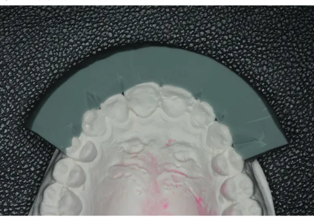
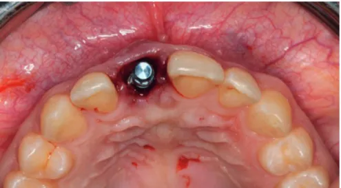
Patient was informed about the treatment procedure and signed a consent form. The unstable crown part of the tooth is stabilized in order to take a precise impression. After taking the impression with irreversible hydrocolloid impression material (Hydrocolor 5, Zhermack SpA, Badia Polesine, Italy), the stone model and a silicon index were prepared (Figure 2). The crown part of the tooth was gently extracted. Following the preparation of the buccal 1/3 of the crown, disinfection procedure was performed and the prepared crown was placed into the silicon index (Zetaplus, Zhermack SpA, Badia Polesine, Italy) (Figure 3a, 3b). The extraction of the fractured root was performed with care to protect the sensitive facial plate of the hard tissue and to evade injury to the interproximal gingival area. Debriding the extraction socket, alveolar bone was promoted to bleed. A notch was created on the palatal wall of the socket and the osteotomy of implant was done in contact with the palatal wall. According to the recommended osteotomy sequence of the implant system, a pilot opening was created by a sharp-pointed pilot drill (4.8mm in diameter, 14mm in length, Bone Level, Straumann Company, Basel, CH). After the enlargement of the osteotomy, the implant was placed to the terminal location with a hand-driven torque wrench with a 35 Ncm of force to restrict stripping of the sensitive bony area (Figure 4). The gap between the implant and the buccal wall was filled with allogeneic graft material (MinerOss Cortical & Cancellous Chips, USA).
194
195 Figure 3. a) The prepared buccal 1/3 of the crown b) placed into the silicon index
196
Figure 4. The placed implant immediately after extraction
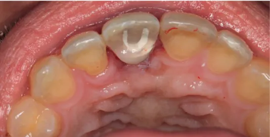
The provisional crown was produced by connecting a provisional abutment (Temporary Peek Abutment-Straumann Company, CH) onto the implant. Using adjacent teeth as landmarks, the abutment was marked 1.5 to 2.0 mm below the assumed incisal edge with a sterile pen. The provisional abutment was removed and reshaped with a high-speed bur (GZ Instrumente, Austria) along with water irrigation. The extracted crown part got cleaned, disinfected and prepared to adapt to the provisional abutment. Following the adaptation of the buccal part of the crown in the silicon index, the screw access tunnel was filled with cotton pellets and afterwards the provisional restoration was achieved with light-curing flowable composite resin (EsFlow, Spident Co., LTD, Korea). The composite was cured for 10 seconds using light curing unit (Valo Cordless, Ultradent). After polymerization, polishing and disinfecting of the provisional crown within chlorhexidine gluconate 0.12% oral rinse was performed (Figure 5). Then, the cotton pellets were removed, after which the interm restoration was checked intraorally.
197 Figure 6. The periapical x-ray after seating of the restoration
Following the final recontouring, the provisional crown was located and the retention screw was hand-tightened and a periapical x-ray was taken to guarantee the precise fit (Figure 6). The abutment screw was condensed over with a plumber’s polytetrafluoroethylene tape (Baytaş, PTFE Teflon Tape, Konya, Turkey) which was presterilized and the screw access tunnel was filled with a composite resin (Charisma, Heraeus Kulzer, Germany) and was cured for 10 second using light curing unit. The interim restoration occlusion was carefully managed to promote implant stability through osseointegration period. Any maximum intercuspal and eccentric contacts were eliminated (Figure 7). The patient was instructed to evade the masticatory load forces of the provisional crown restoration throughout the integration interval.
198
Figure 7. The screw access channel was filled with a composite resin
The patient was recalled 1 week after surgery. Oral care and mastication instructions were reinforced and scheduled future recall appointments (Figure 8). Three months after the surgery, when the patient was recalled, it was seen that the perimplant soft and hard tissues were successfully restored and formed (Figure 9a, 9b).
199 Figure 9. Three months after the surgery, a) the soft tissue b) periapical x-ray of the hard tissue around the implant
200
3. Discussion
Several researches have centered on various sets of preoperative, intraoperative, and postoperative factors. The quality of root canal treatment, periapical situation, and restorability of the tooth may alter the survival of endodontically treated teeth.14 Crown and root fractures are amongst the foremost reasons for tooth extraction after root canal treatment (Landys et al., 2015). This verdict may be defined by a catastrophic sequence that has been associated with reinfection of the root canal system throughout coronal microleakage or total loss of coronal tooth construction after a crown fracture (Vire, 1991). In the esthetic area, the horizontal root fracture of endodontically restored tooth is often a traumatic occurrence for the patient (Singh et al., 2015). Immediate implantation may provide faster prosthetic rehabilitation, reducing treatment time, morbidity, and the number of surgical steps demanded. The decrease in overall treatment time with fewer surgical interventions, diminished soft and hard tissue destruction, and psychological compensation of the patient can be counted as advantages of this procedure (Mathews, 2000).
The presented procedure in this case report demonstrates a technique for manufacturing a single implant-supported provisional restoration in association with implant installation in a fresh extraction area with using the patients extracted crown. Providing a provisional crown in the method defined helps process and preserves the precarious soft tissues around the implant and helps evade several complications correlated with a single-tooth removable provisional denture. Using the crown section of the extracted tooth helped the patient to content. On the other side, fixed provisional crown helped to promote esthetically and biologically relevant soft tissue evolution for the final prosthesis.
Conflict of Interest
The authors declare that they have no conflict of interest.
References
Albrektsson, T., Brånemark, P. I., Hansson, H. A., Lindström, J. (1981) Osseointegrated titanium implants. Requirements for ensuring a long-lasting, direct bone-to-implant anchorage in man. Acta Orthop Scand, 52(2), 155-170.
Chen, C. L., Chang, C. L., Lin, S. J. (2011). Immediate implant placement and provisionalization with simultaneous guided bone regeneration in the esthetic zone. J Dent Sci, 6(1), 53-60.
Chen, S., Wilson, T. G. Jr., Hämmerle, C. H. (2004). Immediate or early placement of implants following tooth extraction: review of biologic basis, clinical procedures, and outcomes. Int J Oral Maxillofac Implants, 19, 12-25.
DeRouck, T., Collys, K., Wyn, I., Cosyn, J. (2009). Instant provisionalization of immediate single-tooth implants is essential to optimize esthetic treatment outcome. Clin Oral Implants Res, 20(6), 566-570.
201 Hämmerle, C. H., Chen, S. T., Wilson, T. G. Jr. (2004). Consensus statements and recommended clinical
procedures regarding the placement of implants in extraction sockets. Int J Oral Maxillofac Implants, 19, 26-28.
Hermann, J. S., Buser, D., Schenk, R. K., Higginbottom, F. L., Cochran, D. L. (2000). Biologic width around titanium implants. A physiologically formed and stable dimension over time. Clin Oral Implants Res, 11(1), 1-11.
Kan, J. Y., Rungcharassaeng, K., Umezu, K., Kois, J. C. (2003). Dimensions of peri-implant mucosa: an evaluation of maxillary anterior single implants in humans. J Periodontol, 74(4), 557-562.
Landys Boren, D., Jonasson, P., Kvist, T. (2015). Long-term survival of endodontically treated teeth at a public dental specialist clinic. J Endod, 41(2), 176-181.
Martin, W. C., Morton, D., Buser, D. (2006). Pre-operative analysis and prosthetic treatment planning in esthetic implant dentistry. In: Buser D, Belser UC, Wismeijer D, eds. ITI Treatment Guide. Implant Therapy in the Esthetic Zone e Single-tooth Replacements, vol. 1. Berlin: Quintessence, 11-20.
Mathews, D. P. (2000). Soft tissue management around implants in the esthetic zone. Int J Periodont Rest Dent, 20(2), 141-149.
McRory, M. E., Cagna, D. R. (2014). A technique for fabricating single screw-retained implant-supported interim crowns in conjunction with implant surgery. J Prosthet Dent, 111(6), 455-459.
Misch, C. E. (Ed.). (2008). Anterior single-tooth replacement: surgical consideration. Contemporary implant dentistry, 3rd ed. St Louis: Mosby, 739-768.
Ng, Y. L., Mann, V., Gulabivala, K. (2010). Tooth survival following non-surgical root canal treatment: a systematic review of the literature. Int Endod J, 43(3), 171-189.
Shor, A., Schuler, R., Goto, Y. (2008). Indirect implant- supported fixed provisional restoration in the esthetic zone: fabrication technique and treatment workflow. J Esthet Restor Dent, 20(2), 82-95.
Singh, M., Kumar, L., Anwar, M., Chand, P. (2015). Immediate dental implant placement with immediate loading following extraction of natural teeth. Natl J Maxillofac Surg, 6(2), 252-255.
Su, H., Gonzales-Martin, O., Weisgold, A., Lee, E. (2010). Considerations of implant abutment and crown contour: critical contour and subcritical contour. Int J Periodontics Restorative Dent, 30(4), 335-343. Vire, D. E. (1991). Failure of endodontically treated teeth: classification and evaluation. J Endod, 17(7), 338-342.