BORONIC ACID CONJUGATED PEPTIDE
AMPHIPHILE SYSTEMS FOR CONTROLLED DRUG
RELEASE
A THESIS SUBMITTED TO
THE GRADUATE SCHOOL OF ENGINEERING AND SCIENCE OF BILKENT UNIVERSITY
IN PARTIAL FULFILLMENT OF THE REQUIREMENTS FOR THE DEGREE OF
MASTER OF SCIENCE IN
MATERIALS SCIENCE AND NANOTECHNOLOGY
By
Hatice Kübra Kara
August 2017
ii
BORONIC ACID CONJUGATED PEPTIDE AMPHIPHILE SYSTEMS FOR CONTROLLED DRUG RELEASE
By Hatice Kübra Kara August 2017
We certify that we have read this thesis and that in our opinion it is fully adequate, in scope and in quality, as a thesis for the degree of Master of Science.
Eda Yılmaz (Advisor)
Mustafa Özgür Güler (Co-advisor)
Bilge Baytekin
Özgül Persil Çetinkol
Approved for the Graduate School of Engineering and Science:
Ezhan Karaşan
iii
ABSTRACT
BORONIC ACID CONJUGATED PEPTIDE
AMPHIPHILE SYSTEMS FOR THE CONTROLLED
DRUG RELEASE
Hatice Kübra Kara
M.Sc. in Materials Science and Nanotechnology Advisor: Eda Yılmaz
Co-advisor: Mustafa Özgür Güler August 2017
Targeted cancer drug delivery is still under investigation and scientists have been focusing on major differences between healthy and cancer tissue to develop novel effective therapies. The cancer microenvironment has different physiological properties than the healthy tissues, for instance, it has more acidic pH, and much of the attention has been given to developing stimuli responsive agents for targeted drug delivery applications. Boronic acid is one of the most well-known stimuli responsive molecule which can form reversible covalent bonds with vicinal diols such as saccharide or catechol, that achieves targeted cancer drug release in a pH dependent manner. At neutral pH, the bond formation is triggered; however, these bonds become weaker at slightly acidic environment. Boronic acid conjugated polymers have been frequently preferred for doxorubicin encapsulation, which is a widely used
iv
acid and DOPA conjugated peptide amphiphiles were used as a biocompatible and biodegradable alternative to polymeric systems. Peptide amphiphiles self assemble to
form peptide nanofibers via noncovalent interactions, such as hydrogen bonding, hydrophobic interactions, van der Waals forces and electrostatic interactions, where boronic acid/DOPA units remain on the exterior part of the nanofibers. In addition to noncovalent interactions, at physiological pH, boronic acid and DOPA moieties on the peptide surface form reversible covalent complexes, resulting in improved hydrogel strength, self-healing capacity and entrapment of doxorubicin inside the 3D-network. On the other hand, under acidic conditions, these interactions weaken and doxorubicin release is accelerated at tumor site. Reversible covalent interaction, secondary structure, morphological, mechanical, release profile analysis were performed on the system. Results showed that this system exhibits promising features that can be used for therapeutic applications.
Keywords: Peptide amphiphiles, self-assembly, boronic acid, hydrogels, controlled
v
ÖZET
BORONİK ASİT KONJUGE EDİLMİŞ PEPTİT
AMFİFİL SİSTEMLERİNİN KONTROLLÜ İLAÇ
SALINIM UYGULAMASI
Hatice Kübra Kara
Malzeme Bilimi ve Nanoteknoloji Programı, Yüksek Lisans Tez Danışmanı: Eda Yılmaz
Tez Eşdanışmanı: Mustafa Özgür Güler Ağustos 2017
Hedeflenmiş kanser ilaç taşıma sistemleri halen araştırılmakta olup, bilim insanları, yeni etkili tedaviler geliştirmek için sağlıklı ve kanserli dokular arasındaki farklılıklara odaklanmaktadır. Kanser dokusu ortamı vücudun geri kalanından daha asidiktir; bu nedenle, uyarıya duyarlı maddeler, hedeflemeli ilaç taşıma uygulama sistemleri için büyük önem taşımaktadır. Boronik asit, sakkarit veya katekoller gibi diyollerle tersinir bağ kurabilen uyarıya duyarlı en popüler moleküllerden biridir. Bağ oluşumu nötr pH'ta tetiklenirken, bu bağlar hafif asidik ortamda zayıflar. En çok bilinen kemoterapik ilaçlardan biri olan doksorubisini kapsüle etmek için boronik asit ve diyol konjuge edilmiş polimerler sıklıkla kullanılır. Bu çalışmada biyolojik olarak uyumlu ve bozunabilir boronik asit ve DOPA‘ya konjuge edilmiş peptit amfifiller polimerik sistemlere alternatif olarak kullanılmıştır. Bu PA'lar, elektrostatik etkileşimler, hidrojen bağları, Van der Waals kuvvetleri ve hidrofobik
vi
gelerek peptit nanofiber yapısını oluştururlar ve oluşan bu yapının dış yüzeyinde boronik asit/DOPA molekülleri bulunur. Kovalent olmayan etkileşimlere ilaveten, nano fiber yüzeyinde bulunan boronik asit ve DOPA, fizyolojik pH’de tersinir kovalent kompleksler oluşturarak, hidrojel kuvvetini, hidrojelin kendi kendine iyileşme kapasitesini ve doksorubisinin jel içinde tutulmasını artırmaktadır. Bununla birlikte, asidik pH'de, bu etkileşimler zayıflar ve doksorubisin salımı tümör bölgesinde hızlanır. İkincil yapı, morfolojik ve mekanik analizler, ilaç salınım profilleri ve tersinir kovalent etkileşimler test edilmiştir. Elde ettiğimiz sonuçlar bu sistemin ileri uygulamalar için uygun olduğunu göstermektedir.
Anahtar kelimeler: Peptit amfifiller, kendiliğinden bir araya gelme, boronik asit,
hidrojel, kontrollü ilaç salımı, kendi kendini iyileştiren biyomateryaller, uyarılara duyarlı sistemler
vii
Acknowledgement
I would like to express my thanks to my MSc advisor Eda Yılmaz for her support throughout my graduate studies. I also thank to Mrs. Zeynep Erdoğan and other engineers for their technical contribution to my thesis. I also would like to thank Bilkent University and The Scientific and Technological Research Council of Turkey (TUBİTAK) for their financial support.
I would like to express my special thanks to my friends for their scientific knowledge, fruitful collaboration and friendship during my MSc and I believe that their endless support and friendship will continue throughout my life.
viii
Contents
1Introduction ... 1
1.1 Self-Assembly ... 1
1.2 Peptide and Their Self-Assemblies ... 2
1.3 Solid Phase Peptide Synthesis ... 7
1.4 Peptide Amphiphile ... 8
1.5 Stimuli Responsive Peptide Systems ... .12
1.6 Boronic Acid ... 16
1.6.1 Saccharides ... 20
1.6.2 Alizarin Red-S ... 21
1.6.3 L-DOPA ... 23
1.7 Application Of Boronic Acid ... 25
2 Material And Methods ... 32
2.1 Introduction ... 32
2.2 Experimental Section ... 35
2.2.1 Chemicals and Reagents ... 35
ix
2.2.3 Synthesis and Characterization of PAs ... 35
2.3.4 Preparative High Performance Liquid Chromatography (Prep-HPLC).37 2.3.5 Zeta Potential Measurements of PAs ... 37
2.3.6 Transmission Electron Microscopy (TEM) ... 38
2.3.7 Scanning Electron Microscopy (SEM) ... 38
2.3.8 CD Analysis ... ....38
2.3.10 ARS Based Spectroscopic Measurements ... 39
2.3.9 Rheological Measurements ... 41
2.3.11 Release Studies... 42
3 Results And Discussion ... 43
4 Conclusion ... 66
x
List Of Figures
1.1 Examples of biological building blocks and their self‐assembled structures (a) Protein folding (b) ds‐DNA (c) tobacco mosaic virus (d) cell
membrane...3
1.2 Peptide bond formation...5
1.3 Schematic representation of different secondary structures of molecular self-assembly of peptides...6
1.4 Schematic representation of Solid Phase Peptide Synthesis...10
1.5 Schematic representation of self-assembly of PAs into nanofibers: a) Chemical structure of PA with four key chemical entities. b) Molecular model of an active epitope-containing PA, their self-assembly into nanofibers, as well as c) SEM image of nanofibers...11
1.6 Peptide amphiphile self-assembly supramolecular nanostructures...12
1.7 Categorization of physical, biological and chemical stimuli...13
1.8 Boronic acid and its reaction with different molecules...18
1.9 Multiple reaction mechanism of boronic acid...19
1.10 Binding and analyte-mediated release Alizarin Red-S with hydrogel-bound boronic acid...22
xi
1.12 Several applications of boronic acid biomaterials...27
1.13 The chemical structure of Bortezomid...30
2.1 Schematic representation of Boronic/Dopa-PA hydrogel network at different pH...34 2.2 Schematic representation of ARS assay...40
2.3 Schematic representation of Dox encapsulation inside the PA network...42 3.1 Schematic representation of Boronic/Dopa nanofiber formation...44
3.2 a) Chemical structure and b) liquid chromatograms and mass spectra of Boronic-PA...47 3.3 a) Chemical structure and b) liquid chromatograms and mass of DOPA-PA...48 3.4 a) Chemical structure and b) liquid chromatograms and mass spectra, of E3
-PA...49 3.5 a) Chemical structure b) liquid chromatograms and mass spectra of K3
-PA...50 3.6 The zeta potential results of individual PAs and their coassembled mixtures...51 3.7 The secondary structure analysis of a) individual PA and b) PA mixtures by circular dichroism...53 3.8 TEM images of the a) Boronic/Dopa-PA b) Boronic/K3-PA c) Dopa/E3-PA
and d) E3/K3-PA nanofibers...54
3.9 SEM images of a) Boronic/Dopa-PA b) Boronic/K3-PA c) Dopa/E3-PA and
xii
3.10 ARS assay of Boronic/Dopa-PA and Boronic/K3-PA...58
3.11 ARS assay of E3/Dopa-PA and E3/K3-PA...59
3.12 Time sweep test of PA mixtures...61
3.13 a) Thixotropic behaviour b) recovery rate of PAs nanofiber gels...62
3.14 Rate of cumulative release profile of Dox through the PA gels at pH 7.4 in HEPES buffer b) pH 5.5 in MES buffer...64
xiii
Abbreviations
ARS Alizarin Red-S
Boc Tert-butoxycarbonyl BTZ Bortezomid CD Circular dichroism DCM Dichloromethane DIEA N,N-diisopropylethylamine DMF N,N-Dimethylformamide Dox Doxorubicin-HCl
ECM Extracellular matrix
ESI Electrospray ionization
Fmoc 9-Fluorenylmethoxycarbonyl
HBTU
N,N,N′,N′-Tetramethyl-O-(1H-benzotriazole-1-yl) uronium hexafluorophosphate
HPLC High pressure liquid chromatography
LC-MS Liquid chromatography-mass spectroscopy
L-DOPA Levo-DOPA (L-3,4-dihydroxyphenylalanine)
Mtt 4-Methyltrityl
NMR Nuclear magnetic resonance
PA Peptide amphiphile
RGD Arginylglycylaspartic acid
QTOF Quadrapole time of flight
SEM Standard error of mean
SEM Scanning electron microscopy
TEM Transmission electron microscopy
TFA Trifluoroacetic acid
TIS Triisopropyl silane
1
Chapter 1
Introduction
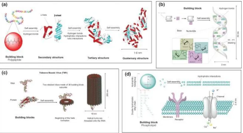
1.1 Self-Assembly:Self-assembly is a spontaneous process, which induces molecules to form a more thermodynamically stable and a well-defined structure via noncovalent forces under equilibrium conditions. In nature, molecular self-assembly is a unique bottom up process to construct with different length scales of complex biological structure using different building blocks. Peptides, lipids, nucleic acids and saccharides that are used to build up lipid bilayer cell membrane, DNA double helix, protein folding, actin filaments and tobacco mosaic viruses (Figure 1.1) are well-known examples [1-7].
In the last centuries, scientists have utilized biomaterials by mimicking or being inspired by the natural organisms. Although noncovalent interactions consist of weak forces such as hydrogen bonding, π-π stacking, van der Waals, hydrophobic electrostatic interactions compared to covalent bonding, they have many advantages in different cases. For instance, they create systems which can self-adapt to
2
environmental changes, reprogram themselves for different stimulus and interactions. These interactions demonstrate reversible characteristics and show the ability to heal themselves under appropriate conditions. Moreover, they can form larger structures via supramolecular chemistry since self-assembly is an enthalpically and entropically driven process. In order to create large nanostructures via supramolecular chemistry, solution phase or lubricious surfaces are needed to obtain required mobility [2, 3, 8-10]. These nanostructures have been utilized in regenerative medicine, drug delivery, and also sensing systems [11].
1.2 Peptide and Their Self-Assemblies
Amino acids are the organic molecules, which play crucial roles in biological systems that contain amine and carboxyl functional groups with side chains. These side chains are specific to each amino acid and determine their chemical and physical properties. Amino acids are classified by their side chain properties such as charged (lysine, histidine, glutamic acid), polar uncharged (serine, asparagine), hydrophobic with an aliphatic (alanine, leucine, valine) or aromatic (tyrosine, phenylalanine)) side chain etc. [9-11].
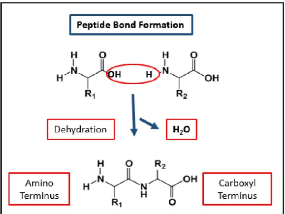
Peptides are formed by the condensation of two amino acid via peptide bonds. In peptide bond formation, the carboxyl group of one amino acid reacts with the amine group of the other amino acid as shown in Figure 1.2. Furthermore, peptides can be composed of natural or synthetic amino acids that can form versatile self-assembled nanostructures by supramolecular chemistry. For instance, hydrophobic side chains are responsible for hydrophobic aggregations via π-π stacking and hydrophobic
3
Figure 1.1 Examples of biological building blocks and their self‐assembled structures . (a) Protein folding (b) ds‐DNA (c) tobacco
4
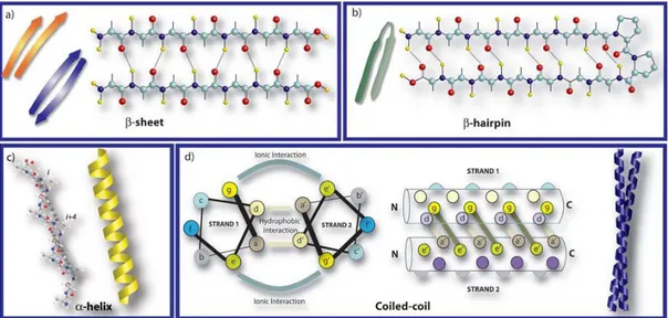
forces. In addition, polar amino acids utilize electrostatic forces or hydrogen bonding and peptide backbone itself promotes hydrogen bonds [12]. These hydrogen bonds result in constructing β-sheets conformation that stabilizes the multiple peptide arrangements. These sheets are further categorized as parallel and antiparallel β-sheets conformation according to the direction of the strands. Parallel conformation is obtained when the strands are placed in the same direction, while antiparallel β sheets are formed if the strands have opposite directions. Antiparallel conformation represents higher stability than parallel β-sheets since their pattern is more thermodynamically preferable. Furthermore, peptide strands can be arranged into right handed helix conformation by the repeating intramolecular hydrogen bonds between the amide groups and the carboxyl groups in same peptide with a pattern,
i,i+4. In some instances, these structural α-helix motifs can produce different
secondary structures by wrapping around each other, named coiled coil structures. The final secondary structure motif is the β-hairpin which is formed by having a short loop segment as a linker between two strands in antiparallel arrangement [12, 15-21]. Different secondary structural motifs of peptides self-assembly are demonstrated in Figure 1.3.
Peptides are biocompatible, biodegradable and bioactive in nature. Furthermore, self-assembly is a unique ability to fabricate a wide range of peptide nanostructures. Their architecture can be modified by changing the building blocks, amino acids, to obtain the desired functionality. Moreover, conjugation of peptides leads to increased diversity in peptides application fields [13]
In general, noncovalent interactions such as hydrogen bonding, hydrophobic, electrostatic and van der Waals forces, π–π stacking and coordination bonds are the
5
main interaction types, which govern the peptide self-assembly. The self-assembly events trigger bonds that form dynamic systems with the modulation of external factors such as pH, light, temperature, counterions, concentrations, and solvents [22-26].
pH is the most crucial external factor which effects the self-assembly of peptide by changing the charge of amino acid residues. Recently, pH change, triggering hydrogel formation has been paid attention for biomaterial studies.
Figure 1.2 Peptide bond formation
Charged amino acid containing systems are neutralized by changing pH of the solution and a decrease in charge repulsion leads to an increase in inter/intra fiber
6
interaction. This results in aggregation and formation of different nanostructures. Furthermore, for charged amino acid containing peptide chains, pH change was also utilized in order to obtain controllable nanostructures [14-16]. For instance, Pandya et al. promoted the system that presented a parallel leucine zipper dimer under reducing conditions; however, when intramolecular disulfide bridge forms, it forms a monomeric helical hairpin architecture [17].
Figure 1.3 Schematic representation of different secondary structures of molecular self-assembly of peptides Adapted from Ref. [21] with permission from The Royal Society of Chemistry.
In addition to pH, effect of light on the self-assembly of peptide has also beem well investigated as well. For instance, Hilvert group claimed that photoinducible β-hairpins could be used for photo-control of peptide nanostructures. They synthesized meta-substituted azobenzene-containing peptides to form β-hairpins. When azobenzene derivative was in thermodynamically preferable trans form, structural
7
determination was not possible; however, after photo-isomerization linker turns into cis form, the structure was obtained as a β-hairpin [19].
Effects of temperature and solvent on peptide nanostructure were studied by Huang et al [20]. In their study, they offered temperature induced reversible diphenylalanine peptide self-assembling nanostructures. Applying external energy such as rising system temperature led to a change in nanostructures such as microtubes, nanowires or organogels in several solvents[20].
The salt concentration is another important factor for peptide self-assembly. In the literature, it is found that salt concentration has effects on nanostructures’ kinetics of formation and architecture. Otsuka and their coworkers [22] studied peptide amphiphile sol-gel transition behavior at different concentration of sodium dihydrogenorthophosphate salt. They concluded that not only gelation speed and gel characteristics but also β-sheet content and nanostructure density were affected by salt concentrations [22].
These factors are the major external factors that influence the organization of peptide building blocks into different nanostructures; however, the control of the self-assembly of these nanostructures is still in progress. Therefore, investigation of the factors to build controllable peptide self-assemblies and tune their architectures is crucial.
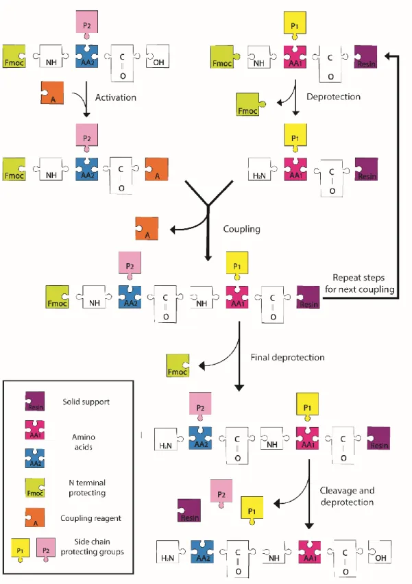
1.3 Solid Phase Peptide Synthesis
Solid phase peptide synthesis (SPPS), which was firstly illustrated by Bruce Merrifield in 1963 [23], is the most suitable method for small to medium size peptide synthesis. This method shows superiority upon other techniques, since it enables fast,
8
simple and economical peptide synthesis, easy functional group coupling, high yields and stability under different conditions. In SPPS, solid support that is also called the resin is one of the most important factors because all peptide chains are constructed on this support. During the peptide synthesis, firstly, peptides are covalently bounded to the beads of resin followed by general coupling procedure. This is based on the repetition of following steps: deprotection of the amino acids-washing-coupling of the amino acid-washing (Figure 1.4). The amino acid, which is unprotected at the C-terminus and protected at the C-terminus are coupled with the unprotected N-terminus of amino acid on the solid support. After each step, in order to remove the remaining chemicals, the beads are washed and filtered followed by the addition of acetic anhydride to block the side reactions. After completion of the synthesis, the peptide is cleaved by the acidic reagent such as trifluoroacetic acid (TFA) which transfers peptide from solid support to solution phase.
1.4 Peptide Amphiphile
A chemical compound, which has both hydrophobic and hydrophilic properties, is called amphiphile and these kind of molecules are called amphiphilic or amphipathic molecules. Detergents, soaps and lipoproteins are the most common examples of amphiphile molecules. Peptide amphiphiles (PA) that are also known as lipidated peptides or lipopeptides are the one of the most well-known amphiphilic molecules existing in living organisms. These molecules consist of a lipid tail which is connected to a peptide and they can be described as peptide based compound that forms self-assembly structure. These amphiphilic molecules can take part in signal transduction pathway, defense mechanism of organisms and protein-protein or protein-lipid interactions [24-28].
9
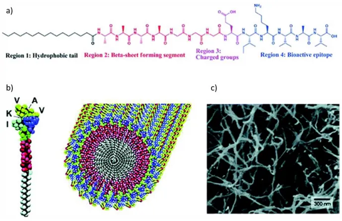
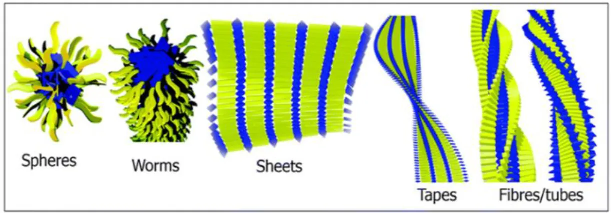
Peptide amphiphiles are comprised of multidomains, these domains can be categorized into hydrophobic and hydrophilic parts. In 2000s, it was explained that peptide amphiphiles (Figure 1.5) are composed of 4 domains. First domain is a hydrophobic tail whose hydrophobicity is one of the most important major forces which triggers the self-assembly of PA into well-defined structures. Second domain is the β-sheet forming region, which typically comprises of short peptides constructing via hydrogen bonds. Third domain has charged residues to increase the water solubility and form self assembled nano-architecture via pH or salt effect. Both type and the number of the charged amino acids are necessary to obtain desired solubility and avoid the aggregation of PAs at physiological medium. The presence and the strength of these noncovalent interactions such as electrostatic forces, hydrogen bonding, hydrophobic interactions, π-π stacking determine the various morphologies of the PA nanostructure such as fibers, micelles, nanotapes, nanotubes, nanosheets (Figure 1.6) and these nanostructures gather to form 3D networks. Finally, bioactive epitope that decide the functionality of the nanostructure can be used for further applications [13, 18, 29-35]. Moreover, bioactive epitope has effects on physical, chemical and mechanical properties of 3D formed network [7, 13, 24, 36]. In the literature, PA containing biomaterials with various epitopes are utilized for several purposes such as adhesion, bioimaging and sensing etc. [7, 23, 29-32, 37].
10
11
Figure 1.5 Schematic representation of self-assembly of PAs into nanofibers: a) Chemical structure of PA with four key chemical
entities. b) Molecular model of an active epitope-containing PA, their self-assembly into nanofibers, as well as c) SEM image of nanofibers [35]
12
Figure 1.6 Peptide amphiphile self-assembly supramolecular nanostructures.
Adapted from Ref. [18] with permission from The Royal Society of Chemistry.
1.5 Stimuli Responsive Peptide Systems
Stimuli responsive or smart/intelligent materials are unique systems which can modify themselves according to the environmental factors. These systems are sensitive to several external factors such as chemical, biological or physical stimuli (Figure 1.7) [38, 39].
Stimuli responsive biomaterials are used for different purposes such as diagnosis, imaging or sensing studies. Therefore, scientists have been developing several systems which can be liposomes, polymer nanoparticles, micelles, dendrimers, and inorganic nanoparticles made up of iron oxide, quantum dots, gold or metal oxide frameworks for a variety of biomedical and bionanotechnology applications [33].
Peptide based materials have been paid attention as stimuli responsive biomaterials due to their intrinsically bioactive, biocompatible, nontoxic and dynamic nature. In the literature, there are several stimuli responsive peptide containing systems designed for biotechnological applications such as drug delivery, tissue regeneration and sensing [13].
13
Figure 1.7 Categorization of physical, biological and chemical stimuli [39].
For instance, the Par-4 which is a pivotal protein for apoptotic pathways and C terminus of this protein is responsible for communicating with proposed effector molecules. The C terminus of Par-4 prefers to stay in monomeric form under physiological conditions; however, the coil coiled secondary structure is formed at low pH and temperature. It was reported that the native unfolded form of the C terminal residues affect the functions of cells and signaling pathways involved in several mechanisms. On the other hand, coil coiled structure is important in binding to effector molecules and forming complexes by the effect of external factors. These systems are effective for specific cancer types such as prostate cancer, various neurodegenerative disease such as Alzheimer's and Huntington's and HIV [40].
14
Furthermore, basic light triggered hydrogelation system was developed by Haines et al which [41] is the alternative of the toxic initiator materials. The 2 wt % solution of photocaged peptide MAX7CNB found in unfolded form in aqueous environment. The photocage was released after exposure to light between 260-360 nm and the peptides were self assembled into the β-hairpin structure which formed viscoelastic hydrogels. After in vitro analyses, it was proven that they possess nontoxic nature and allow adhesion and migration of the cells [41].
Moreover, Hartgerink et al synthesized pH responsive peptide amphiphile molecules that bear an integrin binding sequence RGD with different alkyl lengths [16]. The cysteine residue enables the system to form intramolecular crosslinks by disulfide bonds and a high molecular weight material is formed. They obtained the reversible nanostructure by changing the pH of the system which can be used for several biological and non-biological studies [16].
In another study, Beniash et al used peptide amphiphiles, with RGD sequence design that self assembled into 3D nanofibrillar networks by the aid of polyvalent and monovalent metal ions, which are incubated inside body fluid [42]. Changing the metal concentrations in the solution triggers the gelation of the system. During the gelation process, addition of cells to the solvent leads to the entrapment of cells inside the 3D matrix. This network enables the cell movement and proliferation during entrapment period and the survival of cells did not change for 3 weeks. This biomaterial has a potential to be used in regenerative medicine and cell transplantation studies.
15
Furthermore, Hartgerink et al studied biomineralization by pH induced self-assembly of PAs, where biomimicking biomaterials formed a fibrillar scaffold by reversible crosslinks at a specific pH range [34]. The thiols formed disulfides by oxidation and reverse reaction occured by the reduction of sulfide bond. In their study, suitable environment for direct hydroxyapatite mineralization was created, resulting in aligned composite materials which were similar to bone structure that has collagen fibrils and hydroxyapatite crystals.
Moreover, in another study, Jones et al used peptide conjugation with boronic acid that intrinsically had characteristic pH stimuli responsive features [43]. This system formed a nanoribbon above pH 5.5. Salt addition triggered the self-assembly of FF-boronic acid monomer and desired solubility was achieved by FF-boronic acid group. Furthermore, this assembly is disturbed by polyol addition since boronic acid and catechol forms a boronate complex and the gel structure turns into a homogeneous solution. This material can be a suitable candidate for therapeutics and drug delivery applications.
Another example of a pH based structural transformation was presented by Gologan group [44]. They used heptane bolaamphiphile building blocks to design a stimuli responsive biomaterial. At pH 8, these building blocks formed a helical ribbon conformation; however, at acidic pH, they had tendency to build crystalline tubules. The conversion from helical ribbons to tubules happened in one day; however, reverse transformation happened more slowly [44]. This material can be improved for controlled release studies. In another study, Yang et al designed hydrogelator Nap-FFGEY which forms a supramolecular hydrogel [45]. The hydrogel formation was controlled by a switch between kinase/phosphatase to manage the
16
phosphorylation and dephosphorylation of the peptide. They used liquid form of hydrogelator for subcutaneous injection and observed the hydrogel formation at injected area. This material is a possible candidate for future applications in in vivo models of bioengineering studies. In another study, Zhang et al used dipeptide as a hydrogelator which responded to ligand receptor interactions and showed chiral detection [46]. This kind of specific ligand receptor interactions can be a unique ability to designing hydrogels for biosensing applications [46].
1.6 Boronic Acid
Boronic acid, a member of organoboranes, is an alkyl or aryl substituted boric acid and it is. It has one alkyl substituent and two hydroxyl groups to satisfy the valence shells on the boron atom. In neutral form, it has sp2 hybridization and it has an empty p-orbital. This hybridization leads to obtain a trigonal planar geometry. Boronic acid can be obtained as secondary oxidation products of boranes. Their stability is higher than the first oxidation product, which is borinic acid and comparably less than third oxidation product that is boric acid. Boronic acid has several unique properties, which make it a desirable molecule for different purposes [47, 48].
Boronic acids are utilized as an intermediate and building block for important chemical synthesis reactions such as Suzuki coupling. It acts as a mild Lewis acid, which can accept lone pair electrons so it can react with strong Lewis bases such as fluoride, cyanide or phosphate anions. These properties make the boronic acid suitable material for homogeneous/heterogeneous and bulk/surface material sensing applications. Another unique ability of boronic acid is to form boronate ester via reversible covalent bonds with 1,2 or 1,3 cis-diols. It is a synthetic material that is
17
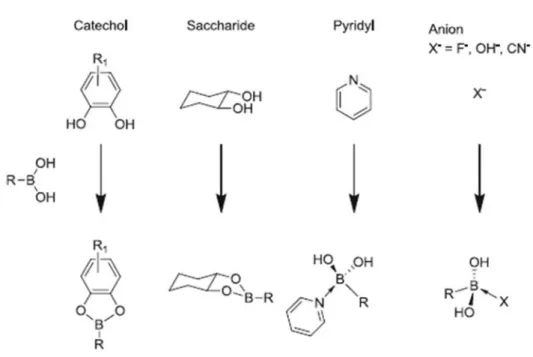
commonly used in biomedical and biotechnological applications due to being environmental friendly, its stability and biodegradability. The most extensively studied biological molecules reacting with boronic acid are saccharides, polysaccharides, nucleic acids, neurotransmitter dopamin and its derivative L-DOPA (Figure 1.8) [49]. The interactions of boronic acid with these molecules are used for detection, signaling pathway identification, enzyme inhibition, purification and cell/gene/drug delivery [47-52].
Although the exact reaction mechanism of boronate ester formation is not completely identified, formation of boronate ester from boronic acid and diol containing ligands are summarized in Figure 1.9 [53].
The boronic acid and their corresponding boronate ester represent sp2 hybridization
and trigonal planar electron domain geometry in neutral form (Figure 1.9a) and tetrahedral electron domain geometry and sp3 hybridization in anionic form (Figure
1.9b). The reaction product between neutral and conjugate base of boronic acid with diol (Figure 1.9c,d) and the thermodynamically preferred structure of the boronic acid or the boronate ester are determined by the pKa value of the boronic acid or the boronate ester. In sp2 hybridization, boron has an empty p orbital and the presence of Lewis bases leads to the production of anion in aqueous media. The pKa value of boronic acid conjugated systems changes between 4.5-10 depending on the substitution of the molecules. For biomedical applications, phenylboronic acid is widely used since phenyl group enables easy modification for the adjustment of pKa of the molecule [48, 51, 54].
18
The reaction tendency is controlled by the environment of the boronic acid such as pH, heat, solvent type, electric field, light and the analyte concentration and in addition to the substitutions of both boronic and ligand molecules. Although all these factors affect the boronic acid affinity, pH of the media and the substitution of the boronic acid are the most investigated parameters in the literature. Since boronate formation is only possible at specific pH range which is determined by the pKa values of the species, these values can be adjusted by the conjugation of boronic acid and diols. The diol/boronic acid reaction pH range has been studied to understand the
Figure 1.8 Boronic acid and its reaction with different molecules. Reproduced from Ref. [49] with permission from Springer.
exact interval. Different studies analyzed the suitable pH interval of boronic acid
(R-B(OH)2) and diol (H2L) interactions under two different conditions. First, if
19
pKa(R-B(OH)2) ≪ pKa(H2L), suitable range can be identified as a pKa(R-B(OH)2) < pH <
pKa(H2L). On the other hand, if pKa(R-B(OH)2) >> pKa(H2L), the crosslink formation is
allowed at pH = pKa R-B(OH)2 ± 1 [54, 55]. There are several diol molecules which can
react with boronic acid; however, saccharides, L-DOPA and Alizarin Red are the most commonly used ones as diol containing molecules for different purposes [49, 55-58]. Their properties, interaction with boronic acid and applications will be explained later in detail in further sections.
Figure 1.9 Multiple reaction mechanism of boronic acid. Reproduced from Ref. [51]
20
1.6.1 Saccharides
One of the most abundant biological molecules inside the body are the saccharides that play crucial roles in events such as cell adhesion, immune response, bacterial invasion and tumor metastasis. However, by noncovalent interactions, it is hard to achieve high efficient sugar sensing. Another challenge for sugar sensing is the selectivity. Although they have different configurations of stereocenters, they represent similar chemical structures so earlier sensors slog on distinguishing saccharide and their immediate sensing. To overcome these problems, new and unique enzyme or lectin (sugar binding proteins) inspired synthetic chemosensor systems have been designed, which have superiority to the biological sensors in terms of stability under biological conditions and cost effectiveness. Therefore, boronic acid containing systems are unique and crucial chemosensors that have been utilized as saccharide sensors. It forms five- or six-membered cyclic esters with saccharides. They can achieve sensing at low concentrations at sub-milimolar levels. However, the most important property of boronic acid that makes it unique for sugar binding is that boronic acid can distinguish the orientation and relative position of hydroxyl groups of sugar and show selective affinity towards different saccharides. For instance, boronic acid has higher affinity toward fructose among monosaccharides when it is in monoboronic form. For phenyl boronic acid, the affinity order is fructose > galactose > mannose > glucose among monosaccharides. However, boronic acid and sugar in different forms such as polymer and self assembled systems exhibit different affinities and due to multivalency they demonstrate higher affinity, which will be, exemplified on section 1.7 [49, 53, 59-64].
21
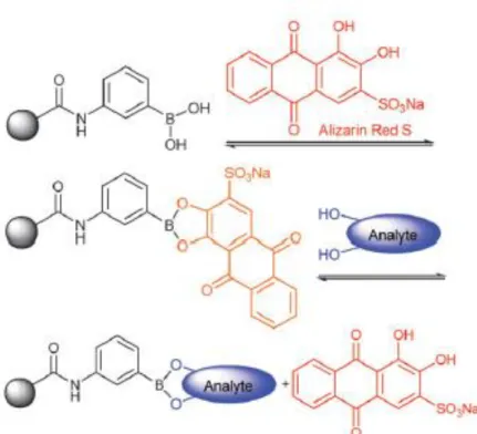
1.6.2 Alizarin Red-S
Alizarin Red-S (ARS) which is a well-known catechol dye can react with boronic acid. This reaction leads to change in fluorescence activity of ARS and solution color. Thus, ARS and boronic acid interaction can be applicable for the concentration detection of boronic acid, recognition of competitive binding of other diol containing molecules and analyzing the affinity of boronic acid towards various molecules, both quantitatively and qualitatively. This assay is based on the transition between fluorescence active compound formation with ARS and its displacement with the competitive diol (Figure 1.10) [50, 52, 57, 65-68]. ARS and boronic acid are both fluorescence inactive; however, their boronate ester complex exhibit fluorescent property. Moreover, boronic acid, ARS and their mixtures possess different colors. Therefore, the formation of boronate ester between boronic acid/ARS and the replacement of ARS with a diol can be monitored by the change in color and using the fluorescence spectroscopy. In general, boronic acid and diol interactions can be achieved at neutral to slightly alkali medium with highly strong covalent bond characteristics. However, ARS can also bind to boronic acid at slightly acidic medium. ARS/Boronic acid complex can be destroyed with the addition of molecules which can bind to boronic acid by displacing ARS. Conclusively, formation of boronic acid/ARS complex leads to an increase in fluorescence intensity of the solution. Fluorescence intensity will decrease when a diol that has higher affinity to boronic acid than ARS is introduced to ARS/boronic acid solution. As a result, ARS based fluorescence assay is a unique method for detecting boronate ester formation. This assay makes the identification of boronic acid and target compounds interaction possible for different systems [50, 52, 54, 65-69].
22
These fluorescence based detection system can be explained by two separate spectra. Firstly, the boronic acid and ARS interactions needs to be examined to understand their dual behavior. At constant boronic acid concentration, increasing ARS amount leads to an increase in fluorescence signal. For the triple component system, another diol containing molecule is introduced into the system. If the molecule has a higher affinity towards boronic acid, the fluorescence active complex (boronic acid/ARS) will dissociate due to the replacement of ARS by the diol. In the literature, this method is utilized generally for the detection of free boronic acid, bond formation and binding constants [50, 66-68].
Figure 1.10 Binding and analyte-mediated release of Alizarin Red-S with
hydrogel-bound boronic acid.Adapted from Ref. [65] with permission from The Royal Society of Chemistry.
23
Furthermore, UV-VIS spectroscopy is another tool for the detection of boronic acid using ARS dye. Boronic acid does not absorb in the UV-Vis region; while, ARS and ARS/boronic acid complex do absorb in UV-Vis region. Addition of boronic acid into the ARS solution leads to blue shift due to formation of boronic acid/ARS complex in the solution. On the contrary, destruction of this complex by adding other diols results in red shift [66].
1.6.3 L-DOPA
L-DOPA (Levo-DOPA) amino acid 3,4-dihydroxyphenylalanine (Figure 1.11) is a valuable molecule which has a wide range of potential applications. L-DOPA, that is called natural glue, is found in the secretion of mussels and responsible for adhesion. Nontoxic nature, quick adhesion process, high resistance to harsh conditions and stability in aqueous environment are the properties of this glue. Furthermore, wide range of temperatures and saline concentrations cannot interfere with adhesion and adhesion can be possible on several surfaces such as wood, metal and glass. However, L-DOPA loses its adhesion ability at a pH triggered by oxidation as opposed to reducing conditions. These properties make L-DOPA a suitable candidate for biomedical and biotechnological applications. Scientists have used the L-DOPA conjugated biomaterials as scaffolds, sensors, or surface coating materials. Its crosslink formation towards the surface are affected by the environmental conditions such as pH, antioxidant and electron withdrawing group functionalization which enables us to precisely control the adhesion [70-75].
In addition to its adhesion ability, DOPA plays an important role for neural system in the body. L-DOPA is a member of class that is called neurohormones like dopamine,
24
Figure 1.11 The chemical structure of L-DOPA
noradrenaline, L-tyrosine. Inside the body, there is a chain conversion between these molecules for instance, L-tyrosine is the precursor of L-DOPA, L-DOPA is converted into dopamine, and dopamine is the precursor of norepinephrine and epinephrine. All these molecules play a vital role for vital reaction inside the body such as heart rate/blood pressure adjustment and Parkinson’s disease/hypertension/schizophrenia treatment. For instance decreasing the level of dopamine in the central nervous system can lead to Parkinson’s disease; however, L-DOPA is used as a Parkinson drug. Although dopamine itself cannot penetrate through the brain barrier, its precursor L-DOPA can pass through the blood-brain barrier and it is converted into dopamine in the blood-brain. L-DOPA, Dopamine, epinephrine noradrenalin and DOPA-P, all have the catechol functional groups which can form a crosslinked structure with boronic acid. The formation of crosslinked structure can be used for real time in vivo molecule sensing, concentration determination and drug delivery. Similar to saccharide sensing, boronic acid have different affinities toward the catechol containing molecules. For instance, noradrenaline ≥ dopamine ≈ L-DOPA > catechol > DOPA-P is the order of affinity towards phenyl boronic acid; however, this order can be changed by the substitution of boronic acid or analyte molecules [70, 76-78].
25
1.7 Application Of Boronic Acid
The key point of boronic acid containing systems for biomedical applications is that it is controllable and forms strong reversible crosslinks with diols which is commonly found in biological molecules. The ability to form reversible and spontaneous interactions, biocompatibility and nontoxic nature are the desired properties of synthetic chemical systems for sensing, diagnostic and proteomics studies which is summarized in Figure 1.12 [50, 64, 79, 80]. However, single molecule systems are feeble, easy to break, and sometimes hard to detect. On the other hand, multivalent systems are much stronger and lean on each other. These systems exhibit superiority about the binding interactions, activity and availability. Therefore, multivalent systems of boronic acid can be used to prepare nano, micro and bulk architectures by using bottom up approach via the self-assembly or polymeric systems for various purposes [53].
In the literature, boronic acid conjugated polymer systems are frequently used as self-healing biomaterials due to their stimuli response and high self recovery capability [79-81]. For instance, boronic acid or diol containing PEG based hydrogel networks were utilized by the aid of boronate ester formation. PEG based mechanically controllable hydrogels that demonstrated high self-healing properties at neutral pH were obtained by Anderson group [79]. The rapid structural recovery was tested by rheological analysis and physical deformation, 100% recovery were exhibited by the reversible covalent bonds between cis-diol and boronic acid moieties [79]. This system showed highly suitable characteristics for tissue engineering as a 3D matrix or drug encapsulation network for the controlled drug release. This kind of examples can be multiplied under physiological conditions with
26
different building blocks and bioactive epitopes. However, at acidic pH the maintenance of self-healing properties of boronic acid containing systems is still problematic.
Sumerlin group published a study that achieved similar self-healing property at acidic pH [82]. They used an intramolecular coordination between boron in 2-acrylamidophenylboronic acid and the carbonyl oxygen that increase the stability of boronate ester structure. They used dopamine acrylamide copolymer and polyvinyl alcohol as diol containing groups. These interactions widen the pH range of the self-healing interval of hydrogels from neutral to acidic environment. The recovery ability of the hydrogels was tested by physically cutting the gel into two pieces. Then, the pieces were put together in contact and almost 1 h later they returned to the original appearance and no scar was observed. Moreover, rheological analyses were also used to understand the self-healing capacity and results demonstrated that both at acidic and neutral pH, hydrogels showed high self-recovery behavior.
Beside this, boronic acid conjugated systems have been recently used for controlled drug release or targeted drug delivery. Briefly, drug delivery systems have been improved to keep the drug release in desired level, sustain desired drug concentration at desired location, decrease side effects of toxic chemicals, and enhance bioavailability [83].
For biomedical and biotechnological applications, biodegradable, biocompatible, nontoxic, multifunctional, shape/size controlled intelligent drug delivery and controlled drug release systems have been developed [48, 50].
27
Boronic acid which is a smart molecule for the controlled drug release systems due to its intrinsic dynamic nature, controllable swelling property and tunable chemical interactions has been widely utilized [84]. Previously, boronic acid based systems were used for controlled insulin release according to glucose level. Kataoka et al [85] investigated smart insulin release system by utilizing P(NIPAM-co-3-AAPBA) hydrogels. This gel system formed a surface barrier layer in the deficiency of glucose and the insulin was encapsulated inside the gel matrix. However, increasing glucose concentration disrupted the layer and insulin can be released from the matrix.
Figure 1.12 Several applications of boronic acid biomaterials [80].
As a result, glucose sensitive on-off release system was designed for self-regulated insulin release. In another study, nearly monodispersed microgels decorated with 3-aminophenylboronic acid were synthesized by Zhang et al [86]. Presence of glucose
28
leads to an increase in mesh size in alkali solution at room temperature because the stability of phenyl boronate ions were supported by the glucose/boronic acid complex and the release was accelerated [86].
Unlikely, in order to design glucose-responsive insulin-delivery system, Siddiqui group [87] proposed the system to increase glucose selectivity rather than fructose, which shows higher affinity towards boronic acid. 2-formyl-3- thienylboronic acid (FTBA) and 4-formylphenylboronic acid (FPBA) decorated chitosan nanoparticles were synthesized and the results showed that FTBA represented relatively higher affinity towards glucose and 1:1 ratio of boronic acid:chitosan showed higher glucose selectivity which can be used as a glucose responsive system that has a possible usage for insulin release under physiological conditions [87]. Furthermore, boronic acid has been used for cancer drug delivery systems. Cancer environment has slightly acidic medium than the physiological pH and boronic acid represented pH based stimuli responsive behavior. Therefore, boronic acid materials have been frequently used for cancer drug delivery [50]. For instance, dual responsive boronic acid and catechol conjugated crosslinked micelles have been demonstrated by Lam’s group [50]. The polymers with boronic acid and catechol built self-assembling structures and intramolecular in situ bond formation of boronic acid/catechol was obtained. Then, drug got encapsulated inside the micelles at physiological pH, where boronate ester crosslink were preferred. However, release of drug accelerated at acidic pH or presence of compete diols due to weakening of the boronic acid-catechol bonds. Therefore, the nanoparticles maintained drug encapsulation in blood circulation; nevertheless, release can be possible at tumor microenvironment that possesses a more acidic ECM. Another example of pH and ligand concentration
29
triggered selective binding was studied on MCF-7 cancer cells by Liu et al [88]. Cells have the glycoproteins on the cell surfaces and by the sialic acid moieties cells can form a reversible bonds with grafted 3-APBA. At pH 6.8, boronic acid had higher tendency to bind sialic acid onto cell surface, which is the approximate pH value of the cancer microenvironment. Furthermore, existence of glucose blocked the sialic acid/boronic acid complexation because boronic acid prefers to bind glucose than sialic acid at physiologic pH. Therefore, the bond between cell and 3-APBA formed at lower pH were released reversibly at higher pH and glucose concentration.
Beside drug delivery systems, there is a boronic acid containing drug called Bortezomid (BTZ) (Figure 1.13) which is used as a proteasome inhibitor [89, 90]. It is composed of N-protected dipeptide with boronic acid that is placed on the C-terminus instead of the carboxylic acid.
However, efficiency of BTZ is not sufficient against several solid tumors. Therefore, scientists have been designing new systems to enhance BTZ activity as a pH triggered smart biomaterial [90]. For example, Cheng group [90] presented a study by using boronic moieties of BTZ. They designed a dendrimer with catechol groups, the boronic acid moieties on the BTZ were conjugated by boronate ester formation, and these compounds were placed in the interior of the nanostructure at physiological pH. However, in vivo and in vitro studies showed that BTZ release was accelerated at tumor microenvironment, which has a slightly acidic medium.
Therefore, designed systems improved the biocompatibility, stability, and blood circulation time of drug and decreased non-specific cellular uptake and these
30
properties make pH triggered drug delivery system a promising candidate for future studies [89-93].
Moreover, Lin et al [64] used boronic acid for selective saccharide sensing. In their study, magnetic iron oxide nanoparticles (Fe3O4 MNPs) modified with
aminophenylboronic acid were designed to separate glycoproteins from the protein mixtures. Boronic acids on the particles formed complexes with glycoproteins such as cellulose and ovalbumin; however, they did not interact with bovine hemoglobin, bovine serum albumin and lysozyme which are non-glycoproteins. After fast and selective binding to glycoproteins, nanoparticles were taken apart from the solution by magnetic separation with glycoproteins. This system was specifically tested to separate ovalbumin from the egg white sample [64].
Figure 1.13 The chemical structure of Bortezomid
Moreover, Shen et al utilized boronic acid for blood sugar detection without any enzymatic reaction [94]. Boronic acid modified C-dots based fluorescent sensors were designed to detect the glucose concentration in the blood. Boronic acid based
31
systems are generally preferred due to easy usage, high efficiency, and selectivity for blood sugar quantification. This chemosensor exhibited selectivity from 9 to 900 μM glucose concentration that exhibited higher sensitivity than the previous fluorescent based studies of boronic acid. Addition of glucose into boronic acid functionalized C-dot systems led to change in fluorescence intensity because diol moieties of glucose bind to boronic acids on the C-dot covalently which resulted in a decrease in distances between C-dots and hence, fluorescence quenching was observed [94].
32
Chapter 2
Material And Methods
2.1 IntroductionTargeted drug delivery in cancer remains one of the major clinical problems. To solve this problem, scientists have tried to find differences between cancerous and healthy tissues. Cancer environment possesses a slightly acidic pH than the healthy tissue; therefore, stimuli responsive materials have drawn attention for targeted drug delivery applications. Boronic acid is one of the most important stimuli responsive molecules which can form a bond with vicinal diols such as saccharide or catechols reversibly. While the bond formation is triggered in neutral pH, these bonds are weakened under slightly acidic conditions [33, 50, 51].
In this study, boronic acid and DOPA conjugated peptide amphiphiles were designed to obtain a controllable drug release system. The oppositely charged boronic acid/DOPA conjugated peptide amphiphiles were mixed to form nanofibers by molecular self- assembly via noncovalent interactions and these molecules are placed
33
on the nanofiber surface. Due to charge neutralization, mixing at higher concentrations forms aggregates and hydrogel formation occurs.
Besides these, boronic acid/DOPA molecules formed intramolecular reversible crosslinks which increase the strength and self-healing capability of hydrogels at neutral pH. These hydrogels represent suitable behavior for controlled drug release systems.
Furthermore, boronic acid/DOPA crosslinks demonstrate pH triggered system and at mildly acidic conditions, the interactions weaken (Scheme 2.1). In this study, Doxorubucin-HCl (Dox) was selected as a release molecule which is a well-known cancer drug that is fluorescence and UV-Vis active. Thus, quantitative and qualitative analysis of Dox can be performed by spectroscopic methods [95, 96]. These make Dox a suitable candidate for various drug release systems.
To test the drug encapsulation and pH dependent release profile, Dox was encapsulated inside the hydrogel network and their behavior was observed at pH 7.4 and 5.5. Moreover, DOPA/E3-PA, boronic/K3-PA and E3/K3-PA groups were also
analyzed as a control group to distinguish the exact effect of the boronic acid/DOPA interaction.
34
35
2.2 Experimental Section 2.2.1 Chemicals and Reagents
Chemicals and reagents that are used during synthesis of peptide amphiphile molecules, hydrogels preparation, and their analysis can be seen below.
2.2.2 Chemical and Reagents to Synthesis Peptide Amphiphile Molecules
[4-[α-(2’,4’-dimethoxyphenyl) Fmoc-aminomethyl] phenoxy] acetamido norleucyl-MBHA resin (Rink amide norleucyl-MBHA resin), 2-(1H-Benzotriazol-1-yl)-1,1,3,3 tetramethyluronium hexafluorophosphate (HBTU), other protected amino acids were purchased from NovaBiochem. 4-carboxylphenylboronic acid, N,N-Diisopropylethylamine (DIAE), dichloromethane (DCM), dimethylformamide (DMF), doxorubicin hydrochloride, piperidine, acetic anhydride, trifluoroacetic acid (TFA) were purchased from Fisher, Merck, Alfa Aesar or Sigma Aldrich. Other chemicals used in characterization such as acetonitrile, 3,4-Dihydroxy-9,10-dioxo-2-anthracenesulfonic acid sodium salt (Alizarin Red S), uranyl acetate, 4-(2-hydroxyethyl)-1-piperazineethanesulfonic acid (HEPES), 2-(N-Morpholino)ethanesulfonic acid, 4-Morpholineethanesulfonic acid (MES) were purchased from Sigma Aldrich and Merck.
2.2.3 Synthesis and Characterization of PAs
By using solid phase peptide synthesis (SSPS) Boronic Acid-EEEGAVVK-(Lauryl)-Am (Boronic-PA), Ac-EEEGAVVK-(Lauryl)-Acid-EEEGAVVK-(Lauryl)-Am (E3-PA)
Lauryl-VVAGKKK-DOPA (Lauryl-VVAGKKK-DOPA-PA), Lauryl-VVAGKKK (K3-PA), was synthesized on the solid
36
(Acetonide)-OH were used as a precursor of boronic acid and DOPA respectively. For amino acid coupling 2 equivalents of F-moc protected amino acid, 3 equivalent of N,N-diisopropylethylamine (DIEA) and 1.95 equivalents of HBTU in DMF were used. The coupling incubation time was 6 h then to acetylate the unreacted amine groups and block the formation of side sequences 10% acetic anhydride/DMF solution had been added for 30 min before Fmoc protecting groups were cleaved by 20% piperidine/dimethylformamide (DMF) solution for 20 min. Trifluoroacetic acid (TFA): triisoproplysilane (TIS): water mixture was administered for 2.5 h in the ratio of 95:2.5:2.5 to cleave the peptide from solid support and transfer toward liquid phase. After each step, the solid support was washed with DCM and DMF to remove the remaining chemicals. To obtain cleaved peptide from solid support, DCM washing was performed and then rotary evaporatory was used to remove excess TFA and DCM. The remaining viscous peptide solution was precipitated by using diethyl ether at -20 °C overnight. The precipitated white pellets were collected by centrifugation followed by dissolving in water then frozen at -80 °C and lyophilized until all ice particles were removed.
The synthesized peptide amphiphiles were characterized by an Agilent 6530 series reverse phase high performance liquid chromatography (HPLC) equipped with high resolution Quadropole Time of Flight (Q-TOF) mass spectrometer with electron spray ionization source (ESI). One mg/mL K3-PA and DOPA-PA molecules
prepared in double distilled water (dd-water) and as a column Agilent Zorbax Extend-C18, 3.5 µm 80A (100 x 4.6 mm) was used as the mobile phase water (0.1% formic acid)/acetonitrile (0.1% formic acid) gradient was performed. On the other hand, an Agilent Zorbax 300SB-C18, 3.5 µm (100 x 4.6 mm) column and water
37
(0.1% NH4OH)/acetonitrile (0.1% NH4OH) gradient was used as a mobile phase for E3-PA and Boronic-PA characterization. Absorbance at 220 nm was used for chromatograms.
2.3.4 Preparative High Performance Liquid Chromatography (Prep-HPLC)
To purify the E3-PA and K3-PA, Agilent 1200 preparative reverse-phase HPLC
system was performed and 10 mg sample was dissolved in 1 ml water (0.1% TFA or NH4OH). For positively charged peptide, Zorbax prepHT 300CB-C8 column
with a water/acetonitrile (0.1% TFA) gradient was used followed by HCl treatment for TFA removal. Furthermore, for negatively charged E3-PA Gemini
5µ C18 110A column with size of 100 × 21.20 mm and water/acetonitrile (0.1% NH4OH) gradient was used. DOPA-PA was pure enough for usage; so in order to
remove excess TFA, only HCl treatment was performed. Moreover, to remove the excess TFA and impurities, dialysis was performed for 2 days.
2.3.5 Zeta Potential Measurements of PAs
2 mM K3-PA, E3-PA, DOPA-PA, boronic-PA and their coassembled combinations
were mixed and the solutions were incubated at pH 7-8 at room temperature in order to observe the charge distribution of negatively and positively charged peptide solutions and their coassembled structures. Further, they were diluted to 250 µM with water and measured by Malvern Nano-ZS Zetasizer by using a dip cell electrode in quartz cuvettes. Standard deviations were calculated from the mean of the data of repetitive experiments (n≥3).
38
2.3.6 Transmission Electron Microscopy (TEM)
2 mM DOPA-PA/boronic-PA, DOPA-PA/E3-PA, boronic-PA/K3-PA and K3/E3-PA
solutions were prepared in water and pH was adjusted to 7.4 (physiological pH) and incubated for 2 h at room temperature. Then the samples were diluted to 125 µM and 10 µL samples were dropped on the copper TEM grid and incubated for 5 min. Solution was removed from the grid and the grid was stained with uranyl acetate (2% (w/w)) for 2 min. After withdrawal of excess dye and washing with dd-water, grids were left to dry at room temperature for 15 min. The TEM instrument that is used for morphological characterization of coassembly systems was FEI Tecnai G2 F30 worked at 100 keV.
2.3.7 Scanning Electron Microscopy (SEM)
Morphological characterization and structural analysis of peptide amphiphile hydrogels were monitored by FEI Quanta 200 FEG scanning electron microscope (SEM) instrument equipped with an ETD detector. 10 mM 20 µL oppositely charged peptide amphiphiles were mixed on the wafer after 30 min incubation at room temperature and for dehydration steps, the sample was incubated in 20%, 40%, 60%, 80% ethanol for 20 min in each solution and finally in 100% ethanol solutions for 3 h to prevent shrinkage of the gels. Tousimis Autosamdri-815B critical-point-drier (CPD) instrument enables to dry dehydrated form of nanofibers gels without damaging the 3D network. Before SEM imaging, samples were sputter coated with 5 nm of a gold-palladium alloy.
2.3.8 CD Analysis
39
2 mM stock solutions of individual PAs were prepared and sonicated. Then oppositely charged PAs were mixed at 1:1 ratio and incubated at room temperature for 1 h. Next, the samples were diluted to 125 µM at physiological pH and pipetted gently not to damage the self assembled structures. Solutions were placed into 1 mm quartz cuvettes and measured by Jasco J-815 CD Spectrophotometer, whose spectra is between 190 to 300 nm with 0.1 nm data pitch, 4 sec DIT, 1 nm band width, 100 nm/min scanning speed with standard sensitivity. For data analysis, the results were converted as mean residue ellipticity and the unit was transformed to deg.cm2.dmol.
2.3.9 ARS Based Spectroscopic Measurements
Formation of boronic acid diol bonds cannot be characterized by direct methods for this study. In the literature examples, instead of direct methods, indirect indicator assays are frequently used to determine the boronic acid-diol interactions. ARS dye is one of the most crucial catechol dye used to identify the interaction between diol groups and boronic acid. As the amount of boronic acid-ARS complex increased, fluorescence intensity was also seen to be increased [65.66].
In our study, it was predicted that boronic acid and ARS forms a fluorescence active complex at neutral and mildly acidic solution. At pH 7.4, introduction of DOPA-PA into solution will break the fluorescence active complex and fluorescence intensity of the systems decreases at neutral conditions. However, at pH 5.5, DOPA-PA addition does not affect the fluorescence active complex since DOPA affinity towards boronic acid at this pH is less than its affinity at neutral conditions which is illustrated in scheme 2.2.
40
41
Samples were prepared by mixing 1.5 mM boronic-PA and 100 µM ARS solutions in HEPES or MES buffers and gently pipetting for providing total entanglement. After 30 min incubation, DOPA-PA molecules prepared in different concentration values, were introduced to the solutions and incubated for 1h. To eliminate the side signals and plate emission, solutions without boronic acid were used as a blank solution. Excitation wavelength was chosen as 440 nm and fluorescence intensity was measured by microplate reader.
2.3.10 Rheological Measurements
To investigate the viscoelastic and self-healing (thixotropic) properties of boronic-PA/DOPA-PA, boronic-PA/K3-PA, DOPA-PA/E3-PA and E3/K3-PA hydrogels,
Anton Paar Physica RM301 Rheometer equipped with a 25 mm parallel plate was used. 10 mM positively and negatively charged peptide amphiphiles were mixed in 1:1 ratio to obtain the charge neutralization on the stage. Total volumes of PAs were determined as 300 µL with 500 µm measuring distance from the stage. After achieving gel stability, time dependent rheological analyses were started while strain (0.01%) and angular frequency (10 rad s−1) were kept constant.
By thixotropic analysis, the differences between self-healing properties of boronic/DOPA-PA group and control groups were analyzed. This test composed of 3 phases, load phase was the first step which was applied for the stabilization of gel, second phase was the decomposition phase. In this phase, to weaken the interaction and damage the gel structure, the strain was enhanced logarithmically from 0.01 to 1000% in 1 min. In the third and final phase, that was the recovery phase, gel tried to heal itself at 0.01% shear rate and 10 rad/min angular frequency values.
42
2.3.11 Release Studies
For cancer drug delivery, the pH differences between the cancerous (slightly acidic pH) and the healthy tissue (neutral pH) were taken into consideration and pH triggered controlled drug release systems were designed. In this study, 10 mM oppositely charged peptide amphiphile and Dox solutions were prepared at physiological pH values. Firstly, positively charged peptide was mixed with Dox solution, vortexed and ultrasonicated to mix the solutions for 5 min. Further, solutions were transferred to cuvettes on the negatively charged peptide solutions by micropipetting in order to form hydrogels via charge neutralization (Figure 2.3). After 12 h incubation at 37 ºC, HEPES buffer (pH 7-7.5) and MES buffer (pH 6-5.5) solutions were gently added onto the gels. As mentioned earlier, Dox molecule is a UV-Vis active molecule and by preparing a standard calibration curve, the amount of the drug released to the buffer was quantified by UV-Vis Spectrophotometer – Cary 100 Bio instrument. Measurements at average time points were taken for 24 h.
43
Chapter 3
Results And Discussion
In this study, peptide amphiphile-conjugated systems were used as an alternative to polymeric systems which are a promising platform due to their biocompatible and biodegradable nature. These PA nanostructures were tailored to exhibit sol-gel transition behaviors in response to external stimuli. This gel like behavior was a result of hydrophobic interaction of carbon tail, electrostatic interactions of oppositely charged residues and hydrogen bonding of backbone [13]. The aim of this study was to form coassembly nanostructures by using oppositely charged boronic acid and DOPA conjugated PA by noncovalent interactions where boronic acid/DOPA units stay on the exterior site of the nanofibers (Figure 3.1). In addition to noncovalent interactions, boronic acid and DOPA moieties present on the peptide nanostructure surface form reversible covalent complexes at physiological pH, which can increase the hydrogel strength, self-healing capacity and entrapment of doxorubicin inside the hydrogel network. However, at acidic pH, these interactions weaken (Figure 2.1) and doxorubicin release accelerates at tumor site.
![Figure 1.7 Categorization of physical, biological and chemical stimuli [39].](https://thumb-eu.123doks.com/thumbv2/9libnet/5617086.111153/26.892.211.741.136.586/figure-categorization-physical-biological-chemical-stimuli.webp)
![Figure 1.9 Multiple reaction mechanism of boronic acid. Reproduced from Ref. [51]](https://thumb-eu.123doks.com/thumbv2/9libnet/5617086.111153/32.892.239.748.490.958/figure-multiple-reaction-mechanism-boronic-acid-reproduced-ref.webp)